
Phytochemical, Antimicrobial and Anti-Inflammatory Studies of Zingiber Nimmonii (J. Graham) Dalzell (Zingiberaceae)
 , Valukattil Ponnachan Thomas2, Jinu John3 and Rogimon Plammoottil Thomas1*
 1Department of Botany, CMS College Kottayam (Autonomous), Kerala, India
2Department of Botany, Catholicate College Pathanamthitta, Kerala, India
3Department of Biotechnology, CMS College Kottayam (Autonomous), Kerala, India
Corresponding author E-mail: rogimon@cmscollege.ac.in
DOI : http://dx.doi.org/10.13005/bbra/3532
Download this article as:
![]()
The present study aimed to investigate the bioactive potential and phytochemical composition of the rhizomes of Zingiber nimmonii (J. Graham) Dalzell, a relatively unexplored species within the Zingiberaceae family. This species, endemic to peninsular India has been traditionally used in indigenous medicine owing to its rich content of secondary metabolites. Phytochemical constituents were extracted from powdered rhizomes using ultrasonic-assisted extraction techniques. The antimicrobial activity of the extracts was evaluated by the agar well diffusion method, while anti-inflammatory effects were assessed through the in vitro sheep red blood cell (SRBC) membrane stabilization assay involving both heat-induced and hypotonicity-induced hemolysis. Preliminary phytochemical screening of petroleum ether and methanolic extracts revealed the presence of flavonoids, reducing sugars, terpenoids, wax and mucilage, carbohydrates, steroids, and amino acids. The antimicrobial assay conducted against four bacterial strains (both Gram-positive and Gram-negative) and two fungal strains demonstrated notable zones of inhibition for Bacillus cereus, Staphylococcus aureus and Candida albicans. The extracts further exhibited significant anti-inflammatory activity by stabilizing erythrocyte membranes and inhibiting hemolysis under both heat and hypotonic stress conditions.
KEYWORDS:Anti-inflammatory; Antimicrobial properties; Phytochemistry; Traditional medicine; Ultrasonication; Zingiber
Introduction
The Zingiberaceae family includes nearly 50 genera and approximately 1,600 species, comprised mainly of aromatic, perennial herbs with tuberous rhizomes, widely distributed across Africa, America, and Asia.1 In regions such as Kerala, these plants are highly valued for their diverse roles in traditional medicine, culinary applications, and religious practices, with the rhizome being the principal part utilized in both native and endemic species. Zingiberaceae rhizomes are recognized for their notable antibacterial and biological properties, supporting their widespread use in pharmaceuticals and food products.2-4
Contemporary studies reveal at least fourteen pharmacological activities attributed to this family, including anti-inflammatory, antioxidant, antifungal, gastroprotective, analgesic, neuroprotective, and renoprotective effects, which substantiate their long-standing use in systems of traditional medicine such as Ayurveda, Chinese, Japanese, and Unani practices5,6.The genus Zingiber, containing about 85 aromatic species native to East Asia and tropical Australia, is particularly valued for its volatile oils and chemical diversity, with more than 447 compounds so far isolated and classified.7-9 Despite these findings, systematic explorations of chemical constituents across the genus remain scarce.
Zingiber nimmonii (Z.nimmonii), one of the least studied species, is endemic to the Peninsular India and has been classified as threatened, highlighting the importance of conservation efforts for both biodiversity and traditional knowledge.10,11 Ethnobotanical and pharmacological research on Z. nimmonii and related species continues to inform sustainable bio resource utilization, aids in the preservation of indigenous practices, and supports the discovery of new therapeutics.12
The rhizomes are fleshy, light yellow with a purplish tinge, having a mild aroma. The height of leafy shoots are up to 110 cm. Size of the leaves are 32-40 X 8-12 cm with oblong-lanceolate in shape, almost sessile. The condensed spike inflorescence is 7 cm directly emerging from the rhizome on short stalk of about 6cm. Flowers are creamy yellow, with a purple spotted lip. Fruits are fleshy capsules, dehiscing into three on maturity, exposing the bright red interior with black seeds covered by white aril.11
Materials and Methods
Collection of plant material
Rhizomes of Zingiber nimmonii were collected between September and October 2023 from Vellarimala, Chooralmala, in the Wayanad District of Kerala (Latitude 11.480778; Longitude 76.167670). The collected specimens were authenticated through expert consultation, and a voucher specimen (No. 11058) was deposited in the herbarium at Catholicate College, Pathanamthitta (CATH) Kerala.
Preparation of extracts
Rhizome samples were thoroughly washed with distilled water to remove surface contaminants, shade-dried at ambient temperature for 7 days, and ground into fine powder using an electrical grinder. Ultrasonic-assisted extraction (UAE) was performed as follows: 10 g of powdered rhizome was extracted with 400 mL of hexane in a conical flask using a probe sonicator (20 kHz, 250 W) maintained at 25 ± 2°C for 10 minutes. The mixture was filtered through Whatman No. 1 filter paper, and the residue was re-extracted with fresh 400 mL hexane under identical conditions. All hexane extracts were pooled together. The remaining residue was subsequently extracted with 400 mL of 90% ethanol using the same UAE conditions. This ethanol extraction was repeated twice with fresh solvent, and all three ethanol extracts were pooled separately. The pooled extracts from each solvent were concentrated under reduced pressure at 40°C using a rotary evaporator (Büchi, Switzerland). The resulting concentrated extracts were stored at 4°C in amber glass vials until further phytochemical and biological analyses.
Analysis of phytochemical constituents
Phytochemical screening of Zingiber nimmonii extracts was carried out using established protocols described by Harborne.13 These methods were employed for the identification of phytochemical constituents present in both hexane and ethanol extracts.
Evaluation antimicrobial property
The antimicrobial activity of the extracts was evaluated using the agar well diffusion technique against four bacterial strains: Klebsiella pneumoniae, Escherichia coli, Staphylococcus aureus and Bacillus cereus, as well as two fungal strains, Candida albicans and Aspergillus niger. All these microorganisms were isolated from natural sources and were characterized by biochemical and molecular studies at Doctor John’s Biotech Centre for Research and Development, Kottarakkara. Mueller Hinton Agar (MHA) served as the growth medium. Extract samples were dissolved in DMSO at a concentration of 1 mg/ml, and 10 µL and 20 µL aliquots were tested. DMSO was included as a negative control, while antibiotic discs of ampicillin (10 μg) and tetracycline (30 μg) were employed as positive controls for bacteria; clotrimazole and nystatin discs were used for fungi.
Each bacterial suspension was evenly spread onto MHA plates using sterile swabs. Wells of 6 mm diameter were aseptically bored into the agar, and the respective samples were introduced. Plates were left at room temperature for 2 hours in a laminar airflow chamber to allow for sample diffusion, then incubated at 37°C for 48 hours. Post-incubation, the diameters of the zones of inhibition were measured in millimetres using an electronic vernier caliper. All experiments were performed in triplicate, and results were reported as mean ± standard deviation.14
Anti-inflammatory studies by SRBC membrane stabilization assay
Preparation of Sheep Red Blood Cells (SRBC) suspension
Fresh whole sheep blood from vein was collected from a local butcher shope in sterile bottles and immediately mixed with an equal volume of sterile Alsever’s solution (containing 2% dextrose, 0.8% sodium citrate, 0.05% citric acid, and 0.42% NaCl). The sheep red blood cells (SRBCs) were separated by centrifugation at 3000 rpm for 10 minutes, followed by three washes with isosaline (0.85% PBS, pH 7.2). Finally, the washed cells were resuspended in isosaline to prepare a 10% v/v suspension based on packed cell volume.
Hypotonicity induced hemolysis
To prepare the stock solution, the alcoholic extract was dissolved in sterile distilled water at a concentration of 1 mg/mL. For the assay, 5 mL of hypotonic solution containing varying concentrations of the extract (25, 50, 100, and 200 μg/mL) were aliquoted into centrifuge tubes. Parallel tubes containing 5 mL of isotonic solution at the same concentration ranges were also prepared in duplicate. Control tubes (5 mL) consisted of either the vehicle (distilled water) or diclofenac sodium at corresponding concentrations as a reference standard.
To each tube, 0.1 mL of the erythrocyte suspension was added and mixed gently. The mixtures were incubated at 37°C for 1 hour, after which they were centrifuged at 3,000 rpm for 5 minutes. The hemoglobin content in the supernatant was measured spectrophotometrically at 540 nm (Systronics UV-Visible spectrophotometer). Hemolysis in distilled water was taken as 100%, and the percentage inhibition of hemolysis by the extracts was calculated accordingly.
% Inhibition of hemolysis = [1 – (A2 – A1) / (A3 – A1)] × 100
Where: A1 = absorbance of test sample in isotonic solution; A2 = absorbance of test sample in hypotonic solution and A3 = absorbance of control in hypotonic solution
Heat induced hemolysis
The extract and standard drug were dissolved in phosphate-buffered saline (PBS), and 5 ml aliquots at varying concentrations (25, 50, 100, and 200 μg/ml) were prepared in triplicate in separate tubes. PBS (5 ml) served as the vehicle control. To each tube, 0.1 ml of the SRBC suspension was added and the contents were gently mixed. The tubes were then incubated at 54°C for 20 minutes in a temperature-controlled water bath, while a parallel set was incubated at -10°C in a refrigerator for 20 minutes. Following incubation, the tubes were centrifuged at 4,000 rpm for 5 minutes, and the hemoglobin concentration in the supernatant was determined by measuring absorbance at 540 nm using a UV-Visible spectrophotometer (Systronics). The percentage inhibition of hemolysis by the extract and standard drug was calculated using the following equation.15
Percentage inhibition of Haemolysis=1-[(A2-A1)/(A3-A1)] ×100
Where A1 = absorbance of test sample unheated, A2 = absorbance of test sample heated and A3 = absorbance of control sample heated.
Results
Preliminary phytochemical studies
Preliminary qualitative analysis revealed the presence of flavonoids, reducing sugars, terpenoids, wax & mucilage, carbohydrates, steroids, and amino acids in the rhizome of Zingiber nimmonii (Table 1).
Table 1. Qualitative phytochemical analysis of Z. nimmonii
| Sl.
No. |
Phytochemicals | Test | Hexane Extract | Ethanol Extract |
| 1 | Alkaloids | Dragendroff’s test | – | – |
| 2 | Tannin | Braymer’s test | – | + |
| 3 | Flavonoids | Shibata’s reaction test | + | + |
| 4 | Carbohydrates | Molisch’s test | – | + |
| 5 | Reducing Sugar | Fehling’s test | + | + |
| 6 | Saponin | Foam Test | – | – |
| 7 | Cardiac glycoside | Keller-Killani test | – | – |
| 8 | Anthraquinone | Borntrager’s test | – | – |
| 9 | Steroids | Liebermann-Burchard Test | – | + |
| 10 | Terpenoids | Salkowski’s Test | + | + |
| 11 | Amino acid | Ninhydrin test | – | + |
| 12 | Wax and Mucilage | Alcohol test | – | + |
‘+’ indicates the presence of phytochemical constituents.
‘-’ indicates the absence of phytochemical constituents.
Antimicrobial potential of Zingiber nimmonii extract
The antimicrobial activity of the rhizome extracts was assessed using the agar well diffusion method. Among the tested microorganisms, the petroleum ether extract exhibited the highest zones of inhibition against Bacillus cereus (8.9 ± 0.96 mm) and Staphylococcus aureus (7.6 ± 1.2 mm). Moderate inhibition of Bacillus cereus (6.3 ± 1.37 mm) was also observed with the methanolic extract, while Klebsiella pneumoniae and Escherichia coli were resistant to both extracts tested. Notably, Staphylococcus aureus displayed resistance to the methanolic extract (Table 2 & Fig 1).
In antifungal assays, only Candida albicans demonstrated sensitivity to both extracts, whereas Aspergillus niger and the positive control, nystatin, showed resistance in all tested preparations (Table 3 & Fig 2).
Table 2: Observations of antibacterial studies
|
Bacterial species |
Diameter of zone of inhibition in mm ± SD; n=3 | ||||
| Z.nimmonii extract (15µg) | DMSO
(-ve ctrl) |
Streptomycin (S)
Control |
Ampicillin (Amp)
Control |
||
| Petroleum
Ether |
Methanol | ||||
| Staphylococcus aureus | 7.6 ±1.2 | — | — | 17.2±0.16 | 16.8±0.86 |
| Bacillus cereus | 8.9 ±0.96 | 6.3±1.37 | — | 17.3±0.82 | 24.1±1.08 |
| Escherichia coli | — | — | — | 24.9±0.24 | 18.9±0.72 |
| Klebsiella pneumoniae | — | — | — | 14.8±0.32 | 14.4±0.46 |
Table 3: Observations of antifungal studies
|
Fungal culture |
Diameter of zone of inhibition in mm ± SD; n=3 | ||||
| Z.nimmonii extract (15µg) | DMSO
(-ve ctrl) |
Clotrimazole (10µg) | Nystatin (10µg) | ||
| Petroleum
Ether |
Methanol | ||||
| Aspergillus niger | 0 | 0 | — | 10.7±1.02 | 0 |
| Candida albicans | 11.9±1.08 | 8.1±1.16 | — | 8.9±1.08 | 0 |
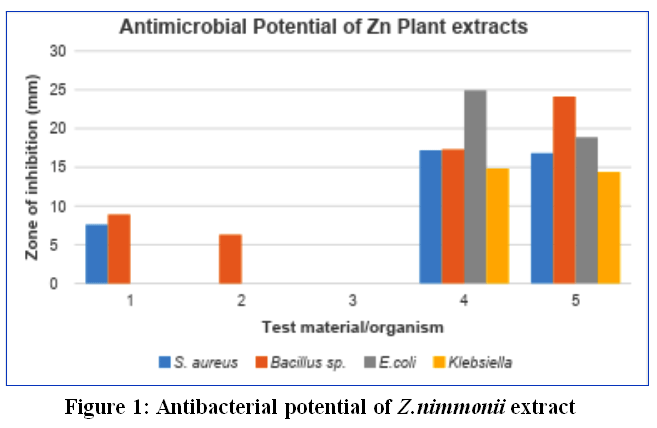 |
Figure 1: Antibacterial potential of Z.nimmonii extract
|
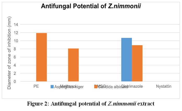 |
Figure 2: Antifungal potential of Z.nimmonii extract
|
Anti-inflammatory Studies
Anti-inflammatory activity was assessed using both heat-induced and hypotonicity-induced sheep red blood cell (SRBC) membrane stabilization assays. The heat-induced and hypotonicity-induced hemolysis models are widely utilized to assess anti-inflammatory activity, as they replicate membrane destabilization in inflammatory conditions. Stabilizing the membranes of red blood cells is thought to be similar to stabilizing the membranes of lysosomes, which prevents the release of lytic enzymes and inflammatory mediators.16,17 Diclofenac is commonly used as a reference standard in models of heat-induced and hypotonicity-induced hemolysis because of its recognized anti-inflammatory properties and its capacity to stabilize biological membranes. Diclofenac inhibits cyclooxygenase enzymes and prevents lysosomal membrane destabilization, which lowers the release of lytic enzymes and inflammatory mediators. This makes it a good standard for testing how well membranes can be stabilized.17 According to the present work the extracts demonstrated concentration-dependent membrane stabilization in both experimental models.
Hypotonicity-Induced Haemolysis
The extracts effectively protected sheep erythrocyte membranes from hypotonicity-induced hemolysis (Table 4, Fig. 3), with percentage inhibition of lysis at each extract concentration surpassing that observed for diclofenac sodium, the reference standard. This data indicates that Zingiber nimmonii extracts exert a membrane stabilization effect by inhibiting erythrocyte lysis under hypotonic stress.
The phytochemical profile of the methanolic extract, particularly its flavonoids and phenolic components, likely contributes to this protective effect by inhibiting the release or activity of lytic enzymes and other inflammatory agents.18
Table 4: Anti-inflammatory properties of Z.nimmonii extract in hypotonicity induced haemolysis model
| Concentration
(µg/ml) |
% Stabilization | |
| Z.nimmonii extract | Diclofenac sodium | |
| 0 | 0.00 | 0.00 |
| 25 | 24.35±1.26 | 10.41±1.01 |
| 50 | 40.64±2.04 | 15.39±2.09 |
| 100 | 60.23±0.94 | 27.47±0.97 |
| 200 | 69.04±1.02 | 64.64±1.62 |
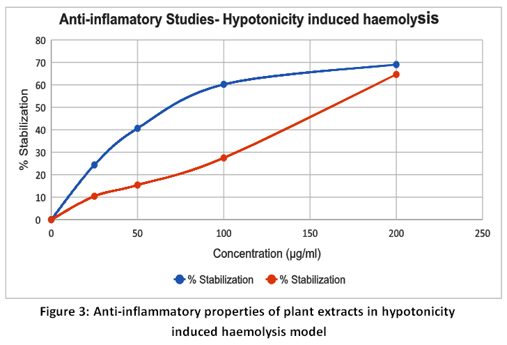 |
Figure 3: Anti-inflammatory properties of plant extracts in hypotonicity induced haemolysis model
|
Heat-induced haemolysis of the SRBC membrane
The extract demonstrated anti-inflammatory potential by protecting sheep red blood cell (SRBC) membranes from heat-induced haemolysis (Table 5, Fig. 4). Both the extract and the reference drug, diclofenac sodium, showed concentration-dependent inhibition of haemolysis in SRBC subjected to heat stress.
Table 5: Anti-inflammatory properties of plant extracts in heat-induced haemolysis model
| Concentration
(µg/ml) |
% Stabilization | |
| Diclofenac | Z. nimmonii | |
| 0 | 0.00 | 0.00 |
| 25 | 9.62±1.62 | 10.81±1.43 |
| 50 | 17.4±1.04 | 24.04±1.19 |
| 100 | 29.64±1.16 | 40.68±1.04 |
| 200 | 59.06±1.02 | 51.94±1.81 |
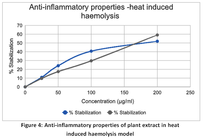 |
Figure 4: Anti-inflammatory properties of plant extract in heat induced haemolysis model
|
Discussion
The major constituents of the rhizome oil of Z. nimmonii differ from those of Zingiber zerumbet and Zingiber officinale. Sabulal et al24 reported that the essential oil from Z. nimmonii possesses significant activity against human pathogenic fungi, including Candida glabrata, Candida albicans, and Aspergillus niger. Additionally, the extracts have demonstrated antioxidant properties, and further research revealed that the essential oil can serve as a novel, safer natural larvicide and repellent against mosquito vectors of malaria, dengue, and filariasis.19 Membrane stabilization is essential in preventing serum protein and fluid leakage into tissues, a hallmark of the inflammatory response. The ability of the extracts to stabilize cellular membranes may be attributed to the inhibition of increased membrane permeability, which is generally mediated by inflammatory mediators.20
Recent scientific literature indicates that studies specifically addressing the anti-inflammatory properties of Zingiber nimmonii are limited. However, the Zingiber genus as a whole, particularly Z. officinale, is well documented for its anti-inflammatory effects. Bioactive constituents such as 6-gingerol, 6-shogaol, and zingerone have been shown to mitigate inflammation by modulating key inflammatory pathways. These compounds exert their effects by inhibiting cyclooxygenase-2 (COX-2) and lipoxygenase (LOX) activities, thereby reducing the synthesis of pro-inflammatory mediators like prostaglandins and leukotrienes.21 Furthermore, other species such as Z. montanum have demonstrated anti-inflammatory potential through the suppression of pro-inflammatory markers, including nitric oxide, inducible nitric oxide synthase, COX-2, interleukin (IL)-1β, IL-6, and monocyte chemoattractant protein-1 in LPS-stimulated macrophages.22 Earlier investigations of Z. nimmonii rhizomes have highlighted their potential as a source of secondary metabolites with anti-infective and antioxidant properties.23 Gas chromatography–mass spectrometry (GC-MS) analyses have confirmed the presence of several chemical constituents of pharmaceutical significance in the rhizome, supporting its potential for the development of novel therapeutic agents.24 In vitro anti-inflammatory tests such as cyclooxygenase and lipoxygenase inhibition, protein denaturation, and membrane stabilization are commonly employed to evaluate the anti-inflammatory potential of phytochemicals, indicating their ability to inhibit major inflammatory mediators.25
Recent studies demonstrated that phytochemicals derived from ethnomedicinal plants such as Zingiber officinale, Ocimum, and Rosmarinus officinalis exhibit significant antimicrobial and anti-inflammatory activities which revealed that phenolic and terpenoid compounds contribute to microbial inhibition through membrane disruption, while anti-inflammatory effects are mediated via cyclooxygenase inhibition and suppression of pro-inflammatory cytokines such as TNF-α and IL-6.26 It was also reported that phytochemicals exhibit multi-target pharmacological effects, including antimicrobial, anti-inflammatory, and antioxidant activities, primarily mediated through phenolic compounds and terpenoids that modulate COX enzymes and pro-inflammatory cytokines.27
Conclusion
The present study highlights the significant phytochemical, antimicrobial, and anti-inflammatory properties of Zingiber nimmonii, a comparatively underexplored member of the Zingiberaceae family. Phytochemical analysis identified the presence of bioactive constituents such as flavonoids, alkaloids, phenolics, and terpenoids, which are likely responsible for the plant’s diverse pharmacological activities. Antimicrobial assays revealed that extracts of Z. nimmonii exhibited pronounced inhibitory effects against various bacterial and fungal strains, underscoring its potential as a natural antimicrobial agent. Additionally, the plant demonstrated notable anti-inflammatory effects, supporting its traditional application in the management of chronic inflammatory disorders. Collectively, these findings suggest that Z.nimmonii may serve as a promising source of novel bioactive molecules for therapeutic development. Nevertheless, comprehensive studies and further experimental validation are warranted to fully establish its medicinal potential. The inclusion of such underutilized medicinal plants into contemporary pharmacological research could facilitate the discovery of safer and more effective drug candidates.
Acknowledgement
We would like to thank the curator and staff of herbarium, Catholicate College, Pathanamthitta (CATH) Kerala. Authors would like to thank the Principal CMS College Kottayam for providing necessary facilities. The authors gratefully acknowledge Doctor John’s Biotech Centre for Research and Development, Kottarakara, Kerala, for providing research facility support and valuable suggestions. : The authors sincerely acknowledge the support, in terms of instrumentation facility, by the DST-FIST.
Funding Sources
The first author is thankful to the financial support (Junior Research Fellowship Order no. 6982/AC 6/2025/MGU) received from the Mahatma Gandhi University, Kottayam, Kerala.
Conflict of Interest
The authors do not have any conflict of interest.
Data Availability Statement
This statement does not apply to this article.
Ethics Statement
This research did not involve human participants, animal subjects, or any material that requires ethical approval.
Informed Consent Statement
This study did not involve human participants, and therefore, informed consent was not required.
Clinical Trial Registration
This research does not involve any clinical trials.
Permission to reproduce material from other sources
Not Applicable.
Author Contributions:
- Therese Jose: Conceptualization, Data Collection, Analysis, Methodology, Writing – Original Draft.
- Valukattil Ponnachan Thomas: Editing, Analysis.
- Jinu John: Editing, Resources.
- Rogimon Plammoottil Thomas: Visualization, Supervision, Project Administration.
References
- Christenhusz MJM, Byng JW. The number of known plant species in the world and its annual increase. Phytotaxa. 2016;261(3):201-217. doi:10.11646/phytotaxa.261.3.1
CrossRef - Skaria BP, Joy PP, Mathew G, Mathew S. Zingiberaceous Plants in Traditional Medicine. Aromatic and Medicinal Plants Research Station; 2005.
- Kumar KM, Asish G, Sabu M, Balachandran I. Significance of gingers (Zingiberaceae) in Indian system of medicine—Ayurveda: an overview. Anc Sci Life. 2013;32(4):253. doi:10.4103/0257-7941.131989
CrossRef - Thomas V, Jose J, Saranya ST, Binoy S. Ethnobotanical significance of Zingiberales: a case study in the Malaipandaram tribe of southern Western Ghats of Kerala. Indian J Tradit Knowl. 2020;19:450-458. doi:10.56042/ijtk.v19i2.35349
CrossRef - Sharifi-Rad M, Varoni EM, Salehi B, et al. Plants of the genus Zingiber as a source of bioactive phytochemicals: from tradition to pharmacy. Molecules. 2017;22(12):2145. doi:10.3390/molecules22122145
CrossRef - Limcharoen P, Sattayasomboon Y, Kongsin S, Sillabutra J. The use of Thai traditional medicine services among type 2 diabetes mellitus patients in community hospitals, Pathum Thani Province, Thailand. J Health Sci. 2022;31(6):975-983.
- Mabberley DJ. The Plant-Book: A Portable Dictionary of the Higher Plants. Cambridge University Press; 1990. doi:10.1017/9781316335581
CrossRef - Destryana R, Estiasih T, Sukardi, Pranowo D. Zingiberaceae rhizome essential oil: a review of chemical composition, biological activity, and application in food industry. IOP Conf Ser Earth Environ Sci. 2024;1299(1):012010. doi:10.1088/1755-1315/1299/1/012010
CrossRef - Kumar VP, Chauhan NS, Padh H. Search for antibacterial and antifungal agents from selected Indian medicinal plants. J Ethnopharmacol. 2006;107(2):182-188. doi:10.1016/j.jep.2006.03.013
CrossRef - Deng M, Yun X, Ren S, Qing Z, Luo F. Plants of the genus Zingiber: a review of their ethnomedicine, phytochemistry, and pharmacology. Molecules. 2022;27(9):2826. doi:10.3390/molecules27092826
CrossRef - Sabu M. Zingiberaceae and Costaceae of South India. Indian Association for Angiosperm Taxonomy, University of Calicut; 2006.
- Gopalan G, Kavunkal HV, Ravindran DS, Mamiyil S, IG S, et al. Chemical constituents and their bioactivities of Zingiber nimmonii: an endemic species in South India. J Med Aromat Plant Sci. 2021;43(1):15-24. doi:10.62029/jmaps.v43i1.Gopalan
CrossRef - Harborne JB. Phytochemical Methods. Chapman and Hall Ltd; 1973:49-188.
- John J, Mehta A, Mehta P. Antimicrobial and phytochemical analysis of Cassia tora leaf extracts. J Basic Appl Biol. 2011;5(1-2):125-131.
- Venkatraman T, Alex M, Andrew R, et al. Evaluation of in vitro anti-inflammatory property of a novel polyherbal formulation containing Sida cordifolia, Solanum xanthocarpum, and Psidium guajava. Adv Biomed Res. 2024;15:157-163. doi:10.15515/abr.0976-4585.15
- Chahardoli A, Qalekhani F, Hajmomeni P, et al. Enhanced hemocompatibility, antimicrobial and anti-inflammatory properties of biomolecules stabilized AgNPs with cytotoxic effects on cancer cells. Sci Rep. 2025;15:1186. doi:10.1038/s41598-024-82349-z
CrossRef - Fathima SN, Alharbi NS, Alharbi SA, Wainwright M. Assessment of in vitro antioxidant and anti-inflammatory activity of plant extracts using membrane stabilization models. J Evid Based Integr Med. 2024;29:1-10. doi:10.1177/2515690X241257127
- Naz R, Ayub H, Nawaz S, et al. Antimicrobial activity, toxicity and anti-inflammatory potential of methanolic extracts of four ethnomedicinal plant species. BMC Complement Med Ther. 2017;17:302. doi:10.1186/s12906-017-1815-z .
CrossRef - Govindarajan M, Rajeswary M, Arivoli S, Tennyson S, Giovanni B. Larvicidal and repellent potential of Zingiber nimmonii essential oil: an ecofriendly tool against mosquito vectors. Parasitol Res. 2016;115:1807-1816. doi:10.1007/s00436-016-4920-x
CrossRef - Yesmin S, Paul A, Naz T, Rahman ABMA, Akhter SF, et al. Membrane stabilization as a mechanism of the anti-inflammatory activity of ethanolic root extract of Choi (Piper chaba). Clin Phytosci. 2020;6(1):1-9. doi:10.1186/s40816-020-00207-7
CrossRef - Truong VL, Manochai B, Pham TT, Jeong WS. Antioxidant and anti-inflammatory activities of Zingiber montanum oil in HepG2 cells and lipopolysaccharide-stimulated RAW 264.7 macrophages. J Med Food. 2021;24(6):595-605. doi:10.1089/jmf.2021.k.0019
CrossRef - Sonam SBV, Samatha A, Mansi S, Shubham RS, Raziya BS. Anti-inflammatory effects of Zingiber officinale: a comprehensive review of its bioactive compounds and therapeutic potential. Medtigo J Pharmacol. 2024;1(1):e3061113. doi:10.63096/medtigo3061113
CrossRef - Nair AR, Ganapathy G. Untargeted gas chromatography–mass spectrometry analysis and evaluation of antimicrobial and antioxidant activity of Zingiber nimmonii rhizome extracts. Pharmacogn Res. 2020;12(4):466-470 doi:10.4103/pr.pr_19_20.
CrossRef - Sabulal B, Dan M, Kurup R, Pradeep NS, Valsamma RK, George V. Chemical composition and antimicrobial activity of essential oils from the rhizomes of Zingiber nimmonii. Nat Prod Commun. 2006;1(6):505-508. doi.org/10.1016/j.phytochem.2006.08.003.
- Sabalingam S. In-vitro approaches to evaluate the anti-inflammatory potential of phytochemicals: A review. J Drug Deliv Ther. 2025;15(1):187-192. doi :10.22270/jddt.v15i1.6956 .
CrossRef - Gawad AMA, Molham F, Tagyan AI, et al. Microbiological and pharmacological investigation of phytochemicals extracted from selected ethnomedicinal plants with their potential against food pathogen. Sci Rep. 2025;15:39017. doi.org/10.1038/s41598-025-05663-0.
CrossRef - Coyago-Cruz E, et al. Antimicrobial, antioxidant, antitumor, and anti-inflammatory properties of Gleichenella pectinata: A bioprospecting of medicinal ferns. Antioxidants. 2025;14(11):1354. doi.org/10.3390/antiox14111354.
CrossRef
Abbreviations
SRBC: sheep red blood cell
DMSO: Dimethyl Sulfoxide
ZN: Zingiber nimmonii
Accepted on: 06-05-2026
Second Review by: Dr. Ban Ali Ahmed
Final Approval by: Dr. Wagih Ghannam


![]()
![]()
