
The Chick Embryo Model: From Developmental Biology to Comprehensive Biomedical Research Insights
 , Kishor Vasant Otari and Ajay Yashvant Kale
 Department of Pharmacology,Navsahyadri Institute of Pharmacy, Pune, India
Corresponding Author E-mail: pallavivallamdeshi8@gmail.com
Download this article as:
ABSTRACT:The chick embryo model is a widely used in vivo experimental system in developmental biology and biomedical research. This review aims to systematically evaluate the applicability of the chick embryo model in studying vertebrate development, disease mechanisms, and therapeutic interventions. The model is characterized by ex-ovo development, rapid organogenesis, cost-effectiveness, and ethical acceptability, making it a valuable alternative to mammalian systems. This review summarizes key aspects including embryonic staging, major developmental processes, and the role of extra-embryonic membranes, particularly the yolk sac membrane and chorioallantoic membrane, in applications such as angiogenesis, tumor biology, pharmacokinetics, and toxicological assessment. Additionally, commonly used experimental approaches, drug delivery routes, and advanced imaging techniques enabling real-time and longitudinal analysis are discussed. The chick embryo model offers significant advantages in terms of accessibility and experimental flexibility; however, physiological and anatomical differences from mammals limit direct translation to human systems. Therefore, findings obtained from this model should be interpreted cautiously and validated using higher animal models. In conclusion, the chick embryo remains a reliable and ethically acceptable platform for preliminary in vivo investigations, with ongoing technological advancements expected to further enhance its relevance in translational biomedical research.
KEYWORDS:Angiogenesis; Biomedical research; Chick embryo; Chorioallantoic membrane (CAM); Developmental biology; Embryogenesis
Introduction
The chick embryo model has long been recognized as a valuable system in developmental biology, congenital abnormalities, and translational biomedical research. Its scientific relevance dates back more than 2000 years, with early observations recorded in ancient Egypt around 300 BCE. Systematic studies on chick embryo development were later conducted by Aristotle around 330 BCE, who documented embryonic stages and morphological changes in his work Historia Animalium, laying the foundation for modern embryology. During the Renaissance, Leonardo da Vinci further advanced anatomical and developmental understanding through detailed embryological illustrations.1
Over the past five decades, significant advancements have established the chick embryo as a versatile model across multiple domains of developmental biology, particularly in the evaluation of drug safety and in studies of limb, nervous system, and cardiac development.2,3 The development of chick embryo-based teratogenic models has contributed substantially to understanding the mechanisms underlying congenital abnormalities, reinforcing its importance in developmental toxicology. The transition of chick embryo research from descriptive observations to sophisticated experimental systems highlights its growing relevance in both fundamental and applied biomedical sciences.
Recent studies have further expanded the application of the chick embryo model in cancer research, pharmacology, and translational studies due to its accessibility, cost-effectiveness, and experimental flexibility.2,4 The chorioallantoic membrane (CAM), in particular, serves as an effective interface between in vitro and in vivo systems and has been widely utilized for assessing nanoparticle stability, biocompatibility, circulation, and therapeutic potential.5The domestic chicken (Gallus gallus domesticus) remains the most commonly used avian species in research, and its embryos provide an efficient platform for studying dynamic developmental and pathological processes, including tumor progression and cardiovascular biology.
The development of modern immunological and molecular techniques has further enhanced the utility of chick embryos as experimental models, particularly in cancer research and vertebrate morphogenesis.1 The relative immunodeficiency of early-stage embryos enables tumor cell implantation on the CAM, facilitating the study of tumor growth, angiogenesis, invasion, and extracellular matrix remodeling.6 Additionally, within the framework of the 3Rs (Replacement, Reduction, and Refinement), the chick embryo model offers a less ethically contentious and more cost-effective alternative to conventional mammalian models. Its rapid development, reproducibility, and independence from maternal influence further support its widespread use. However, despite these advantages, certain alternative models still lack the developmental and metabolic complexity of vertebrate systems, reinforcing the continued relevance of the chick embryo model.7
Furthermore, the chick embryo model has demonstrated significant utility in preclinical drug distribution and pharmacokinetic studies, offering precise control over experimental parameters and exposure conditions.8 Therefore, this review aims to comprehensively evaluate the chick embryo model as an in vivo experimental platform in biological and biomedical sciences. It covers embryonic development, key developmental stages, and extra-embryonic membranes, along with their relevance to experimental applications. Additionally, this review discusses various experimental approaches, including imaging techniques, and highlights applications in pharmacology, toxicology, cancer biology, and regenerative medicine. Importantly, while the chick embryo model provides valuable insights, physiological and anatomical differences between avian and mammalian systems limit direct extrapolation to humans, necessitating further validation in higher animal models.
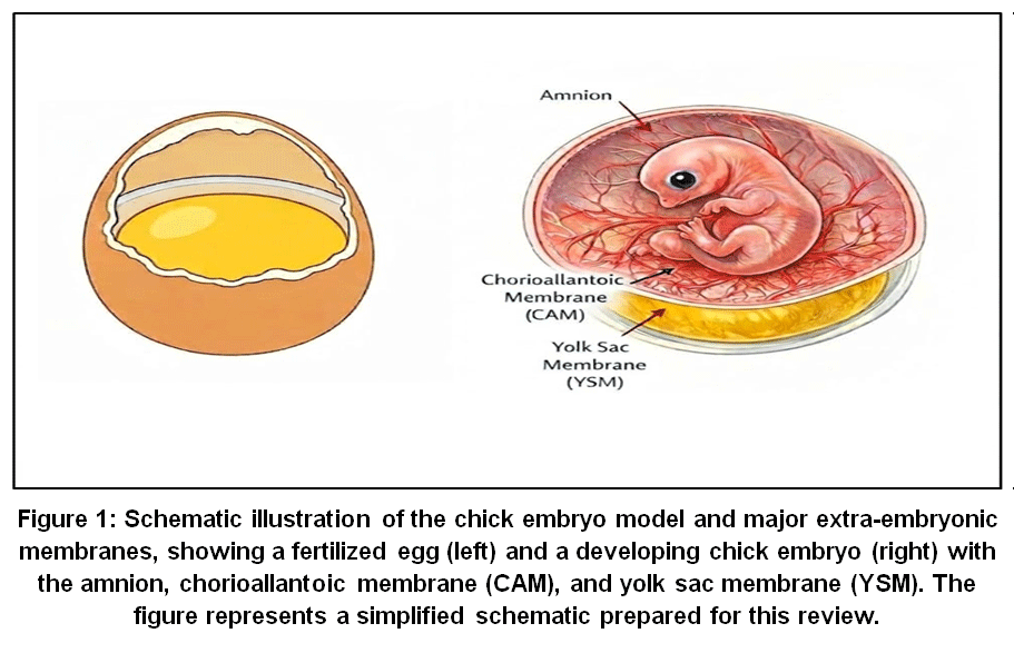 |
Figure 1. Schematic illustration of the chick embryo model and major extra-embryonic membranes, showing a fertilized egg (left) and a developing chick embryo (right) with the amnion, chorioallantoic membrane (CAM), and yolk sac membrane (YSM).
|
Developmental Biology of chick Embbryo
Fertilization and Early Embryogenesis
Fertilization in the chick embryo occurs in the infundibulum of the oviduct, where multiple spermatozoa penetrate the inner perivitelline layer and enter the germinal disc, the cytoplasmic region of the yolk where embryonic development is initiated. The first cleavage division begins shortly after fertilization and coincides with the formation of shell membranes. Cleavage in chick embryos is meroblastic and discoidal, resulting in blastomeres that remain confined to the germinal disc. Subsequent cleavage divisions become increasingly asynchronous, reflecting the complexity of early embryonic organization. Despite species-specific variations, the sequence of nuclear events during fertilization and early cleavage shows similarities with other vertebrates.9,10
Early embryogenesis also involves the formation of protective and supportive structures, including the vitelline membrane, eggshell, and extra-embryonic membranes, which play essential roles in providing mechanical protection, gas exchange, and nutrient supply. Embryonic development is commonly described using the Hamburger and Hamilton staging system, which correlates morphological changes with developmental time during incubation. In addition, dynamic changes occur in the yolk, albumen, amniotic fluid, and eggshell, including alterations in structural integrity and biochemical composition, all of which are critical for sustaining normal embryonic growth.
Embryonic development is a highly coordinated three-dimensional process, and advanced imaging techniques such as micro-computed tomography (micro-CT) have emerged as valuable tools for studying both normal and abnormal development. Micro-CT enables high-resolution, non-destructive visualization of developing organs and tissues, making it particularly useful for assessing organogenesis and teratogenic effects. This is especially important during early developmental stages, when critical structures such as the neuroectoderm and thoracoabdominal regions are formed and are highly susceptible to developmental disturbances. However, despite its advantages, the application of micro-CT in early chick embryogenesis is limited by the lack of suitable contrast agents, highlighting the need for further methodological advancements to improve imaging accuracy andapplicability.11
Chick Embryonic Developmental Stages and Experimental Relevance
Key developmental stages and their experimental relevance are summarized in Table 1.
Table 1: Overview of chick embryonic developmental stages, key biological events and experimental relevance.
| Incubation Day
|
HH Stage Range
|
Key Developmental Events
|
Experimental Relevance
|
| Days 1-2
|
HH
1-10
|
Gastrulation, neural tube formation, somite development
|
Cell fate mapping, early toxicity studies.12,13
|
| Days 3
|
HH
11-17
|
Heart looping, initiation of circulation
|
Cardiotoxicity, early angiogenesis studies.13,1
|
| Days 4-5
|
HH
18-24
|
Brain regionalization, limb bud formation | Teratogenicity, limb development.12
|
| Days 6-7 | HH
25-28
|
Sensory organ and visceral organ development | Sensory and metabolic toxicity studies.13
|
| Days 8-10
|
HH
29-34
|
Cartilage formation, feather germ appearance | Skeletal and respiratory studies.13
|
| Days
11-13
|
HH
35-37
|
Ossification neuromuscular development
|
Bone and neuromuscular studies.1,14
|
| Days
14-16
|
HH
38-40
|
Gut maturation, yolk sac absorption
|
Nutrient metabolism, pharmacokinetics.13,1
|
| Days
17-19
|
HH
41-43
|
Lung maturation, embryonic positioning
|
Late-stage developmental studies.12
|
| Days
20-21 |
HH
44-46
|
Pipping, hatching
|
Post-hatch adaptation.12,14
|
Organogenesis and Growth Phases
Organogenesis in the chick embryo involves the differentiation and maturation of major organ systems, including the nervous, cardiovascular, and musculoskeletal systems. This stage is characterized by rapid cell proliferation, migration, and tissue differentiation, leading to the formation of structurally and functionally distinct organs. Key developmental events such as neurulation, cardiac development, and limb formation occur in a sequential and tightly regulated manner. Vascularization plays a crucial role during this phase by ensuring adequate nutrient and oxygen supply to growing tissues. Due to its well-defined developmental stages and accessibility, the chick embryo serves as a valuable model for studying organ development and developmental abnormalities.However, despite its advantages, variations in developmental patterns between avian and mammalian systems must be considered when extrapolating findings to higher vertebrates.7
Extra-Embryonic Membranes
Yolk Sac Membrane
The yolk sac membrane (YSM) is a highly specialized extra-embryonic structure responsible for nutrient digestion, absorption, and transfer to the developing chick embryo. Originating from the embryonic hindgut, it progressively surrounds the yolk sac during early incubation and undergoes significant structural and functional changes throughout development. The YSM is characterized by a well-developed vascular network, a mesodermal layer, and a folded epithelial surface with microvilli, which together enhance its absorptive capacity and facilitate efficient nutrient transport into the embryonic circulation.15,16
Beyond its structural role, the YSM functions as a metabolically active organ with similarities to the embryonic gut. It expresses a wide range of genes encoding digestive enzymes and nutrient transporters involved in the uptake of lipids, proteins, carbohydrates, and micronutrients. This activity is developmentally regulated, with peak functional capacity observed during mid-embryogenesis, followed by a gradual decline as the embryo prepares for post-hatch intestinal feeding.16,17
Structural adaptations of the YSM, including increased surface area and enhanced lipid-processing capacity, are closely associated with efficient nutrient utilization and rapid embryonic growth. These features make it a valuable model for studying in vivo nutrient absorption, metabolism, and developmental physiology. However, despite its functional similarities to mammalian systems, species-specific differences should be considered when extrapolating findings beyond avian models.17
Chorioallantoic Membrane
The chorioallantoic membrane (CAM) is a highly vascularized extra-embryonic structure that develops around Hamburger-Hamilton (HH) stage 8 and becomes fully functional by approximately HH stage 15. It plays a critical role in embryonic development by facilitating gas exchange, calcium absorption from the eggshell, and nutrient transport to the growing embryo.12 Owing to its dense vascular network and easy accessibility, the CAM serves as an effective in vivo platform for studying angiogenesis, tumor growth, metastasis, and host–tumor interactions.The experimental utility of the CAM is further enhanced by the ability to create a window in the eggshell, allowing direct and real-time observation of vascular responses, drug effects, and tissue interactions under microscopic conditions. These features make it a widely used model in cancer research, pharmacology, and biomaterial testing. However, despite its advantages, careful handling and precise experimental conditions are required to maintain embryo viability and ensure reproducibility of results.14,5In contrast to the yolk sac membrane, which primarily supports nutrient absorption, the CAM is more specialized for gas exchange and vascular studies, highlighting the complementary roles of extra-embryonic membranes in chick embryo development.
Experimental Platform and Techniques
In-ovo Manipulation
In-ovo manipulation is a widely used experimental approach in chick embryo research, typically performed during embryonic days 3-4, when access to the embryo and extra-embryonic membranes is achieved by creating a small window in the eggshell. This technique allows the embryo to remain within its natural environment, thereby preserving physiological conditions while enabling real-time observation and experimental intervention. A wide range of substances, including drugs, hormones, nucleic acids (DNA and RNA), proteins, and dyes, can be administered using this method to investigate gene function, cellular behavior, and developmental processes. In-ovo manipulation has been extensively applied in studies of angiogenesis, tumor growth, and tumor–host interactions, as well as in pharmacological and toxicological evaluations. The approach enables in vivo analysis of dynamic biological processes with minimal disruption to embryonic development. However, despite its advantages, in-ovo manipulation has certain limitations. The presence of eggshell and membrane barriers can restrict accessibility, making repeated interventions and long-term longitudinal studies more challenging.14
Conditions for Incubation of Eggs
Successful chick embryo development depends on tightly controlled incubation conditions, including temperature, humidity, ventilation, and appropriate egg handling. During early development, the embryo originates from the area pellucida, while surrounding regions contribute to the formation of extra-embryonic structures. Between days 2 and 11 of incubation, key extra-embryonic membranesnamely the yolk sac, amnion, allantois, chorion, and sub-embryonic fluidare formed. These structures support embryonic growth by facilitating gas exchange, nutrient absorption, waste elimination, and mechanical protection.10 Avian eggs provide a self-contained system for embryonic development, with yolk and albumen supplying nutrients and maintaining osmotic balance. This system has also been utilized in biotechnological applications such as in-ovo feeding and immunization to improve embryonic health and hatchability. However, in ex-ovo culture systems, where the embryo is removed from the eggshell, strict environmental control is required to support normal development. Critical factors such as oxygen availability, humidity, and calcium supplementation must be carefully regulated. Temperature plays a central role in embryonic development, with optimal incubation typically maintained between 37-38 °C. Humidity must be controlled to prevent dehydration, particularly after the development of the chorioallantoic membrane, while adequate ventilation ensures proper oxygen supply and heat dissipation. Since the eggshell serves as the primary source of calcium for skeletal development, ex-ovo systems require external calcium supplementation to prevent developmental abnormalities. Additionally, factors such as airflow, egg size, and spacing influence heat distribution and embryonic metabolism. Deviations from optimal incubation conditions can adversely affect embryonic growth, viability, and hatchability.15
Routes of Administration
In the chick embryo model, multiple routes of administration are employed to deliver drugs, biomolecules, or experimental agents at specific developmental stages. These routes allow precise control over the site, timing, and dosage of exposure, making the model highly suitable for pharmacological, toxicological, and developmental studies. Common routes include the air cell, yolk sac, allantoic sac, amniotic cavity, and chorioallantoic membrane (CAM). The choice of administration route depends on the developmental stage of the embryo and the objective of the study. In early stages, the air cell route is often used as a minimally invasive approach for initial exposure. For example, in-ovo administration via the air cell has been utilized for the delivery of compounds such as escitalopram.15 The yolk sac route facilitates interaction with nutrient reserves, while the amniotic cavity allows direct exposure of the embryo to administered substances. In contrast, the allantoic sac is commonly used during mid to late developmental stages, as it enables systemic distribution of administered agents through the embryonic circulation. This route has been widely applied in studies evaluating drug effects, toxicity, and developmental responses, particularly during later embryonic days.8 Additionally, CAM-based administration provides a direct interface with a highly vascularized system, making it especially useful for angiogenesis, tumor biology, and drug delivery studies.
Overall, the route of administration plays a critical role in determining bioavailability, absorption kinetics, and the overall biological response of the embryo. Therefore, careful selection of the administration route is essential for ensuring experimental accuracy and reproducibility. The major routes of administration, their delivery sites, and advantages are summarized in Table 2.
Table 2: Overview of administration routes in the chick embryo model, delivery sites, and advantages.
| Route of Administration | Site of Delivery | Advantages
|
| Air Cell | Air chamber between shell membranes at blunt end | Minimally invasive, simple access |
| Yolk Sac | Yolk sac membrane surrounding yolk | Systemic exposure via nutrient absorption |
| Amniotic Cavity | Amniotic fluid surrounding embryo | Efficient delivery to embryo |
| Allantoic Sac | Allantoic cavity connected to embryonic circulation | Systemic circulation access |
| Chorioallantoic Membrane (CAM) | Highly vascular membrane beneath eggshell | Highly vascular, real-time observation |
| Albumen | Egg white surrounding embryo | Easy administration, non-invasive |
Imaging and Monitoring
Advanced imaging technologies have significantly enhanced the utility of the chick embryo model by enabling non-invasive and real-time monitoring of embryonic development, tissue regeneration, and tumor progression. Various imaging modalities, including magnetic resonance imaging (MRI), micro-computed tomography (micro-CT), ultrasound, and optical imaging, are widely used depending on the objective of the study.Micro-CT is particularly valuable for high-resolution three-dimensional visualization and quantitative analysis of embryonic structures, skeletal development, and tissue engineering applications. In contrast, MRI is well suited for soft tissue imaging and organ development studies, providing detailed anatomical and functional information without damaging the embryo. Advanced MRI techniques, such as T2-weighted imaging and diffusion tensor imaging, have further supported investigations of nervous system development and structural organization.
In addition, optical imaging methods allow dynamic observation of developmental processes and vascular changes. Nuclear and molecular imaging techniques, particularly positron emission tomography (PET) combined with computed tomography (CT), have expanded the application of chick embryo models in tumor angiogenesis, metabolism, and drug distribution studies, especially in chorioallantoic membrane-based tumor models. Ultrasound imaging also enables non-invasive real-time assessment of tumor growth and tissue characteristics during embryonic development.Collectively, these imaging approaches enhance the preclinical and translational value of the chick embryo model by providing repeatable, high-resolution structural, functional, and molecular data across different stages of development.18
Ethis and Regulatory Considerations
The chick embryo model aligns well with the principles of the 3RsReplacement, Reduction, and Refinementwhich aim to minimize animal use and suffering in scientific research. Compared to mammalian models, chick embryos are often considered a more ethically acceptable alternative, particularly during early developmental stages when the nervous system is not fully developed.7,1 This allows researchers to investigate complex developmental, pharmacological, and toxicological processes without the need for higher vertebrates. In many regulatory frameworks, early-stage chick embryos are not classified as “protected” animals, supporting their use in preliminary biomedical research.19,7
The principle of reduction is supported by the high experimental yield and accessibility of the chick embryo model, enabling multiple experimental conditions and repeated measurements within a single cohort. Similarly, refinement is achieved through the use of minimally invasive techniques such as egg windowing, localized administration, and real-time imaging, which reduce procedural stress. Additionally, standardized incubation conditions and defined experimental endpoints contribute to improved data quality while maintaining embryo welfare.
Regulatory requirements for chick embryo research vary across countries and institutions. In many jurisdictions, experiments conducted before late embryonic stages (commonly before embryonic day 14) may not require formal ethical approval, provided that embryos are not allowed to hatch or survive beyond defined limits. However, adherence to institutional and national guidelines remains essential, and best practicesincluding minimizing unnecessary manipulation, preventing contamination, and ensuring proper disposalmust be followed.
Beyond ethical considerations, the chick embryo model offers significant practical advantages. Fertilized eggs are inexpensive and do not require specialized housing or animal care facilities, reducing overall research costs. The simplicity of incubation systems, combined with ease of experimental access through in-ovo techniques, supports rapid, reproducible, and scalable experimentation. These features make the chick embryo a valuable and efficient model for developmental biology and translational research, particularly in early-stage screening and hypothesis testing.7,20
Biomedical and Translational Applications
Cancer and Metastasis
The chick embryo model, particularly the chorioallantoic membrane (CAM), is widely used in cancer and metastasis research due to its dense vascular network, rapid development, and relative immunodeficiency. Tumor cells can be xenografted onto the CAM, where they readily induce angiogenesis and invade surrounding tissues, enabling detailed investigation of tumor growth, vascular remodelling, and tumor microenvironment interactions. In addition, the CAM model supports the study of metastatic behavior, as implanted human cancer cells can intravasate into the embryonic vasculature and disseminate to distant sites, allowing evaluation of organ tropism and metastatic potential. This system has also been applied to assess tumor metabolism, anti-angiogenic therapies, and drug responses in a controlled in vivo environment. Due to its accessibility, cost-effectiveness, and compatibility with imaging and molecular techniques, the CAM model serves as a valuable preclinical platform for cancer research. However, despite its advantages, differences between avian and mammalian physiology should be considered when translating findings to higher vertebrate systems.21
Angiogenesis and Vascular Biology
The chorioallantoic membrane (CAM) of the chick embryo is widely used for studying angiogenesis and vascular biology due to its rapid vascular development, dense capillary network, and high accessibility. The CAM supports both physiological and pathological angiogenesis, making it a suitable model for investigating neovascularization and vascular remodelling. It has been extensively applied to evaluate the effects of various substances, including growth factors, biomaterials, nanoparticles, and both pro-angiogenic and anti-angiogenic agents. Quantitative analysis of vascular parameters such as vessel density, length, and branching can be performed using real-time imaging techniques, enabling dynamic assessment of vascular development.
Furthermore, the CAM model allows the study of endothelial cell function, blood flow regulation, vascular permeability, and interactions between blood vessels and the extracellular matrix in a living system. Due to its experimental accessibility, reduced ethical constraints, and translational relevance, the CAM remains a valuable in vivo platform for vascular research. However, differences between avian and mammalian systems should be considered when extrapolating findings to human studies.4
Drug Screening and Toxicity
The chick embryo model is increasingly recognized as a reliable in vivo platform for drug screening and toxicity assessment, serving as an intermediate system between in vitro studies and mammalian models. Its rapid development, external growth, and ease of experimental access allow evaluation of key endpoints such as survival, organogenesis, and developmental toxicity, including teratogenic effects and structural abnormalities. Multiple routes of administration, including the yolk sac, chorioallantoic membrane (CAM), allantoic cavity, and albumen, enable controlled assessment of drug biodistribution, exposure, and dose-dependent toxicity. Whole-embryo exposure is particularly useful for evaluating developmental safety and teratogenicity, whereas localized application to the CAM allows real-time analysis of angiogenesis, tissue response, and drug efficacy. Due to its cost-effectiveness, experimental flexibility, and reduced ethical constraints, the chick embryo model is well suited for early-stage drug development and preliminary toxicity screening. However, while it provides valuable biological insights, differences in metabolism and physiology between avian and mammalian systems should be considered when extrapolating findings to humans.4,1
Studies on Regeneration and Tissue Engineering
The chick embryo represents a valuable model for studies in regenerative biology and tissue engineering due to its rapid development, accessibility, and conserved molecular signaling pathways. It is particularly useful for investigating processes such as spinal cord regeneration, which exhibits stage-dependent regenerative capacity during embryonic development. The chorioallantoic membrane (CAM) provides a suitable platform for studying key aspects of wound healing, including angiogenesis, inflammation, and tissue remodelling.In addition, chick embryos have been utilized in studies of cardiac repair, liver development, and progenitor cell dynamics, offering insights into mechanisms relevant to human regenerative medicine. Regenerative processes in limb and inner ear development have also revealed conserved signaling pathways, supporting the relevance of this model in stem cell research and tissue engineering applications. Collectively, these features highlight the utility of the chick embryo as a preclinical platform for investigating regeneration and repair mechanisms.15
Furthermore, the chick embryo model provides a versatile system for studying complex three-dimensional (3D) tissue constructs in biologically relevant environments. Decellularized chick embryo matrices can serve as natural scaffolds that mimic native tissue architecture, facilitating the study of cellmatrix interactions, tissue integration, and remodelling. These 3D systems are also useful for modelling early tumor microenvironments, supporting cell survival, invasion, and angiogenic responses.Moreover, grafting of biomaterials or engineered tissues onto the CAM enables real-time evaluation of vascularization, host integration, and tissue responses. This approach offers a cost-effective and scalable alternative to mammalian models and is increasingly applied in drug testing, regenerative biomaterial evaluation, and nanomedicine.22,23
Limitations
Despite its broad applicability and ethical advantages, the chick embryo model has several limitations that must be considered when interpreting experimental outcomes. One major limitation is the immature immune system during early embryonic development. Functional adaptive immunity develops only around embryonic days (ED) 12 to 14, with incomplete maturation of T and B lymphocytes. While this immunodeficiency facilitates xenograft studies, it limits investigations involving immune responses, immunopharmacology, and host–pathogen interactions.7,14 Another constraint is the restricted experimental time window. Ethical and regulatory considerations typically limit experiments to pre-hatching stages (commonly before ED14 to17), which restricts studies on long-term drug exposure, chronic toxicity, disease progression, and postnatal outcomes. Additionally, later developmental stages may introduce variability due to increased inflammatory responses and non-specific tissue reactions, complicating data interpretation.Genetic manipulation in chick embryos also remains challenging compared to established mammalian models. Although genomic resources have improved, the availability of stable transgenic lines is limited, and gene-editing approaches such as CRISPR/Cas9 are not yet fully standardized in this system. As a result, mechanistic studies requiring targeted gene modification are less feasible.7,14
Furthermore, physiological and metabolic differences between avian and mammalian systems can affect translational relevance. Drug absorption routes in chick embryossuch as via the yolk sac, amniotic cavity, or chorioallantoic membrane (CAM)do not always correlate with mammalian pharmacokinetics. Therefore, findings from chick embryo studies require validation in mammalian models before clinical extrapolation (1).Finally, the CAM model is highly sensitive to environmental conditions, including temperature, humidity, oxygen levels, and experimental handling. Minor variations in incubation or shell windowing techniques can significantly influence outcomes, particularly in vascular studies. In addition, distinguishing true angiogenesis from transient vascular changes such as vessel dilation may require careful experimental design and histological validation.
Future Perspectives
Advances in analytical techniques and experimental technologies are expected to further expand the applications of the chick embryo model in pharmacology and biomedical research. High-resolution imaging modalities, including micro-computed tomography, fluorescence microscopy, and real-time live imaging of the chorioallantoic membrane (CAM), now enable detailed spatial and temporal analysis of embryonic development, angiogenesis, and tissue dynamics. These approaches allow precise monitoring of developmental processes and the effects of bioactive compounds in vivo.12,13 Progress in molecular biology has also enhanced the utility of the chick embryo model. Emerging gene-editing technologies, particularly CRISPR/Cas9, along with in vivo electroporation and viral vector-based gene delivery systems, are improving the ability to manipulate gene expression during embryonic development. These advancements are expected to overcome previous limitations in genetic studies and enable more detailed mechanistic investigations. The chick embryo model is increasingly recognized as a translational bridge between in vitro systems and mammalian in vivo models. In line with the 3Rs principle, it offers a reduction in the use of higher vertebrates while supporting early-stage evaluation of drug efficacy, biodistribution, and safety. In particular, CAM-based models are anticipated to play a growing role in areas such as tumor biology, angiogenesis, biomaterials testing, and personalized medicine, including patient-derived xenograft studies.6,1
Future progress will depend on the standardization of experimental protocols, including incubation conditions, routes of administration, and outcome measures, to improve reproducibility and comparability across studies. In addition, integration with bioinformatics and systems biology approaches is expected to enhance data interpretation and model predictability. Overall, driven by technological advancements and increasing emphasis on ethical research practices, the chick embryo model is likely to gain wider acceptance and application in developmental biology, preclinical research, and alternative testing strategies.14,24
Conclusion
The chick embryo model represents a valuable and widely used in vivo system in developmental biology and biomedical research. Its key advantages, including ethical acceptability, cost-effectiveness, ease of manipulation, and experimental accessibility, make it a practical alternative to higher vertebrate models for early-stage investigations. The model has demonstrated broad applicability across multiple domains, including organogenesis, angiogenesis, pharmacology, toxicology, cancer research, and regenerative medicine. However, important limitations must be acknowledged. Differences in immune system development, metabolic processes, and overall physiology limit the direct extrapolation of findings to human systems. Therefore, results obtained using chick embryos should be interpreted cautiously and validated in mammalian models when necessary. With ongoing advancements in imaging technologies, molecular approaches, and gene-editing tools, the scope and utility of the chick embryo model continue to expand. Overall, it serves as a reliable, ethically acceptable, and translationally relevant platform for early-stage in vivo studies, complementing rather than replacing higher vertebrate models in biomedical research.
Acknowledgement
The authors would like to acknowledge the Department of Pharmacology, Navsahyadri Institute of Pharmacy, for providing the necessary facilities to conduct this review.
Funding Sources
The author(s) received no financial support for the research, authorship, and/or publication of this article.
Conflict of Interest
The authors do not have any conflict of interest.
Data Availability Statement
This statement does not apply to this article.
Ethics Statement
This research did not involve human participants, animal subjects, or any material that requires ethical approval.
Informed Consent Statement
This study did not involve human participants, and therefore, informed consent was not required.
Clinical Trial Registration
This research does not involve any clinical trials.
Permission to reproduce material from other sources
Not Applicable.
Author Contributions
- Pallavi Vallamdeshi: Literature search, data interpretation and manuscript writing
- Ajay Yashvant Kale: Manuscript revision
- Kishor Vasant Otari: Supervision and reviewed the manuscript
References
- Wachholz GE, Rengel BD, Vargesson N, Fraga LR. From the farm to the lab: how chicken embryos contribute to the field of teratology. Front Genet2021; 12: 666726. doi:10.3389/fgene.2021.666726
CrossRef - Kain KH, Miller JW, Jones‐Paris CR,et al. The chick embryo as an expanding experimental model for cancer and cardiovascular research. Dev Dyn2014; 243(2): 216–228. doi:10.1002/dvdy.24093
CrossRef - Tona K, Voemesse K, N’nanlé O, et al. Chicken incubation conditions: role in embryo development, physiology and adaptation to the post-hatch environment. Front Physiol 2022; 13: 895854. doi:10.3389/fphys.2022.895854
CrossRef - Wan Z, Hirche C. Chick chorioallantoic membrane as an in vivo model for the study of angiogenesis and lymphangiogenesis. J Vasc Res 2025; 62(2): 109–120. doi:10.1159/000542875
CrossRef - Palumbo C, Sisi F, Checchi M. CAM model: intriguing natural bioreactor for sustainable research and reliable testing. Biology 2023; 12(9): 1219. doi:10.3390/biology12091219
CrossRef - Miebach L, Berner J, Bekeschus S. In ovo model in cancer research and tumor immunology. Front Immunol2022; 13: 1006064. doi:10.3389/fimmu.2022.1006064
CrossRef - Kaplan‐Arabaci O, Dančišinová Z, Paulsen RE. The chicken embryo: an alternative animal model in development, disease and pharmacological treatment. Pharmacol Res Perspect 2025; 13(2): e70086. doi:10.1002/prp2.70086
CrossRef - Zosen D, Hadera MG, Lumor JS, Andersen JM, Paulsen RE. Chicken embryo as animal model to study drug distribution to the developing brain. J Pharmacol Toxicol Methods 2021; 112: 107105. doi:10.1016/j.vascn.2021.107105
CrossRef - Perry MM. Nuclear events from fertilisation to the early cleavage stages in the domestic fowl (Gallus domesticus). J Anat 1987;150: 99–109
- Nangsuay A, Meijerhof R, van den Brand H, Kemp B. Effects of breeder age, egg storage duration, and incubation conditions on embryo development and post-hatch performance. Poultry Science 2016; 95(11): 2726–2733. doi:10.3382/ps/pew240
CrossRef - Rzhepakovsky I, Piskov S, Avanesyan S, et al. High-performance microcomputing tomography of chick embryo in the early stages of embryogenesis. Appl Sci 2023; 13(19): 10642. doi:10.3390/app131910642
CrossRef - Hamburger V, Hamilton HL. A series of normal stages in the development of the chick embryo. J Morphol 1951; 88(1): 49– doi:10.1002/jmor.1050880104
CrossRef - Bellairs R, Osmond M. Atlas of chick development. Amsterdam: Elsevier; 2005
- Sarnella A, Ferrara Y, Terlizzi C, et al. The chicken embryo: an old but promising model for in vivo preclinical research. Biomedicines 2024; 12(12): 2835. doi:10.3390/biomedicines12122835
CrossRef - Sukparangsi W, Thongphakdee A, Intarapat S. Avian embryonic culture: a perspective of in ovo to ex ovo and in vitro studies. Front Physiol 2022; 13: 903491. doi:10.3389/fphys.2022.903491
CrossRef - Yadgary L, Yair R, Uni Z. The chick embryo yolk sac membrane expresses nutrient transporter and digestive enzyme genes. Poultry Sci 2011; 90(2): 410–416. doi:10.3382/ps.2010-01075
CrossRef - Yadgary L, Kedar O, Adepeju O, Uni Z. Changes in yolk sac membrane absorptive area and fat digestion during chick embryonic development. Poultry Sci2013; 92(6): 1634–1640. doi:10.3382/ps.2012-02886
CrossRef - Khare RS, Radkar TV. Studies on the evaluation of teratogenic potential of SSRI (escitalopram) on chick embryogenesis (Gallus gallus domesticus). J Drug Deliv Ther 2025; 15(3). doi:10.22270/jddt.v15i3.7050
CrossRef - Augustine R, Alhussain H, Hasan A, et al. A novel in ovo model to study cancer metastasis using chicken embryos and GFP expressing cancer cells. Bosn J Basic Med Sci 2020; 20(1): 140–148. doi:10.17305/bjbms.2019.4372
CrossRef - European Commission. Directive 2010/63/EU on the protection of animals used for scientific purposes. Official Journal of the European Union. 2010.
- Chen L, Yuan M, Zhang X, et al. Exploration of chick embryo and chorioallantoic membrane on imaging-navigated platforms for anticancer pharmaceutical evaluations. Mol Imaging. 2023. doi:10.1177/15330338231206985
CrossRef - Ribeiro LNM, Schlemper AE, da Silva MV, Fonseca BB. Chicken embryo: a useful animal model for drug testing? Eur Rev Med Pharmacol Sci 2022; 26(13).
- Guller A, Kuschnerus I, Rozova V, et al. Chick embryo experimental platform for micrometastases research in a 3D tissue engineering model: cancer biology, drug development, and nanotechnology applications. Biomedicines2021; 9(11): 1578. doi:10.3390/biomedicines9111578
CrossRef - Ribatti D. The chick embryo chorioallantoic membrane (CAM): a multifaceted experimental model. Mech Dev2016; 141: 70– doi:10.1016/j.mod.2016.05.003
CrossRef
Abbreviations
3Rs: Replacement, Reduction and Refinement
CAM: Chorioallantoic membrane
CT: Computed tomography
DNA: Deoxyribonucleic acid
ED: Embryonic day
GFP: Green fluorescent protein
HH: Hamburger–Hamilton
MRI: Magnetic resonance imaging
Micro-CT: Micro-computed tomography
PET: Positron emission tomography
RNA: Ribonucleic acid
SSRI: Selective serotonin reuptake inhibitor
YSM: Yolk sac membrane
Accepted on: 08-05-2026
Second Review by: Dr. Omar Kiydar Hassan
Final Approval by: Dr. Ali Mohamed Elshafei


![]()
![]()
