
Micropropagation and Tissue Culture Approaches in Cyclea peltata (Lam.) Hook. f. and Thoms. and Tiliacora acuminata (Lam.) Hook. f. and Thoms.
 , Jinu John2 and Rogimon Plammoottil Thomas1
 1Department of Botany, CMS College Kottayam (Autonomous), Kerala, India.
2Department of Biotechnology, CMS College Kottayam (Autonomous), Kerala, India.
Corresponding Author E-mail: renjivarghese1977@gmail.com
Download this article as:
ABSTRACT:Plant tissue culture serves as a vital biotechnological approach for conserving and micropropagating medicinally significant plant species in India. This study investigates the in-vitro regeneration potential of Cyclea peltata (Lam.) Hook.f. & Thoms.and Tiliacoraacuminata (Lam.) Hook. f. & Thoms. using standard Murashige and Skoog (MS) media. Leaf explants from C. peltata on MS medium fortified with 2,4-dichlorophenoxyacetic acid (2,4-D) and kinetin (1:1 ratio) produced callus within 14 days of culture. Subsequent transfer of this callus to MS with indole-3-acetic acid (IAA) and kinetin (0.5:5 ratio) induced indirect organogenesis, featuring meristemoid formation and shoot primordia. Additional subculturing on MS supplemented with benzylaminopurine (BAP) and IAA (10:1 ratio) promotedshoot multiplication and average 6 cm elongation in 50% of cultures. Direct shoot regeneration from stem explants with apical meristems on MS + BAP and kinetin (10:1) was delayed, occurring after 5 months. For T. acuminata, nodal explants on MS + BAP and IAA (10:1) exhibited initial morphogenic responses and meristematic activity asnodal swelling of 0.7 cm by 28 days, though mature shoots had not yet formed. These findings demonstrate efficient callus-based organogenesis in C.peltata through phased 2,4-D-kinetin, IAA-kinetin and BAP-IAA regimens, alongside preliminary responsiveness in T. acuminata that warrants refinement of hormone balances and culture parameters. The outlined protocols lay the groundwork for scalable micropropagation and germplasm preservation of these valuable medicinal climbers in conventional Indian tissue culture facilities.
KEYWORDS:Cyclea peltata; Micropropagation; MS medium; Tiliacora acuminata;Tissue culture
Introduction
Plant tissue culture represents a potent biotechnological technique for quick clonal replication, germplasm preservation, and genetic enhancementof economically important plants. In India, protocols based on Murashige and Skoog (MS) medium, augmented with auxins and cytokinins, are widely employed in academic and research settings to achieve micropropagation, organogenesis, and callus induction. These reliable in vitro platforms enable large-scale production of superior genotypes and safeguard endangered or heavily exploited species.1 Indeed, mass propagation via tissue culture stands out as one of the most commercially viable applications of this technology, opening avenues for preserving and multiplying rare medicinal plants. Plants are rich reservoirs of primary and secondary metabolites such as alkaloids, terpenoids, flavonoids, saponins, coumarins, glycosides, phenolics, carboxylic acids, amino acids, sugars, and proteins that underpin their biological roles and distinctive traits. The therapeutic potential of plants stems primarily from these bioactive compounds, making their analysis crucial for advancements in pharmacology, antimicrobial studies, and clinical applications.2
Tiliacora acuminata (Lam.) Hook. f. & Thoms. and Cyclea peltata Hook. f. & Thoms. are perennial medicinal climbers integral to traditional Ayurvedic medicine. C. peltata exhibits antilithiatic, anti-ulcer, anti-inflammatory, and antimicrobial properties, while T. acuminata demonstrates potent antioxidant and antimicrobial activities. Overharvesting for medicinal purposes has led to habitat degradation and population declines, threatening their long-term viability.3
Although phytochemical and pharmacological investigations of C. peltata and T. acuminata exist, standardized and reproducible in vitro protocols for their micropropagation and organogenesis remain limited. In particular, systematic evaluations of callus induction, shoot regeneration, and rooting under conventional Indian tissue culture conditions are lacking. This study aims to optimize surface sterilization techniques and assess the effectiveness of 2,4-Dichlorophenoxyacetic acid, kinetin, 6-benzylaminopurine, and IAA for callus and shoot induction in MS-based cultures of these species. Traditionally, these medicinal plants are propagated through seeds, but low seed production and poor germination significantly limit its conventional propagation. Hence,development of an efficient micropropagation method is necessary for the rapid multiplication of elite clones.4The outcomes will establish a robust in vitro system for mass propagation, germplasm preservation, and broader biotechnological applications of these pharmacologically valuable climbers within Indian research frameworks.5
Materials and Methods
Plant material and explant selection
Healthy, mature vines of Cyclea peltata and Tiliacora acuminata were gathered from natural habitat of Kottayam, Kerala, India. Collected materials were rinsed extensively under running tap water before transport to the laboratory for tissue culture initiation. Explants for C. peltata included (i) leaf discs, (ii) stem sections with apical meristems and (iii) nodal stem segments with axillary buds. Nodal stem explants served as the main source for T. acuminata. All explants were trimmed and surface-sterilized under laminar airflow conditions prior to inoculation.
Surface Sterilization
Explants underwent initial washing in liquid detergent Tween 20 for 2-5 minutes and multiple distilled water rinsing then. Sterilization proceeded in a laminar airflow cabinet with a 45-60 seconds immersion in 70%ethanol, then 0.1%mercuric chloride (HgCl₂) for 1-2 min under gentle shaking. Then the explants were rinsed 3-4 times in sterile distilled water to eliminate residual sterilant, followed byblot drying on sterile filter paper and immediately inoculated onto media.
Culture Media and Conditions
All experiments employed Murashige and Skoog (MS) basal mediumsupplemented with 3 % sucrose and 0.8-1.0 %agar. The pH of the medium was adjusted to 5.8 and then the medium was autoclaved at 121°C and 15 psi for 15-20 min. Aliquots of 20-25 mL were poured into 25 × 150 mm glass tubes. Cultures were incubated at 25 ± 2°C under a photoperiod of 16 hours with cool-white fluorescent illumination at 40-60 µmol m⁻² s⁻¹.
Hormone Treatments for Cyclea peltata
Leaf explants were placed on MS medium with 2,4-dichlorophenoxyacetic acid (2,4-D) and kinetin at 1:1 ratios (1.0 mg L⁻¹ of each) to initiate callus. For direct shoot organogenesis, apical stem explants were cultured on MS containing benzylaminopurine (BAP) and kinetin at 10:1 ratios (e.g., 10.0:1.0 mg L⁻¹). In callus-mediated protocols, stem explants with axillary buds or apices were first exposed to MS + 2,4-D and kinetin (1:1) for callus proliferation; resultant calli were subcultured onto MS + BAP and indole-3-acetic acid (IAA) at 10:1 ratio for shoot regeneration. Each treatment included 6-12 replicates in a randomized block design.
Response Assessment for Cyclea peltata
Cultures derived from leaf and stem explants of C. peltata were monitored every 7-10 days for contamination, callus development and shoot organogenesis. Key parameters included the percentage of responsive explants, average number of shoots per explant and shoot length measured after 4-6 weeks of incubation. Emergent shoots were routinely subcultured onto fresh medium to support elongation and proliferation.
Culture Response of Tiliacora acuminata
Nodal explants of T. acuminata were established on MS basal medium (hormone-free control) or with optimized plant growth regulator combinations, maintained under identical growth conditions. Observations at 14 and 28 days post-inoculation documented morphogenic responses such as nodal swelling, axillary meristem activation, callus proliferation, and bud primordia formation. Response frequency was quantified as the proportion of explants exhibiting meristematic activity, though complete shoot development was not achieved during this initial phase.
Results
Tissue culture studies of C. peltata
Callogenesis from leaf explants
Leaf explants of Cyclea peltata cultured on Murashige and Skoog (MS) mediumwith 2,4‑Dichlorophenoxyacetic acidand Kinetin in a 1:1 ratio showed successful callus induction. Callus formation was first observed 14 days after inoculation, with compact, friable callus developing from the cut margins and surfaces of the leaf explants. The callus tissues were light green to cream‑coloured and remained highly proliferative on subculture.
Table 1: Callus formation in MS media after 14 days of incubation of the leaf explant of Cyclea peltata
| Medium + Hormone | Hormone Concentration and Ratio | Explant Type | Number of tubes inoculated | Days to response | Percentage of Callus induction | Nature of Callus |
| MS + 2,4-D and Kinetin | 5 mg/L, 1:1
|
Leaf | 6 | 14 days | 33%
(2/6 contamination, 2/6 no response) |
Light green to cream coloured, compact, friable callus |
| MS + BAP and Kinetin | 5 mg/L, 1:1
|
Axillary bud | 6 | 14 days | 0%
(3/6 contamination, 3/6 dead) |
Nil |
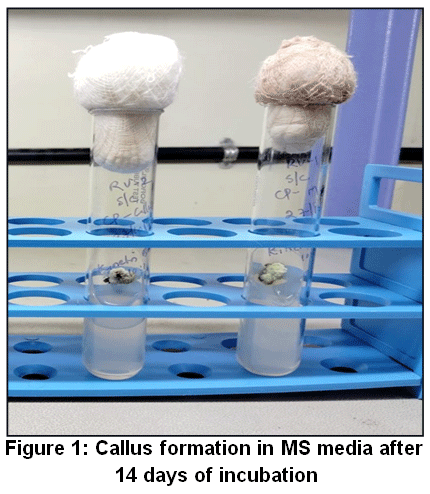 |
Figure 1: Callus formation in MS media after 14 days of incubation
|
Shoot initiation from stem with apical meristem
Stem segments bearing apical meristems of C. peltata were cultured on MS medium with 10:1 ratio of benzyl aminopurine (BAP) and Kinetin, Under these conditions, shoot primordia began to differentiate after 5 months of continuous incubation. The emerging shoots were small, axillary, and showed slow elongation, indicating a relatively low rate of direct organogenesis from the apical meristem without prior callus induction.
Table 2: Shoot initiation in MS media after 5 months of incubation of the stem explant of Cyclea peltata
| Medium + Hormone | Hormone Concentration and Ratio | Explant Type | Number of tubes inoculated | Days to response | Percentage of Shoot regeneration | Shoot length (cm) |
| MS + 2,4-D and Kinetin | 5 mg/L,1:1
|
Leaf | 6 | 14 days | 0%
(2/6 contamination, 4/6 dried and no response) |
Nil |
| MS + BAP and Kinetin | 5 mg/L, 10:1
|
Stem | 6 | 5 months | 17%
(2/6 contamination, 3/6 no response) |
3 cm |
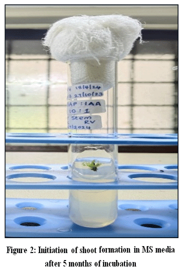 |
Figure 2: Initiation of shoot formation in MS media after 5 months of incubation
|
Shoot regeneration via BAP and IAA treatment
Stem segments carrying axillary buds or apical meristems were first cultured on MS mediumwith 1:1 ratio of 2,4‑D and Kinetin to induce callus. Subsequent subculture of callus‑bearing explants onto MS medium containing BAP and indole‑3‑acetic acid (IAA) in a 10:1 ratio resulted in the initiation of shoot formation within 6 weeks of incubation. Multiple shoots emerged from the callus–meristem interface, suggesting that the combination of prior callus induction with an optimized cytokinin–auxin balance (BAP: IAA, 10:1) promoted efficient organogenesis and shoot bud activation.
Table 3: Shoot initiation in MS media on sub culture, after 6 weeks of incubation, of the stem explant with axillary buds of Cyclea peltata
| Medium + Hormone | Hormone Concentration and Ratio | Explant Type | Number of tubes inoculated | Days to response | Percentage of Shoot regeneration | Shoot length (cm) |
| MS + 2,4-D and Kinetin and sub culture on MS + BAP and IAA | 5 mg/L, 1:1.
5 mg/L, 10:1. |
Stem segments carrying axillary buds | 6 | 6 weeks | 50%
(3/6 contamination) |
5-7 cm |
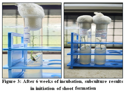 |
Figure 3: After 6 weeks of incubation, subculture results in initiation of shoot formation
|
Callus formation and subculture on IAA and kinetin
Leaf explants of Cyclea peltata cultured on MS medium supplemented with 2,4‑D and kinetin in the ratio 1:1 showed callus formation within 2 weeks of incubation. The callus developed from the cut margins and adaxial surfaces of the leaf explants, forming compact, light‑green, proliferative tissue. Subculture of this callus‑bearing explants onto MS medium supplemented with IAA and kinetin in the ratio 0.5:5 resulted in continued callus growth along with morphological changes suggestive of early organogenic potential.
Table 4: Callus formation from leaf explant of Cyclea peltata after 2 weeks of incubation on MS medium supplemented with 2,4‑D and kinetin in the ratio 1:1
| Medium + Hormone | Hormone Concentration and Ratio | Explant Type | Number of flasks inoculated | Days to response | Percentage of Callus induction | Nature of Callus |
| MS + 2,4-D and Kinetin | 5 mg/L, 1:1 | Leaf | 1 | 2 weeks | 100%
(0/1 contamination) |
Green coloured, compact, friable callus. |
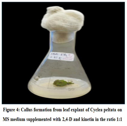 |
Figure 4: Callus formation from leaf explant of Cyclea peltata on MS medium supplemented with 2,4‑D and kinetin in the ratio 1:1
|
Indirect organogenesis from callus subculture
Callus obtained on subculture in MS medium with IAA and kinetin in the ratio 0.5:5 showed indirect organogenesis, with the development of meristematic swellings and shoot primordia arising from the callus tissue. Further subcultures were carried out on MS medium with BAP and IAA in10:1 ratio, which promoted the emergence and elongation of multiple shoots from the organogenic callus, indicating that the high‑cytokinin, low‑auxin environment (BAP: IAA, 10:1) is favourable for shoot organogenesis in Cyclea peltata The cultures were initially established on MS medium containing 2,4‑D and kinetin in 1:1 ratio, which induced compact, proliferative callus from leaf and stem explants.
Table 5: Callus formation and Indirect organogenesis in Cyclea peltata from sub cultured callus
| Medium + Hormone | Hormone Concentration and Ratio | Explant Type | Number of flasks inoculated | Days to response | Percentage of Callus induction / indirect organogenesis | Response |
| MS +2,4-D and Kinetin. | 5 mg/L, 1:1 | Leaf | 1 | 14 days | 100%
(0/1 contamination) |
Brown, friable callus |
| MS +IAA and Kinetin.
|
5 mg/L, 0.5:1
|
Callus | 1 | 2 months | 100% (0/1 contamination) | Indirect organogenesis (shoot formation from callus) |
| MS +BAP and IAA and Kinetin. | 5 mg/L, 10:1 | Callus | 2 | 3 months | 100% (0/2 contamination) | Indirect organogenesis (shoot formation from callus) |
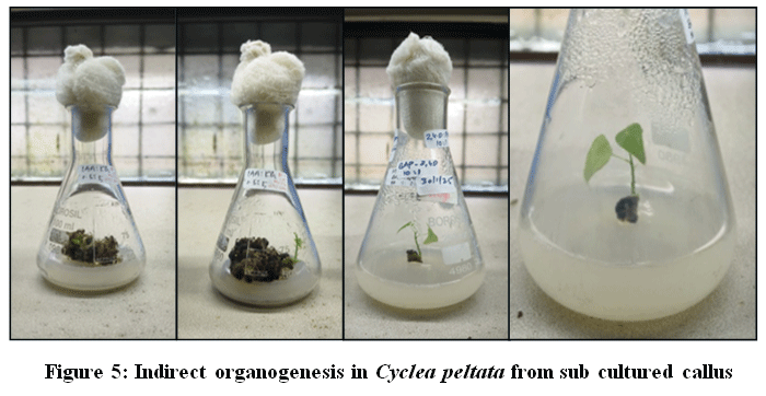 |
Figure 5: Indirect organogenesis in Cyclea peltata from sub cultured callus
|
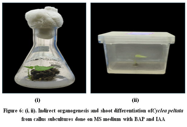 |
Figure 6: (i, ii) Indirect organogenesis and shoot differentiation ofCyclea peltata from callus subcultures done on MS medium with BAP and IAA
|
Tissue culture studies of T. acuminata
Nodal stem explants of Tiliacora acuminata were cultured on MS medium supplemented with benzyl aminopurine (BAP) and indole‑3‑acetic acid (IAA) in a 10:1 ratio. Under standard growth‑room conditions, the explants showed visible morphological changes after 28 days of incubation. Slight swelling at the nodes and axillary regions, along with the development of meristematic swellings, was observed, indicating early in vitro responsiveness. However, well‑developed shoots were not yet evident within this period, suggesting that the BAP–IAA (10:1) treatment, while inducing meristematic activity, may require further optimization for efficient shoot organogenesis in Tiliacora acuminata.
Table 6: Nodal stem explants of Tiliacora acuminata on MS medium with BAP and IAA in 10:1 ratio showing meristematic swellings.
| Medium + Hormone | Hormone Concentration and Ratio | Explant Type | Number of tubes inoculated | Days to response | Percentage of Nodal swelling | Nodal swelling (cm) |
| MS + BAP and IAA | 5 mg/L, 10:1. | Nodal stem | 6 | 28 days | 50%
(3/6 contamination) |
0.5 – 0.7 cm |
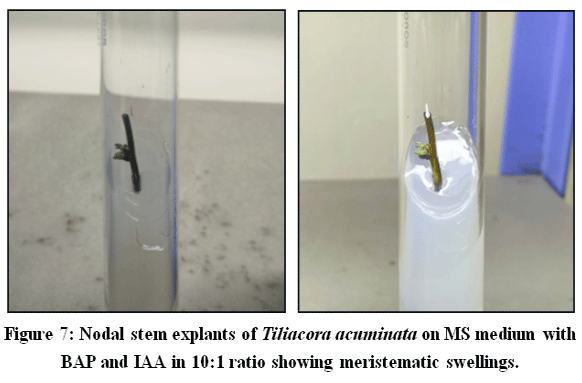 |
Figure 7: Nodal stem explants of Tiliacora acuminata on MS medium with BAP and IAA in 10:1 ratio showing meristematic swellings.
|
Discussion
This study confirms that leaf explants of Cyclea peltata exhibit robust callogenesis when cultured on MS medium with 2,4-D and kinetin at equal concentrations. The 1:1 auxin-to-cytokinin balance effectively triggers cell dedifferentiation and proliferation, a pattern well-documented in tissue culture where auxins like 2,4-D drive mitotic activity while cytokinins sustain tissue expansion. Initial callus appearance at cut edges after 14 days reflects typical wound-induced competence, enhanced by localized accumulation of endogenous hormones at injury sites.6,7
The resulting friable, light green callus demonstrates high totipotency, ideal for suspension cultures, metabolite extraction, or indirect organogenesis. Friable textures facilitate biomass scaling, while chlorophyll pigmentation signals photosynthetic viability under photoperiodic lighting. Sustained subculturing viability underscores the protocol’s potential for conserving this medicinal climber amid habitat pressures.8-10
Nodal explants of T. acuminata on MS + BAP: IAA (10:1) displayed nodal swellings by day 28, marking cytokinin-driven meristem activation and axillary bud priming. High BAP levels typically override apical dominance to stimulate lateral shoots, with minimal IAA aiding cell expansion without excessive callusing.11,12Yet the lack of elongated shoots indicates this regimen supports only de novo meristemoids, not full organogenesis. Such delays align with recalcitrance in woody lianas, where intrinsic hormone profiles or explant maturity demand extended timelines or phased media shifts. Excessive cytokinin may promote proliferation over differentiation, a common challenge in antioxidant-rich medicinal species.13,14
Refinements like lower BAP doses, TDZ substitution, or sequential rooting media could accelerate shoot maturation beyond 28 days. These findings validate leaf callus protocols for C. peltata biomass and nodal induction for T. acuminata as starting points for micropropagation.15-18Both systems advance sustainable sourcing of bioactive climbers, bridging conservation with pharmaceutical applications in Indian biotechnology frameworks.19-21
Conclusion
This study establishes efficient tissue culture protocols for Cyclea peltata using standard MS media prevalent in Indian laboratories, achieving both callus-mediated and indirect organogenesis. Leaf explants rapidly formed callus on 2,4-D: kinetin (1:1), with subsequent subculturing on IAA: kinetin (0.5:5) and BAP: IAA (10:1) yielding meristemoids and shoot development, outperforming slow direct shoot induction from apical stem segments on BAP: kinetin (10:1).
Tiliacora acuminata nodal explants on BAP: IAA (10:1) exhibited initial nodal swellings by 28 days, signalling morphogenic potential but requiring extended optimization for complete shoot regeneration. These findings offer scalable micropropagation for C. peltata conservation and a promising framework for refining T. acuminata protocols within Indian biotechnology systems.
Acknowledgement
The author gratefully acknowledges Department of Botany and Department of Biotechnology, CMS College Kottayam (Autonomous), Kerala, India for providing the necessary facilities to carry out this study.
Funding Sources
The author(s) received no financial support for the research, authorship, and/or publication of this article.
Conflict of Interest
The authors do not have any conflict of interest.
Data Availability Statement
This statement does not apply to this article.
Ethics Statement
This research did not involve human participants, animal subjects, or any material that requires ethical approval.
Informed Consent Statement
This study did not involve human participants, and therefore, informed consent was not required.
Clinical Trial Registration
This research does not involve any clinical trials.
Permission to reproduce material from other sources
Not Applicable.
Author Contributions
- Renji Varghese: Conceptualization, Methodology, Data Collection,Writing – Original Draft
- Jinu John: Analysis, Writing – Review & Editing
- Rogimon Plammoottil Thomas: Writing – Review
References
- Manda R, Seru G. Evaluation of hypoglycaemic and wound healing activities of Tiliacora acuminata. J Chem Pharm Res. 2016;8(5):494-497.
- Thomas TD, Shankar S. Multiple shoot induction and callus regeneration. Plant Biotech. 2009;3(2):67-74.
CrossRef - Shine VJ, Anuja GI, Suja SR, Raj G, Latha PG. Bioassay-guided fractionation of Cyclea peltata using in vitro RAW 264.7 cell culture, antioxidant assays and isolation of bioactive compound tetrandrine. J Ayurveda Integr Med. 2018; 4(3):5-10.
- Jyothi A, Meena KC, Bince M, Thomas DT. A rapid in vitro multiplication system for commercial propagation of pharmaceutically important Cyclea peltata (Lam.) Hook. and Thoms. based on enhanced axillary branching. Ind Crops Prod. 2010;31(1):92-98.
CrossRef - Jahan R, Khatun MA, Nahar N, Jahan FI, Chowdhury AR, Nahar A, et al. Use of Menispermaceae family plants in folk medicine of Bangladesh. Adv Nat Appl Sci. 2010;4(1):1-9.
- Kundu S, Ahmed KMMM, Mahavidyalaya RA. Traditional Indian herbs and their medicinal importance: an Ayurvedic approach with contemporary view. Int J Ayurvedic Herb Med. 2016;4(2):2260-2267.
- Bala M, Pratap K, Verma PK, Singh B, Padwad Y. J Ethnopharmacol. 2015;22(1):61-99.
- McMurry JE. Secondary metabolites: an introduction to natural product chemistry. In: Organic Chemistry with Biological Applications. 2009;12(3):1016-1046.
- Paul A, AVASR, SRM. Preliminary phytochemical screening of six medicinal plants of Menispermaceae. Int J Pharma Bio Sci. 2016;7(1):77-81.
- Müller H, Brackhagen O, Brunne R, Henkel T, Reichel F. Natural products in drug discovery. Ernst Schering Res Found Workshop. 2000;(32):205-216.
CrossRef - Bhagya N, Chandrashekar KR. Effect of growth regulators on callus induction from Cyclea peltata (Lam.) Hook. f. and Thoms. Asian J Pharm Clin Res. 2013;6(4):412-442.
- Gururaj HB, Giridhar P, Ravishankar GA. Micropropagation of Tinospora cordifolia (Willd.) Miers ex Hook. f. and Thoms.: a multipurpose medicinal plant. Curr Sci. 2007;92(2):23-26.
- Abraham J, Cheruvathur MK, Mani B, Thomas TD. A rapid in vitro multiplication system for commercial propagation of pharmaceutically important Cyclea peltata (Lam.) Hook. and Thoms. based on enhanced axillary branching. Ind Crops Prod. 2010;31(1):92-98.
CrossRef - Kuo CL, Chang JY, Kumar GS. In vitro production of benzylisoquinoline from Stephania tetrandra through callus culture under the influence of different additives. Bot Stud. 2011;52(2):285-294.
- Jyothi A, Meena K, Cheruvathur M, Thomas DT. A rapid in vitro multiplication system for commercial propagation of pharmaceutically important Cyclea peltata (Lam.) Hook. and Thoms. based on enhanced axillary branching. Ind Crops Prod. 2010;31(1):92-98.
CrossRef - Danya U, Udhayasankar MR, Arumugasamy K, Punitha. An efficient protocol devised for rapid callus induction from leaf explants of Stephania wightii (Arn.) Dunn., an endemic medicinal plant. 2012;12(1):92-98.
- Bhagya N, Chandrashekar K. In vitro production of tetrandrine from callus culture of Cyclea peltata Hook. f. Thoms. Proc Natl Acad Sci India Sect B Biol Sci. 2021;91(2):102-148.
CrossRef - Bhaskaran S, Dakshinamoorthi A, Kutralingam K, Ramasamy K. Evaluation of ethanolic extract of Tiliacora acuminata leaves for pancreatic lipase inhibition and lipid modulation: in silico and in vitro studies. Asian J Pharm Clin Res. 2025;18(1):175-183.
CrossRef - Fang K, Liu C, Gom B, Benrkia R, et al. Unveiling the mystery of the genus Tiliacora: a holistic view on the ethnomedicinal uses, phytochemical richness and pharmacological potentials. Fitoterapia. 2025;14(1);323-380.
CrossRef - De Wet H, Van Wyk BE. An ethnobotanical survey of southern African Menispermaceae. S Afr J Bot. 2008;74(1):64-78.
CrossRef - Jordan M. Seed plants of southern Africa: families and genera. In: Leistner OA, ed. Strelitzia. 2000;10(1)356-359.
Accepted on: 28-05-2026
Second Review by: Dr. Karpagam S
Final Approval by: Dr. Wagih Ghannam


![]()
![]()
