
Cytotoxic Effects of Chlorhexidine on Gingival Mesenchymal Stem Cells and their Implications for Regenerative Dentistry
 , Kishor Vasant Otari and Ajay Yashwant Kale
 Department of Pharmacology, Navsahyadri Institute of Pharmacy, Pune, India.
Corresponding author E-mail:shitalshinde9600@gmail.com
Download this article as:
ABSTRACT:Chlorhexidine (CHX) is one of the most widely used antiseptic agents in dentistry because of its broad antimicrobial activity and long-lasting substantivity. It is routinely used in mouth rinses, periodontal therapy, root canal irrigation, and surgical disinfection. However, increasing evidence suggests that chlorhexidine may exert cytotoxic effects on oral cells, including gingival fibroblasts, epithelial cells, and mesenchymal stem cells. Gingival mesenchymal stem cells (GMSCs) have attracted significant interest in regenerative dentistry due to their high proliferative capacity, multipotent differentiation potential, and immunomodulatory properties. These stem cells contribute to tissue repair and regeneration in periodontal tissues and are considered promising candidates for cell-based regenerative therapies. Despite its antimicrobial benefits, chlorhexidine exposure has been reported to impair stem cell viability, proliferation, migration, and differentiation. The cytotoxicity of chlorhexidine is believed to occur through multiple mechanisms, including oxidative stress, mitochondrial dysfunction, apoptosis induction, and disruption of cellular membranes. These adverse cellular responses may negatively affect the regenerative potential of gingival stem cells and compromise periodontal healing after clinical procedures. The present review summarizes current scientific evidence regarding the cytotoxic effects of chlorhexidine on gingival mesenchymal stem cells and examines the molecular mechanisms underlying these effects. Furthermore, the review discusses the implications of chlorhexidine-induced cytotoxicity in regenerative dentistry and highlights potential strategies to minimize its harmful impact while maintaining antimicrobial efficacy. Understanding the balance between antimicrobial activity and cellular toxicity is essential for optimizing clinical protocols in regenerative dental therapies.
KEYWORDS:Chlorhexidine; Cytotoxicity; Gingival Mesenchymal Stem Cells; Periodontal Regeneration; Regenerative Dentistry
Introduction
Chlorhexidine is a cationic BI-biguanide antiseptic that has been widely used in dentistry for more than five decades. The concept of antimicrobial and fungal properties of chlorhexidine has made it the gold standard of chemical agents for plaque control in the oral care business due to its broad-spectrum against Gram-positive bacteria, Gram-negative bacteria, fungi, and some viruses. In the periodontal disease prevention and treatment, it is issued as a routine prescription as mouth rinses, gels, sprays, and varnishes. Chlorhexidine is used in endodontics as well as in operating rooms as an irrigating and disinfecting agent.
The efficacy of chlorhexidine is highly dependent to bind and influencing the membrane integrityand bacterial cell walls. The motion leads to the leakage of the intracellular contents and, ultimately, cell death by microorganisms. The other commendable quality given to chlorhexidine is its substantivity, which refers to its ability to bind to the mouth regions. This method disables gradually with time, hence providing the chlorhexidine its long-lasting antimicrobial impacts.1
The recent past has recorded an increased interest in stem cell-based therapy in repair dentistry. Gingival mesenchymal stem cells represent a species of population of multipotent stem cells of the gingival connective tissue. These cells are able to grow and form various forms of cells, such as the osteoblasts, the chondrocytes, and the adipocytes. In addition to the ability to differentiate, the gingival stem cells are also immunomodulatory with tissue repair capability, albeit via paracrine signalling. They exist, can produce tremendous growth, and contain little or no ethical constraints; hence, they are the most promising sources of regenerative use in periodontal therapy and tissue engineering.2
However, in dental surgery, clinical use of chlorhexidine causes certain serious doubts regarding its influence on the viability and activity of the gingival stem cells. In vitro studies have indicated that chlorhexidine treatment will cause cell death, reduce cell growth, and cause cell death in mesenchymal stem cells. These effects are capable of inhibiting the process of remaining of the gingival tissues in addition to influencing the outcomes of the regenerative procedures in a negative way. In light of the increasing enthusiasm for the application of the stem cell-based regenerative dentistry, one needs to be aware of what the popular antiseptics, such as chlorhexidine, could do to stem cell biology. This review will provide a general overview of the cytotoxic effect of chlorhexidine on the gingival mesenchymal stem cells and also discuss the molecular pathway through which chlorhexidine may exert its effects on cellular damage. These findings applied to the regenerative dentistry and clinical practice are also discussed.3
Gingival Mesenchymal Stem Cells in Regenerative Dentistry
Biological Characteristics of Gingival Stem Cells
The unique type of adult stem cell present in the connective tissues of the gingiva is Gingival mesenchymal stem cells (GMSCs). These are cells largely derived from the lamina propria layer of the gingivitis tissue, and fall under a broad category, mesenchymal stem cells (MSCs). The mesenchymal stem cells are undifferentiated cell stroma that are self-renewing and multipotent in nature and can be differentiated into a wide range of differentiated cell types. Gingival tissue is one of the most attractive and convenient sources of stem cells used in regenerative medicine and dentistry, among many other sources of MSCs the human body.
The gingival mesenchymal stem cells have several distinct biological qualities which enable differentiation of these cells with other examples of cells present in the oral tissues. The morphological characteristics of such cells are defined by a fibroblast-like spindle-like figure in in vitro cell culture. They cultivate successfully in plastic culture properties and multiply in the anticipated environments in the laboratories. This is one of the relevant adherence properties that is utilized to identify mesenchymal stem cells in cell culture platforms. The Gingival stem cells produce colonies of adhesive fibroblast-like cells that are normally referred to as colony-forming unit fibroblasts, indicative of their clonogenic strength, besides replication abilities out of just one progenitor cell.4
Gingiva mesenchymal stem cells have a range of common surface markers, and these are usually associated with mesenchymal stem cells, molecularly. These are the markers on the surface of the cells, namely CD73, CD90, and CD105, that greatly mediate the cell adhesion, signalling, and differentiation. CD73 is an enzyme that is referred to as ecto-5′-nucleotidase and is involved in the metabolism of purines and immune system regulation. Thy-1 or CD90 is a glycoprotein that has been shown to mediate cell-linked communications and take part in signalling pathways that govern cell growth and differentiation. Endogenous or CD105 is transforming growth factor – beta receptor and is a significant determinant of angiogenesis and vascular growth.5
In addition to the mesenchymal markers expressed, hematopoietic markers, such as CD34, CD45, and CD14, were not expressed in the gingival stem cells. The lack of these markers demonstrates that the blood-forming tissues fail to form the gingival stem cells and destroy them in comparison to the hematopoietic stem cells. The provided marker pattern has been widely used as the identification/characterization method of gingival mesenchymal stem cells in the laboratory environment and clinical practices.6
One of the most prominent biological properties of the latter is the high level of proliferative abilities of the mesenchymal stem cells of the gingiva. A significant number of cell divisions can be performed on such cells without affecting the stemness properties and genetic stability. In fact, the spread rate of the gingival stem cells is higher, and the population doubling time is lower compared to other sources of mesenchymal stem cells such as the bone marrow-derived stem cells. This speedy replication rate allows scientists to obtain vast numbers of cells in a relatively short period, applicable in tissue engineering of cells and the application of the regenerative method of this type of medication.7
The other interesting aspect of the gingival stem cell is that it is highly stable on the genetic aspect and does not change even in long-term cultures. When grown over large volumes, a large number of stem cell populations are susceptible to genetic abnormalities. However, the use of gingival mesenchymal stem cells has been proven to preserve the constant chromosomes composition as well as limited genetic alterations even with the repeated passages in cell cultures. Their safety profile in the potential clinical use is enhanced by the stability.8
One more important advantage, which is associated with the use of gingival stem cells, is the availability. An example of the least invasive way to acquire gingivitis tissues during the execution of routine dental interventions is the removal of a tooth or performing a periodontal procedure or gingivitis. As opposed to oncologists who get the bone marrow through aspirating or other violent means of getting the stem cells, harvesting of the gingival tissue causes minimal harm to patients and has very minimal rates of side effects. To a greater degree, the fact that the gingival tissues can heal wounds effectively increases their capacity for rapid wound healing, rendering the process of harvesting tissues as safe and feasible to be carried out in clinical practice.9
Differentiation Potential
One of the features of the cell is the multipotent differentiation of the mesenchymal stem cell that constitutes the gingiva of human beings. These cells have the potential to become a number of types of specialized cells in the presence of the right environment and cues. The potential of distributing the gingival stem cell is much better than that of mesenchymal stem cells from other tissues, such as bone marrow, adipose tissue, and dental pulp.In addition to the osteogenic differentiation, Gingival stem cells may be adipogenic. With culturing in adipogenic media containing growth factors and lipase hormones, they will contain intracellular lipid droplets and develop adipocyte specific proteins in response to adipogenic media. This characteristic demonstrates that these are multipotent properties, and it confirms their mesenchymal stem cell properties.10
There is also potential for condrogenic differentiation of gingival mesenchymal stem cells. In such cases, these cells would produce chondrocyte-like cells when subjected to a set of conditions if tested under the conditions of chondrogenic induction, where cells release extra cells to the environment in the form of type II collagen and proteoglycans. Another example of the differentiation variety of the gingival stem cells is the cartilaginous-like structures.Besides being able to differentiate, these gingival stem cells also play an important role in the healing of tissues in virtue of the fact that it secretes bioactive substances. The cells produce a wide range of growth factors, cytokines, and signalling molecules that influence the surrounding cells and trigger their tissue regeneration. Vascular endothelial growth factor, transforming growth factor-beta, and fibroblast growth factors are some of the most vital growth factors secreted by the gingival stem cells.11
The growth factor of vascular endothelium plays a crucial role in angiogenesis, whereby new vessels are formed with the preexisting ones. The regeneration tissues require new blood vessels to supply oxygen and nutrients to them. The growth factor- beta transformation is seen in controlling the cell growth, cell differentiation, and cell production of extracellular matrix. Fibroblast growth factors also have the role of healing tissues due to their role in increasing fibroblasts and collagen production.These paracrine signalling pathways enable the effect of gingival stem cells on the behaviour and development of the surrounding microenvironment that facilitates tissue healing and the process of regeneration of the cells in the environment.12
Role in Periodontal Tissue Regeneration
The regenerative potential of gingival mesenchymal stem cells has been demonstrated in most experiments and animal models. Different specialized structures constitute periodontal tissues, and they include the gingiva, periodontal ligament, cementum, and alveolar bone. Periodontal disease has the potential to ruin these tissues rendering teeth to move and subsequent loss of teeth. Therefore, one of the goals of regenerative periodontal therapy is the multiplication of these structures.
Gingival dermal stem cells have a regenerative effect on periodontal tissues in several ways. Firstly, one can distinguish between them into osteoblasts, and thus, they can take an immediate part in the production of new alveolar bone. The bone regeneration is imperative to FABG just to restore the structural integrity of the periodontally diseased teeth.13
Second, the gingival stem cells may also be converted into fibroblast-like cells that contribute to the process of periodontal ligament tissue regeneration. The periodontal ligament also plays a significant role in the tooth’s function in bonding and anchoring the tooth to the surrounding bone, and in the diminishing the mechanical load in the mastication activity.In addition to the fact that they are differentiating with their properties, the gingival stem cells exhibit high immunomodulatory properties. When interacting with the immune cells, such as T lymphocytes, Macrodendritic cells, and dendritic cells, these cells are capable of regulating the immune responses. The release of anti-inflammatory cytokines and immunoregulatory molecules with the assistance of gingival stem cells, helps with the suppression of inflammation and the formation of optimal tissue repair around it.14
Gingival stemcells anti-inflammatory capability has significance farce of great intensity, particularly in periodontal disease, wherein chronic inflammation is a fundamental trigger of tissue-brokerage. There is a possibility that with the regulation of immune responses, gingival stem cells can suppress the action of inflammation and support the restoration of periodontal tissue.The abundance, ease, and accessibility of gingival mesenchymal stem cells have caused growing concern in exploring their application as a future therapeutic measure in regenerative dentistry because of their high level of proliferation, multipotent differentiation potential, and immunomodulatory ability. They are also investigating their potential use in other forms of regeneration, including periodontal regeneration, bone tissue engineering, and soft tissue reconstruction.The role of the gingival stem cells in biomaterials, scaffold design, and tissue engineering is expanding further due to new developments in these two fields. These cells can be used alongside bioactive scaffolds and growth factorsto developnew forms of more efficient regenerative therapies, which can replace the damaged tissues in the oral cavity.Generally, the gingival mesenchymal stem cells provide a good source of regenerative dentistry. Their biological properties and the ways of their treatment may be further studied, and it is likely that new strategies of treating periodontal diseases and acquiring new methods of restoring the oral tissues regeneration.15
Chlorhexidine in Dentistry
Chemical Structure and Mechanism of Action
Chlorhexidine is an antiseptic agent which is a synthetic product and belongs to the antimicrobial agents of the bisbiguanide group. Since chlorhexidine has a wide antimicrobial spectrum and half-life, the introduction of chlorhexidine into the field of dentistry and medicine, in general, has permeated the field of chlorhexidine establishment, due to its widespectrum of activity. Chlorhexidine is a chemical compound that consists of a pair of the same chlorophenyl rings bonded together through six hexamethylene rings and two sets of biguanide. The resultant molecular structure provides the molecule with a high level of positive charge that is highly essential in its antimicrobial action of action.16
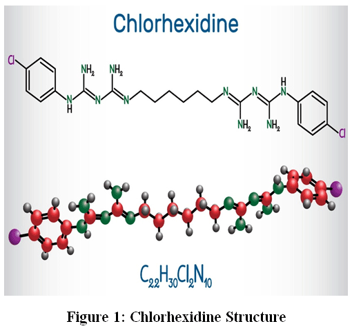 |
Figure 1: Chlorhexidine Structure
|
Fig.1, The antimicrobial mechanism and chemical structure of chlorhexidine. Chlorhexidine is a cationic bisbiguanide antiseptic, which consists of two chlorophenyl rings with a hexamethylene bridging between them. The positively charged molecule interacts with the negatively charged bacterial cell membranes, causing membrane integrity disruption, intracellular component leakage, and microbial disruption.The molecule of chlorhexidine is cationic, and it can strongly interact with negatively charged surfaces. The proteins, bacterial cell walls, and membranes are negatively charged phospholipids which attract the positively charged chlorhexidine molecules. In the presence of bacterial cells, the electrostatic interaction enables the antiseptic to be sticky to the cells due to the contact with the chlorhexidine. This communication disrupts the cell membrane integrity and modifies the cell membrane permeability.17
The anti-microbial effect of the chlorhexidine is concentrationdependent. Chlorhexidine primarily exhibits bacteriostatic properties at rather low concentrations. The compound affects the oncotic metabolism of the bacteria in this state, causing an increase in permeability of cell membranes. This causes the intracellular leakage of part of the essential components in the cell, such as potassium ions and low molecular weight compounds, due to loss of membrane permeability. This causes suppression of metabolic activities of bacteria; therefore, no cell growth and reproduction. Althoughby this stage bacteria cells may still be structurally intact, their reproductive ability is reduced considerably.Chlorhexidine exhibits a bactericidal effect at very high levels of concentration. In this case, the compound will cause extensive rupture of the cell membrane of the bacterial cell as well as cellular proteins coagulation. It is the greater permeability of the membrane that allows the chlorhexidine to enter the cytoplasm on a deeper level, where the nucleic acids and other important enzymes are found. The outcome of this contact is irreversible precipitation of cell contents and cell death. Chlorhexidine bactericidal actions are therefore associated with structural damage to the microorganism cells and irreversible damage to important elements in the organism.18
Chlorhexidine possesses another exclusive characteristic known as substantivity, besides having direct antimicrobial effects. Substantivity is the ability through which the compound adheres to oral tissues such as enamel, dentin, mucosal surface, and salivary proteins and stays for longer periods of time. Chlorhexidine is washed off the surface where it is deposited onto the oral cavity after its attachment, and it develops antimicrobial activity hours after application. Such duration of action enhances the efficacy of chlorhexidine regarding the control of microbial groups in the mouth cavity to a significant level.Chlorhexidine has Gram-positive, Gram-negative, and facultative anaerobic activity, in addition to some fungi, and against all microorganisms within the spectrums of Gram-positive and Gram-negative. Chlorhexidine tends to increase the susceptibility of gram-positive bacteria more because the cell walls of these bacteria contain higher concentrations of negatively charged molecules that aid in the association of chlorhexidine. Gram-negative sensitivity is not as high as that of bacteria have an outer membrane, where the penetration of the antiseptic agents may be contained. However, chlorhexidine is effective against most of the dental plaque and periodontal disease related medical pathogens.19
In addition to being antibacterial, chlorhexidine is antifungal, particularly against Candida species. It is a clinically meaningful property which finds application in treating oral fungi and oral microbial balance. Other studies also reported antiviral action against certain enveloped viruses, which also demonstrate the versatility of chlorhexidine as an antiseptic agent.The other mechanism through which chlorhexidine acts as an antimicrobial agent is through interference with thedevelopment of biofilm. Multifactorial microbial biofilm, dental plaque, is attached to the tooth surfaces, and it is of the foremost importance in the pathogenesis of periodontal disease and dental caries. Chlorhexidine deters attachment and forms biofilms as well as prevents their attachment. The plaque of the oral cavity is impeded by chlorhexidine based on the surfaces and development of the oral illnesses.Although chlorhexidine is very antimicrobial, it is not discriminating against the host cell compared to microbial cells. The membrane-perturbing effects that are causing harm to the bacterial membrane are just like the ones that can be done to the mammalian cell membrane under certain conditions. This indiscriminate practice has resulted in the challenge of a potential cytotoxic propinquity on the tissues of the mouth, particularly after a considerable amount of chlorhexidine is used and a period of usage.20
Clinical Applications
The application of chlorhexidine in dental practice has been highly embedded due to its huge success in controlling the mouth microorganisms, and also in the prevention of dental diseases. In the form of a mouth rinse, chlorhexidine is among the most commonly used drugs clinically as a plaque control agent and in the treatment of gingivitis. It is common to recommend the use of chlorhexidine mouthwashes to patients with issues with proper oral hygiene, with the aid of pure mechanical cleaning of the plaque. Chlorhexidine mouth rinses enable the minimization of the impact of plaque in the mouth and the avoidance ofgingival inflammation through reduction of the number of gastric organisms in the mouth.In particular, chlorhexidine is particularly applicable as a mouth rinse in the oral cavity in cases of gingivitis that is an inflammatory disease of the gamma of gel bacterium and is primarily caused by plaque of these bacteria. It has been observed that routine preparation of the chlorhexidine mouthwash is essential in lessening the intensity of the plaque and generating gingivectomy bleeding. This is because the antimicrobial protection with chlorhexidine is substantive and therefore the chlorhexidine would appear to be a more effective protection of antimicrobial compared to most antimicrobial mouth rinses.21
The other clinical application of chlorhexidine is in endodontics. Root canal infections occur as a result of microorganisms entering the pulp chamber as well as the root canal system. The treatment of root canal involves good removal of these microorganisms to yield good treatment. The chlorhexidine root canal irrigant is the most frequently used irritant root canal irrigant since it is an antimicrobial against endodontic pathogens such as Enterococcus faecalis. In addition to the ability to kill bacteria, chlorhexidine can also penetrate dentinal tubules and sustain an antimicrobial effect in the root canal system.Chlorhexidine is used to dress the oral and maxillofacial surgery as both a reoperative and postoperative antiseptic. Preoperative chlorhexidine mouth rinsing is commonly prescribed before dental extractions and surgical procedures in order to reduce the percentage of bacterial contamination of the surgical site. Chlorhexidine has the potential to minimize the occurrence of postoperative infections by a reducing the microbial load of the mouth cavity to improve the effectiveness of surgery.22
The implant industry has also received the application of chlorhexidine. The colonization of bacteria during the healing process of dental implants may expose the implant to vulnerability, thus leading to peri-implantitis and implant failure. The patients are normally administered with chlorhexidine oral rinses after undergoing the implant surgery in an attempt to maintain a clean environment in the mouth and to minimize the possibilities of oral contamination of the surgery site. The antimicrobial properties of chlorhexidine are effective in offering the protection of the operative area and assist in the adhesion of the implant and the bone tissues that are adjacent.
The application of chlorhexidine, however, has numerous adverse effects, irrespective of all its numerous clinical benefits in cases of its use at high concentration or during prolonged usage. Extrinsic staining of teeth and restorative materials is considered as being one of the most commonly occurring side effects. Chlorhexidine can react with foodstuffs and other chromogens in beverages, and the appearance of brownish stains on the surface of teeth. Although these are not permanent stains, and they can be eliminated during the process of professional cleaning, they may affect the patient’s adherence to chlorhexidine use in the long-term.23
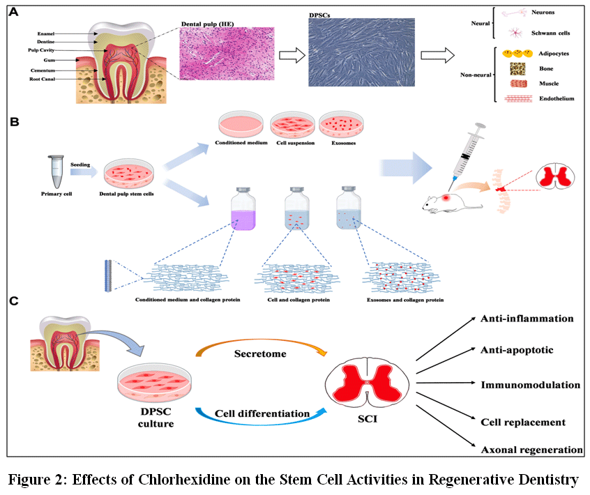 |
Figure 2: Effects of Chlorhexidine on the Stem Cell Activities in Regenerative Dentistry
|
Fig 2, Another adverse effect that has been widely reported is known as taste change, also known as dysgeusia. The other phenomenon in some patients undergoing the use of chlorhexidine mouth rinses is taste perception. The effect of such a change can also be felt in the recognition of salty or bitter taste and may persist up to several hours after the process of rinsing.The issue of cytotoxic activity of chlorhexidine on host cells has received increased attention in recent years. It is documented in research that there could be a negative effect of chlorhexidine usage on oral fibroblasts, epithelial cells, and mesenchymal stem cells. These cell killer activities may impact the healing and regeneration of tissues of the wound, especially when chlorhexidine is administered in direct contact with the point of surgery or the uncovered areas.Therefore, despite the fact that chlorhexidine is an indispensable antimicrobial agent when applied in the world of dentistry, it must be utilized in a well-balanced way such that it is applied in the most appropriate manner with the minimal possible side effects. Practicing dental treatment must also be conducted with good considerations since the right level of concentration, duration of use, and clinical manifestations would yield the best results.24
Cytotoxic Effects of Chlorhexidine on Gingival Mesenchymal Stem Cells
Effects on Cell Viability
The exposure of the cells to the chlorhexidine was found to have one of the probable and most reliable implications, which included finding that the viability of the cells was diminished considerably with respect to the type of cells that were affected, the gingiva mesenchymal stem cells (GMSCs). Cell viability is the capacity of the cells to stay alive, be metabolic and structural, in contact with the external substances. Within the framework of the sphere of regenerative dentistry, stem cell conservation is especially required since the latter is the epicenter of the process of the tissue repair, regeneration, and wound healing. Nevertheless, numerous experimental research studies have demonstrated the fact that even at concentration levels practically utilized as a dentally appropriate agent, chlorhexidine was capable of generating cytotoxicity to the oral cell, including the gingival stem cell.25
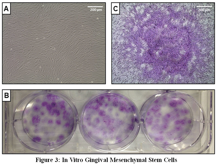 |
Figure 3: In Vitro Gingival Mesenchymal Stem Cells
|
Fig 3, The in vitro experiment of the cultured gingival mesenchymal stem cell experiment has proven that there is a decreased survivability of the cell with increasing exposure of chlorhexidine to the cell in a dose-dependent way. The treatment of the stem cells with the chlorhexidine solutions also reacts with the mammalian cell membrane. There is a chance that this contact would destabilize the lipid biological layer cell membrane, resulting in the greater permeability of the cell membrane and destruction of the cell. Chlorhexidine concentration that causes it and the length of exposure are mainly important when considering the amount of damage.It is found that despite the benefits, experimental studies also discovered pretty harsh effects that chlorhexidine can even exert on the viability of the stem cell when it is exposed for a short time. As an indication, exposure durations of one minute have been considered even to produce influences on cell survival, which is being measured. The findings are specific to clinical dentistry, which employs the use of chlorhexidine in the carrying out of periodontal irrigation, root canal debridement, and scalpel-to-scalpel site debridement. The clinical environments may fall within the short range of the exposure time ; however, owing to its substantivity, chlorhexidine may possess long-term biological effects on the oral tissues.26
The other aspect is the cell viability, and that is the generation of oxidative stress. Reactive oxygen species have been linked to the exposure of the cell to chlorhexidine. These reactive molecules are able to damage such constituents of the cell as proteins, lipids, and nucleic acids. Oxidative stress could alter the workability of mitochondria and result in the loss of energy stores by cells. The event of damaging the mitochondria may possess negative implications on the existence of the cell since mitochondria are involved in the production of adenosine triphosphate that is energy utilized incellular functions.Chlorhexidine can only cause apoptotic processes that result in programmed death, other than oxidative stress. The well-constructed process of cell destruction is termed apoptosis, the process whereby the maladjusted or damaged cells are erased. The process of apoptosis is a normal physiological process, but in case the emptying process of apoptosis is over-expressed, there would be a loss of viable cells on a large scale. The exposure of chlorhexidine has been mentioned to cause caspase enzyme, Bax pro-apoptotic protein, and a reduction in the Bcl-2 anti-apoptotic protein. Such patterns of molecular manipulations help in the invigoration of the process of apoptosis and also help in decrease in viability of the stem cells.27
The effects on periodontal tissue healing are titanic when the cell viability following the use of chlorhexidine is diminished. Gingival mesenchymal stem cells support the healing and restoration of degenerated tissues because the cells are able to multiply, differentiate, and secrete growth factors. On the one hand, the regenerative ability of a tissue can be impaired, assuming that exposure tochlorhexidine can lead to a decline in the number of viable stem cells. This is viewed as alarming as far as regenerative dental care, whereby the outcome success of an outcome is dependent on the population of the stem cells in the tool-and-die.
More so, the chlorhexidine sensitivity of the gingival stem cells may be affected by the condition of the environment and plant properties. The cytotoxicity of chlorhexidine can depend on such variables as the pH, the temperature, and the presence of serum proteins in solution. Elsewhere, the affairs of chlorhexidine may be neutralized to some extent by binding with serum proteins to the molecule, and may prevent the occurrence of the interaction of the latter with the cell membranes. However, despite the protective conditions, chlorhexidine has been demonstrated to exert its cytotoxic effects on stem cells.Overall, among the worst effects associated with exposure to chlorhexidine in the instance of gingival mesenchymal stem cells is the loss ofcell viability. The mechanisms implicated in this cytotoxicity should be familiar so as to ensure the maximum is extracted from the safe uses of chlorhexidine in clinical dentistry.28
Effects on Cell Proliferation
The reason is thatcell proliferation is an important biological process that assists the stem cells to split into new ones that have the capacity to sustain, heal, and fix tissues. Regenerative dentistry proposes that the cell immobility development of the gingivalisomes mesenchymal stem cell is essential in structuring the periodontal tissues, and the metamorphosis of the structures. This has, however, been imparted by exposure to the chlorhexidine that was also observed to severely suppress the proliferation ability of these stem cells.The second implementation of decreased proliferation is because of the generation pathways of stress-signalling. The intracellular signalling cascades that are triggered by the use of chlorhexidine may be triggered by the presence of oxidative stress, which will influence the manner in which the cell will respond to the injury. They may cause the activation of the genes of the stress-response that suppress cell multiplication and trigger their restoration. Although this type of response can be used in a protective fashion when applied in a long-term course of action, a long-term activating response can lead to long-term cell growth release.29
The damaged regeneration of the gingival stem cells is quite critical to tissue regeneration. The periodontal therapy will demand the stem cells to multiply and produce enough cells which can be utilized to replace the damaged tissues. The passive regeneration of the tissue is also exposed to jeopardy as long as the exposure to chlorhexidine postpones the formation of the stem cells. This lack would have a negative impact on the effectiveness of such operations as periodontal regeneration, bone grafting, and the positioning of implants.Regardless of these issues, it must be stated that the level of chlorhexidine concentration may also differ in terms of proliferation inhibiting. The chlorhexidine level below the high level might not be as damaging as the high level of cell proliferation is. Consequently, the concentration and exposure times of chlorhexidine can be streamlined in order to minimize the negative cellular responses to chlorhexidine and preserve the antimicrobial properties of chlorhexidine.30
Effects on Stem Cell Differentiation
The exposure to chlorhexidine also has the ability to moderate the differentiation of the gingival mesenchymal stem cells, besides interfering with the cell viability and proliferation. The cell differentiation process of stem cells is very regulated to the extent that the undifferentiated cells further differentiate to become particular cell types with particular functions within the body. When it comes to the replacement of damaged tissues, the possibility of the gingival stem cells to differentiate into osteoblasts, fibroblasts, and any other form of cell in case of periodontal regeneration is absolute.Osteogenic differentiation is one of the differentiation programs that are important and relevant differentiation programs in the treatment of periodontal therapy. The stem cell can be transformed into cells that produce the bone-forming cells, and this can be used to produce an extracellular matrix that is mineralized, which is possible by the conversion of the stem cell to an osteogenic differentiated cell. It is an extremely delicate task in the process of the regeneration of alveolar bone that supports the teeth.Nevertheless, exposure to chlorhexidine has been found to lead to the alteration of the osteogenic differentiation ability of the gingival stem cells.The evidence taken as a whole indicates that chlorhexidine may have negative impacts on the many aspects of the biology of gingival stem cells, such as viability, proliferation, and differentiation. These are the impacts that reveal why one should be cautious concerning the use of chlorhexidine as regards its use as a part of the clinical processes pertaining to the adoption of a procedure linked to the process of tissue regeneration.31
Molecular Mechanisms of Chlorhexidine-Induced Cytotoxicity
Oxidative Stress
The reactive oxygen species is considered one of the main mechanisms that explain why chlorhexidine is cytotoxic. Oxidative stress is caused by excessive production of reactive oxygen species, and it causes cellular protein, lipid, and DNA damage.
Mitochondrial Dysfunction
Exposure to chlorhexidine is linked to mitochondrial digestion and destabilization of the mitochondrial membrane potential. The impairment of the mitochondrial extracts decreases the generation of cellular energy and thus plays a role in the generation of the apoptotic pathways.32
Apoptosis and Programmed Cell Death
Caspase signalling pathway activation is not the only way that chlorhexidine can affect apoptosis. It has also raised and decreased the expression of Bax and Bcl-2 when subjected to chlorhexidine as a pro and anti-apoptotic protein respectively.33
Implications for Regenerative Dentistry
Even the application of chlorhexidine in the dental regenerative practice is a cause of concern, considering that the substance is a cytotoxic agent to the gingival stem cell, too. The ability of stem cells to repair tissues is lower, and this aspect might be detrimental in periodontal healing and regeneration, since it has been established that stem cells are important in repairing tissues.
Treatment of cells is the number one principle in preserving cells in regenerative dentistry. High concentrations of chlorhexidine can slow down the regeneration of the gingival tissues and decrease the healing duration.34
Use of Lower Concentrations
The reduction of the cytotoxic impacts and the maintenance of the antimicrobial effect of the chlorhexidine dilution can be realised in clinical practice.
Limited Exposure Time
Presently, the harm caused by chlorhexidine on the cell can also be reduced by such a method as the extension of the exposure duration of the chlorhexidine, hence, increasing the survival volume of the stem cells.
Alternative Antimicrobial Agents
Some other alternative antiseptic agents are also in the offing that can replace chlorhexidine. They are herbal extracts, essential oils, and antimicrobial peptides that can reduce their cytotoxicity in relation to host cells.
Future Directions
Further studies would develop better antimicrobial activities that will be less harmful, in addition to the outcomes that will ensure that the integrity of the stem cells is not compromised, but would successfully inhibit the microbes. Innovations within the trends of tissue engineering and biomaterials can form the solution to the simultaneous administration of antimicrobial agents and regenerative therapies.
It demands further research and clinical trials in the future to understand more about the long-term impacts of chlorhexidine on the regeneration of tissues, which may be through the mediation of the stem cells.35
Discussion
Chlorhexidine (CHX) is commonly used in dentistry because of its strong antimicrobial action; however, this review emphasizes its harmful effects on gingival mesenchymal stem cells (GMSCs), which are essential for periodontal regeneration. The reviewed studies consistently show that CHX decreases cell viability in a dose- and time-dependent manner, even at concentrations typically used in clinical practice. The cytotoxicity of CHX is largely linked to oxidative stress, where excessive production of reactive oxygen species (ROS) disturbs normal cellular balance and causes damage to lipids, proteins, and DNA. This is accompanied by mitochondrial dysfunction, leading to reduced ATP production and impaired cellular activity. At the molecular level, CHX triggers intrinsic apoptotic pathways by increasing pro-apoptotic factors such as Bax and caspases, while reducing anti-apoptotic proteins like Bcl-2. These changes accelerate programmed cell death and decrease the number of viable regenerative cells. Moreover, CHX inhibits cell proliferation and disrupts osteogenic differentiation, which limits the capacity of GMSCs to support tissue repair and bone formation. These effects are particularly important in regenerative dental procedures, where proper stem cell function is crucial for healing. The severity of these effects depends on factors such as concentration and duration of exposure, suggesting that even slight variations in clinical use can influence outcomes. Hence, optimizing CHX application is important to reduce its negative impact on stem cells while preserving its antimicrobial benefits.
Conclusion
Chlorhexidine is another antimicrobial, which has not been scrutinized as not working effectively in the dentistry arena. Nevertheless, there is growing to have more and more evidence that it is capable of cancerogenic effects on the mesenchymal stem cells of the gingiva. Such effects are the diminishing cell viability, the harmful impact on the proliferation, and the suppressed differentiation. The pathophysiological pathways include oxidative stress and mitochondrial dysfunctions and activate the apoptotic pathways. As the major wound repairing of the tissues is carried out by the gingival stem cells, the recovery of the regenerative dental therapy can be affected by the cytotoxicity of chlorhexidine. Thus, the clinical practice should carefully consider the process of chlorhexidine concentration and time measures. The study needs to be done further so that an antimicrobial solution can be developed to allow regulation of the microbes in order to ensure death and activities of the stem cells are avoided.
Acknowledgement
The authors would like to acknowledge the Department of Pharmacology, Navsahyadri Institute of Pharmacy, for providing the necessary facilities to conduct this review.
Funding Sources
The author(s) received no financial support for the research, authorship, and/or publication of this article.
Conflict of Interest
The authors do not have any conflict of interest.
Data Availability Statement
This statement does not apply to this article.
Ethics Statement
This research did not involve human participants, animal subjects, or any material that requires ethical approval.
Informed Consent Statement
This study did not involve human participants, and therefore, informed consent was not required.
Clinical Trial Registration
This research does not involve any clinical trials.
Permission to reproduce material from other sources
Not Applicable.
Author Contributions
- Shital Ajit Shinde: Conceptualization, literature survey, writing – original draft, and final manuscript preparation.
- Kishor Vasant Otari: Supervision, critical revision of the manuscript, and academic guidance.
- Ajay Yashwant Kale: Overall supervision, administrative support, and final approval of the manuscript.
References
- Bayirli AB, Uytun M, Ekinci B, Genc D. In vitro evaluation of chlorhexidine and oleuropein: antibacterial activity and effects on gingival mesenchymal stem cell viability. S Afr J Bot. 2026;190:338-345. doi:10.1016/j.sajb.2024.12.015
CrossRef - Brunello G, Becker K, Scotti L, Drescher D, Becker J, John G. Effect of three chlorhexidine-based mouthwashes on human gingival fibroblasts: an in vitro study. Appl Sci. 2022;12(5):2417. doi:10.3390/app12052417
CrossRef - Fiorillo L, D’Amico C, Mehta V, Cicciu M, Cervino G. Chlorhexidine cytotoxicity on oral tissues: a systematic review. Oral Oncol Rep. 2024;9:100245. doi:10.1016/j.oor.2023.100245
CrossRef - Cunha G, Saugo GDA, Gabrielli MAC, et al. Cytotoxicity evaluation of chlorhexidine and Blue® M applied to human gingival fibroblasts and keratinocytes. J Stomatol Oral Maxillofac Surg. 2024;125(5):101923. doi:10.1016/j.jormas.2024.101923
CrossRef - Saberikia H, Rashno M, Babadi F, Rakhshan V. Preliminary cytotoxicity assessment of Jaftex versus chlorhexidine mouthwashes on human gingival fibroblasts. BMC Oral Health. 2025;25:379. doi:10.1186/s12903-025-0379-5
CrossRef - Danila AI, Rominu M, Munteanu K, et al. Development of solid nanosystems for delivery of chlorhexidine with increased antimicrobial activity and decreased cytotoxicity. Molecules. 2025;30(1):162. doi:10.3390/molecules30010162
CrossRef - Hashim NT, Babiker R, Dasnadi SP, et al. The impact of ozone on periodontal cell line viability and function. Curr Issues Mol Biol. 2025;47(2):72. doi:10.3390/cimb47020072
CrossRef - Santos CA, Pessoa ADS, Vilhena FV, et al. Less cytotoxic phthalocyanine derivative promotes in vitro wound healing compared to chlorhexidine. J Appl Biomater Funct Mater. 2025;23:22808000251314630. doi:10.1177/22808000251314630
CrossRef - Steckiewicz KP, Cieciorski P, Barcinska E, et al. Silver nanoparticles as chlorhexidine and metronidazole drug delivery platforms for treating periodontitis. Int J Nanomedicine. 2022;17:495-517. doi:10.2147/IJN.S339144
CrossRef - Sepahdar A, Kordestani M, Karkhane M, et al. Gum-assisted magnesium oxide nanoparticles using guar extract focusing on their bioactivities. IET Nanobiotechnol. 2025;2025:9924353. doi:10.1049/nbt2.9924353
CrossRef - Bhandi S, Patil S, Boreak N, et al. Effect of different intracanal medicaments on the viability and survival of dental pulp stem cells. J Pers Med. 2022;12(4):575. doi:10.3390/jpm12040575
CrossRef - Inchingolo F, Inchingolo AM, Latini G, et al. Oxidative stress and natural products in orthodontic treatment: a systematic review. Nutrients. 2023;16(1):113. doi:10.3390/nu16010113
CrossRef - Caruso S, Valenti C, Marinucci L, et al. Systematic review of zinc’s benefits and biological effects on oral health. Materials. 2024;17(4):800. doi:10.3390/ma17040800
CrossRef - Umapathy VR, Natarajan PM, Swamikannu B. Regenerative strategies in dentistry: harnessing stem cells, biomaterials and bioactive materials for tissue repair. Biomolecules. 2025;15(4):546. doi:10.3390/biom15040546
CrossRef - Brunello G, Becker K, Scotti L, Drescher D, Becker J, John G. Chlorhexidine mouthwashes and fibroblast cytotoxicity. Appl Sci. 2022;12:2417. doi:10.3390/app12052417
CrossRef - Fiorillo L, Cicciu M, Cervino G, D’Amico C. Cytotoxic potential of antiseptics in oral tissues. Oral Oncol Rep. 2024;9:100245. doi:10.1016/j.oor.2023.100245
CrossRef - Cunha G, Gabrielli M, Pereira-Filho V. Cytotoxic effects of dental antiseptics. J Stomatol Oral Maxillofac Surg. 2024;125:101923. doi:10.1016/j.jormas.2024.101923
CrossRef - Saberikia H, Rashno M, Babadi F. Mouthwash cytotoxicity in gingival fibroblasts. BMC Oral Health. 2025;25:379. doi:10.1186/s12903-025-0379-5
CrossRef - Santos CA, Damante CA, Zangrando MS. Phthalocyanine derivatives versus chlorhexidine. J Appl Biomater Funct Mater. 2025;23:22808000251314630. doi:10.1177/22808000251314630
CrossRef - Steckiewicz KP, Bauer M, Kamysz W, Inkielewicz-Stepniak I. Nanoparticles as antimicrobial drug delivery systems. Int J Nanomedicine. 2022;17:495-517. doi:10.2147/IJN.S339144
CrossRef - Bhandi S, Patil S, Raj AT, Testarelli L. Dental pulp stem cell survival with intracanal medicaments. J Pers Med. 2022;12(4):575. doi:10.3390/jpm12040575
CrossRef - Inchingolo F, Latini G, Ferrante L, Dipalma G. Natural antioxidants and oxidative stress in dentistry. Nutrients. 2023;16:113. doi:10.3390/nu16010113
CrossRef - Caruso S, Pagano S, Valenti C. Zinc and oral tissue regeneration. Materials. 2024;17:800. doi:10.3390/ma17040800
CrossRef - Umapathy VR, Natarajan PM, Swamikannu B. Biomaterial strategies in regenerative dentistry. Biomolecules. 2025;15:546. doi:10.3390/biom15040546
CrossRef - Hashim NT, Islam MS, Rahman MM. Ozone effects on periodontal cells. Curr Issues Mol Biol. 2025;47:72. doi:10.3390/cimb47020072
CrossRef - Danila AI, Rominu M, Popovici R. Chlorhexidine nanosystem development. Molecules. 2025;30:162. doi:10.3390/molecules30010162
CrossRef - Bayirli AB, Uytun M, Ekinci B, Genc D. Stem cell viability and chlorhexidine exposure. S Afr J Bot. 2026;190:338-345. doi:10.1016/j.sajb.2024.12.015
CrossRef - Sepahdar A, Kordestani M, Bahadori S. Magnesium oxide nanoparticles in biomedical applications. IET Nanobiotechnol. 2025. doi:10.1049/nbt2.9924353
CrossRef - Brunello G, Becker J, John G. Cytotoxicity of antiseptic mouthwashes. Appl Sci. 2022;12:2417. doi:10.3390/app12052417
CrossRef - Cunha G, Bufalino A, Pereira-Filho V. In vitro cytotoxicity of antiseptics. J Stomatol Oral Maxillofac Surg. 2024;125:101923. doi:10.1016/j.jormas.2024.101923
CrossRef - Santos CA, Oliveira RC, Damante CA. Antimicrobial derivatives and wound healing. J Appl Biomater Funct Mater. 2025;23:22808000251314630. doi:10.1177/22808000251314630
CrossRef - Steckiewicz KP, Kamysz W, Inkielewicz-Stepniak I. Nanomedicine approaches for periodontal therapy. Int J Nanomedicine. 2022;17:495-517. doi:10.2147/IJN.S339144
CrossRef - Fiorillo L, Cervino G, Cicciu M. Oral antiseptics and cytotoxicity review. Oral Oncol Rep. 2024;9:100245. doi:10.1016/j.oor.2023.100245
CrossRef - Bhandi S, Patil S, Raj AT. Intracanal medicaments and stem cell survival. J Pers Med. 2022;12:575. doi:10.3390/jpm12040575
CrossRef - Inchingolo F, Latini G, Dipalma G. Oxidative stress and dental therapy. Nutrients. 2023;16:113. doi:10.3390/nu16010113
CrossRef
Abbreviations
CHX: Chlorhexidine
GMSCs: Gingival Mesenchymal Stem Cells
MSCs: Mesenchymal Stem Cells
ALP: Alkaline Phosphatase
Runx2: Runt-Related Transcription Factor 2
VEGF: Vascular Endothelial Growth Factor
TGF-β: Transforming Growth Factor Beta
FGFs: Fibroblast Growth Factors
ROS: Reactive Oxygen Species
ATP: Adenosine Triphosphate
Bax: Bcl-2 Associated X Protein
Bcl-2: B-Cell Lymphoma-2 Protein
DNA: Deoxyribonucleic Acid
RNA: Ribonucleic Acid
CD: Cluster of Differentiation
CD73: Ecto-5′-Nucleotidase
CD90: Thy-1 Cell Surface Marker
CD105: Endoglin Marker
HGEPp: Human Gingival Epithelial Progenitor Cells
HGF-1: Human Gingival Fibroblast Cell Line
PLGA: Poly (Lactic-co-Glycolic Acid)
PVA: Polyvinyl Alcohol
MSC-CFU: Mesenchymal Stem Cell Colony Forming Unit.
Accepted on: 09-05-2026
Second Review by: Dr. Randa Salah Gomaa Mahmoud
Final Approval by: Dr. Eugene A. Silow


![]()
![]()
