
Apocynin in Diabetes: Modulation of Oxidative Stress and Inflammatory Signaling
 , Kishor Otari, and Ajay Kale*
 Department of Pharmacology, Navsahyadri Institute of Pharmacy, Pune, India.
Corresponding Author E-mail: ajaykalephd@gmail.com
Download this article as:
ABSTRACT:Diabetes mellitus is a chronic metabolic disorder characterized by persistent hyperglycemia and progressive development of microvascular and macrovascular complications. Increasing evidence identifies oxidative stress and redox-sensitive inflammatory signaling as central mechanisms underlying diabetes-associated organ injury. Apocynin, a naturally occurring methoxy-substituted phenolic compound, has gained attention as a redox-modulating agent with potential therapeutic relevance in diabetes. The present review summarizes the chemical characteristics, mechanistic actions, and protective effects of apocynin in experimental models of diabetic complications. Apocynin primarily acts through inhibition of NADPH oxidase assembly, thereby limiting excessive reactive oxygen species generation and suppressing downstream activation of redox-sensitive pathways. Through modulation of oxidative and inflammatory cascades, apocynin demonstrates protective effects in diabetic nephropathy, neuropathy, cardiomyopathy, and retinopathy. In addition to reducing oxidative burden, apocynin supports endogenous antioxidant defenses and improves cellular and metabolic stability. However, despite these promising findings, no clinical studies have yet evaluated apocynin in patients with diabetes or its associated complications. Although preclinical findings are promising, translational challenges, including pharmacokinetic limitations and limited clinical evaluation, remain to be addressed. Collectively, apocynin emerges as a promising candidate targeting convergent redox-inflammatory pathways implicated in diabetic complications.
KEYWORDS:Apocynin; Diabetes Mellitus; NADPH Oxidase; Oxidative Stress; Reactive Oxygen Species
Introduction
Diabetes mellitus (DM) is a chronic metabolic disorder characterized by persistent hyperglycemia resulting from impaired insulin secretion, insulin action, or both, and remains one of the most prevalent non-communicable diseases worldwide.1 Recent estimates from the International Diabetes Federation indicate a rapidly increasing global burden, with approximately 536.6 million individuals affected in 2021 and projections suggesting an increase to 783.2 million by 2045. Notably, nearly half of these cases remain undiagnosed, reflecting the insidious and progressive nature of the disease.2,3 This expanding prevalence is accompanied by a substantial increase in disease burden, driven largely by the development of chronic complications that significantly impair quality of life and increase the risk of premature mortality.4 Collectively, these epidemiological trends and clinical factors underscore the growing challenge posed by diabetes and highlight the need for improved management strategies as well as a deeper understanding of the molecular mechanisms underlying disease progression.
At the molecular level, chronic hyperglycemia triggers multiple pathogenic processes, with oxidative stress (OS) acting as a key mediator of cellular dysfunction and tissue injury.5 OS arises from an imbalance between the production of reactive oxygen species (ROS) and the capacity of endogenous antioxidant defense systems to neutralize them.6 In diabetes, persistent hyperglycemia enhances ROS generation through several mechanisms, including glucose autoxidation, protein glycation, mitochondrial dysfunction, and activation of enzymatic sources such as NADPH oxidase (NOX).5,7 Excess ROS disrupts cellular redox homeostasis and activates stress-responsive inflammatory signaling pathways.8 As a result, this redox imbalance contributes to metabolic dysregulation, progressive tissue injury, and the development of major diabetes-associated complications, including nephropathy, neuropathy, cardiomyopathy, and retinopathy, which collectively account for significant morbidity and mortality in individuals with diabetes.5,9
Despite advances in understanding the role of OS in diabetes pathogenesis, current pharmacological strategies for DM management primarily focus on achieving glycemic control through insulin therapy and oral antidiabetic agents.10 While effective in lowering blood glucose levels, these interventions do not adequately address the underlying oxidative imbalance and chronic low-grade inflammation that drive disease progression.11 Notably, OS and inflammatory signaling often persist even in patients with well-controlled glycaemia, partly due to metabolic memory, a phenomenon in which prior hyperglycemic exposure induces sustained cellular and molecular damage, and consequently patients remain vulnerable to progressive tissue injury and long-term complications.¹² In addition, conventional therapies are associated with limitations such as adverse effects, declining efficacy, and incomplete protection against diabetes-related organ dysfunction.¹³ These challenges highlight the need for complementary therapeutic approaches that target redox-dependent and inflammatory pathways involved in diabetes pathogenesis.
In this context, increasing attention has been directed toward plant-derived bioactive compounds with antioxidant and anti-inflammatory properties as potential modulators of OS–driven pathology. These compounds often exhibit multi-targeted actions and favorable safety profiles, enabling simultaneous regulation of key pathogenic pathways.14 Among these, apocynin has emerged as a promising candidate due to its ability to inhibit NOX–mediated ROS generation and modulate redox-sensitive signaling cascades. Accumulating evidence suggests that apocynin confers protective effects across multiple diabetes-associated complications by attenuating OS, suppressing inflammatory responses, and preserving cellular and tissue integrity.15,16 This review therefore aims to critically evaluate the mechanistic role of apocynin in modulating OS–driven inflammatory pathways and to highlight its therapeutic potential in the prevention and management of diabetes-associated complications.
Apocynin: Chemical Properties, Botanical Sources, and Ethnomedicinal Relevance
Apocynin is a naturally occurring phenolic compound belonging to the class of methoxy-substituted catechols and is predominantly isolated from plant species traditionally used in diverse systems of medicine.17Picrorhiza kurroa is widely acknowledged as the primary botanical source of apocynin.18 Additionally, there are reports of apocynin being present in other plants used in Ayurveda, traditional Chinese medicine, and various folk medicinal systems. The plants mentioned have a long history of use in managing inflammatory, hepatic, respiratory, and metabolic disorders. Their traditional applications have offered early insights into the therapeutic potential of apocynin, which in turn has led to systematic pharmacological investigations.18-22
Structurally, apocynin possesses a well-defined phenolic framework characterized by a hydroxyl group, a methoxy substituent, and an acetyl side chain attached to an aromatic ring (figure 1).17 The phenolic hydroxyl group contributes to ROS scavenging activity, whereas the methoxy group enhances molecular stability and lipophilicity, thereby facilitating cellular uptake.23 Together, these structural attributes underpin its redox-modulating capacity and contribute to its biological activity in OS–driven conditions. The defined chemical framework of apocynin has also enabled its synthesis and structural modification, facilitating further exploration of structure–activity relationships relevant to pharmacological applications.24On the basis of these chemical and ethnomedicinal characteristics, subsequent investigations have focused on elucidating the molecular mechanisms through which apocynin exerts its biological actions, particularly its capacity to modulate OS and inflammation-related pathways central to DM and its complications.23,24
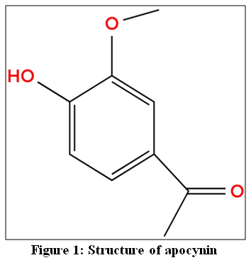 |
Figure 1: Structure of apocynin
|
Mechanism of Action of Apocynin in Diabetes
Apocynin is widely recognized as an inhibitor of NOX, a family of membrane-associated enzyme complexes that represent a major source of ROS under diabetic conditions.25 NOX enzymes catalyze the transfer of electrons from NADPH to molecular oxygen, resulting in the formation of superoxide and related reactive intermediates. Structurally, these enzymes consist of membrane-bound catalytic subunits and cytosolic regulatory components, such as p47phox and p67phox, which translocate to the membrane upon activation to assemble the functional oxidase complex.26 Sustained activation of NOX isoforms contributes to excessive ROS production and amplification of OS and inflammatory signaling pathways in diabetes.27 Apocynin has been reported to inhibit NOX activity by interfering with the assembly of this active enzyme complex, particularly by preventing the translocation of cytosolic subunits such as p47phox, thereby limiting superoxide generation. On this basis, apocynin has been regarded as a pharmacological inhibitor of NOX-derived ROS and a potential therapeutic agent for mitigating OS–mediated damage in diabetes.17,25
However, subsequent experimental evidence has challenged the view of apocynin as a classical direct NOX inhibitor. The precise mechanism of apocynin remains a subject of active investigation and appears to be highly context-dependent.28 Apocynin is proposed to function as a prodrug requiring peroxidase-mediated activation, particularly by myeloperoxidase, to generate reactive dimeric species such as diapocynin, which interfere with enzyme assembly.29 This mechanism is most prominent in peroxidase-rich phagocytic cells.30 In contrast, in vascular endothelial and smooth muscle cells, which exhibit limited peroxidase activity, apocynin often fails to directly inhibit NOX and instead exerts its effects primarily through antioxidant mechanisms, including modulation of ROS-dependent signaling pathways.31 Consistent with this cell-type specificity, apocynin has been reported to exhibit relatively weak direct free radical scavenging activity while demonstrating greater efficacy in neutralizing non-radical oxidants such as hydrogen peroxide (H2O2) and hypochlorous acid.30 Its effects also appear to vary across cellular systems, with studies reporting both increases and reductions in ROS levels in non-phagocytic cells, likely reflecting differences in cellular redox environments, enzyme expression, and experimental conditions.28
In addition, apocynin has been associated with preservation of endogenous antioxidant defenses, including superoxide dismutase and catalase.32 It has also been reported to influence multiple signaling pathways associated with diabetic complications, including modulation of eNOS-dependent superoxide production and suppression of nuclear factor-κB (NF-κB) and nod-like receptor pyrin domain-containing 3 (NLRP3) inflammasome activation.28,33 By modulating these interconnected oxidative and inflammatory pathways, apocynin may enhance the efficiency of insulin signaling, regulate cellular stress responses, and support metabolic homeostasis.34 Apocynin thus acts as a context-dependent modulator of OS, which may contribute to its protective effects in diabetes-associated complications.
Table 1: Natural sources of apocynin and their traditional medicinal uses
| Plant source | Plant part used | Traditional medicinal uses |
| Picrorhiza kurroa | Roots and rhizomes | Used in Ayurveda for diabetes, liver disorders, fever, asthma, and inflammatory conditions.18 |
| Apocynum cannabinum | Roots | Traditionally used as a cardiotonic, diuretic, and anti-inflammatory agent.19 |
| Apocynum venetum | Leaves and stems | Used in traditional Chinese medicine for hypertension, anxiety and cardiovascular disorders.20 |
| Iris germanica | Roots and rhizomes | Employed in folk medicine for antimicrobial and anti-inflammatory purposes.21 |
| A. androsaemifolium | Roots | Traditional remedies for circulatory disorders and in cold remedies.22 |
Apocynin in Diabetic Complications
Apocynin in Diabetic Nephropathy
Diabetic nephropathy (DN) is a chronic microvascular complication of DM characterized by progressive damage to the glomeruli, mesangial expansion, injury to podocytes, thickening of the glomerular basement membrane, interstitial fibrosis, and a gradual decline in renal function. This decline ultimately leads to chronic kidney disease and, in some cases, end-stage renal failure. Clinically, patients with DN exhibit albuminuria, a declining glomerular filtration rate, hypertension, and progressive proteinuria as the disease progresses. This condition significantly diminishes the quality of life and elevates mortality rates among individuals with diabetes.35,36Globally, DN accounts for a significant proportion of chronic kidney disease cases. Nearly one-third of individuals with DM develop clinically significant nephropathy during their lifetime. This condition represents a major public health burden due to its associated risks of cardiovascular disease and the necessity for renal replacement therapy.37 The pathogenesis of DN is multifactorial, involving metabolic, hemodynamic, oxidative, and inflammatory mechanisms. Chronic hyperglycemia leads to excessive generation of reactive oxygen species (ROS), formation of advanced glycation end-products, activation of protein kinase C, and deregulation of growth factor signaling. These processes contribute to endothelial dysfunction, proliferation of mesangial cells, accumulation of extracellular matrix, and tubulointerstitial fibrosis.38,39
Preclinical studies indicate that apocynin mitigates structural and functional renal injury in DN through multiple mechanisms (figure 2). In streptozotocin-induced diabetic rats, OS is characterized by increased renal H2O2 levels, elevated lipid peroxidation products, and significant proteinuria, reflecting oxidative injury and renal dysfunction. This oxidative imbalance is partly driven by activation of the NOX system and the associated overproduction of ROS. These alterations are associated with mesangial expansion and increased expression of fibronectin and type I collagen, contributing to extracellular matrix accumulation and progressive glomerular injury.40,41 Apocynin attenuates these changes by reducing ROS generation, normalizing p47phox translocation and gp91phox expression, and limiting mesangial matrix expansion, thereby preserving renal architecture and function.42
It has also been reported that innate immune and inflammatory mechanisms contribute significantly to the progression of DN, with pattern recognition receptors (PRRs) playing central roles. Among these, nucleotide-binding oligomerization domain-like receptors (NLRs), particularly NLRP3, are critically involved.43 In the diabetic milieu, PRR activation together with excessive ROS generation sustains renal inflammatory signaling. NLRP3 forms the inflammasome complex, promoting activation of caspase-1 and release of pro-inflammatory cytokines such as interleukin-1β and interleukin-18, thereby driving inflammation and fibrotic remodeling.44 X-linked inhibitor of apoptosis protein (XIAP) is functionally linked to this pathway.45 Apocynin has been shown to attenuate this process by reducing the diabetes-induced upregulation of NLRP3 and XIAP, thereby suppressing inflammasome activation and associated renal injury.46
In addition, modulation of pro-inflammatory mediators and activation of HO-1/Nrf2 cytoprotective signaling contribute to its renoprotective effects.47 In vitro studies using renal mesangial cells demonstrate that apocynin suppresses key inflammatory mediators, including inducible nitric oxide synthase (iNOS), cyclooxygenase-2 (COX-2), and monocyte chemoattractant protein-1 (MCP-1). This effect is associated with activation of the HO-1/Nrf2 pathway, a major endogenous antioxidant defence system. Apocynin-induced activation of Nrf2 and subsequent induction of HO-1 enhance antioxidant capacity and suppress MCP-1 expression, thereby attenuating inflammatory signaling and contributing to cytoprotection in diabetic renal injury.48
Apocynin further exerts protective effects by preserving glomerular structure and preventing renal apoptosis.49 In diabetic rats subjected to contrast-induced nephropathy, apocynin significantly reduced plasma urea and creatinine levels, indicating improved renal function. Histopathological findings demonstrated reduced tubular degeneration, diminished interstitial oedema, decreased vascular congestion, and preservation of glomerular architecture. In addition, apocynin markedly reduced renal apoptosis, highlighting its protective role against superimposed renal injury in diabetic conditions.50Collectively, these findings indicate that apocynin exerts multifaceted renoprotective effects by targeting OS, inflammatory signaling, and structural remodeling in DN.
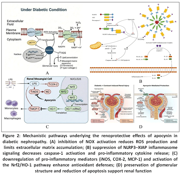 |
Figure 2: Mechanistic pathways underlying the renoprotective effects of apocynin in diabetic nephropathy.
|
Apocynin in Diabetic Neuropathy
Diabetic neuropathy (Dn) is a common microvascular complication of DM, affecting a substantial proportion of patients over the course of the disease. It is characterized by progressive damage to peripheral nerves and typically presents with sensory loss, neuropathic pain, tingling, and altered pain perception, often beginning in the distal extremities in a characteristic stocking–glove distribution.51 The development of Dn is closely associated with chronic hyperglycemia–induced OS, which disrupts neuronal and glial function, impairs nerve blood flow, and compromises nerve conduction. In addition, hyperglycemia-induced metabolic disturbances, including activation of the polyol pathway and accumulation of advanced glycation end products, further exacerbate neuronal injury. Excessive oxidative and nitrosative stress is recognized as a central driver of nerve damage, positioning redox-modulating pathways as important therapeutic targets.51–53
Preclinical studies indicate that apocynin exerts neuroprotective effects in Dn primarily through attenuation of hyperglycaemia-induced oxidative and nitrosative stress, which underlies both functional and structural nerve damage, as depicted in figure 3. In streptozotocin-induced diabetic models, apocynin exerts neuroprotective effects by modulating OS–related mechanisms. In addition to inhibiting NOX activity, it improves nociceptive pain thresholds, indicating attenuation of hyperalgesia. Catalase expression is reduced, suggesting diminished oxidative burden rather than impaired antioxidant defence. Histopathological analysis of sciatic nerve tissue shows reversal of extracellular matrix expansion, axonal and myelin degeneration, and Schwann cell loss, indicating preservation of peripheral nerve architecture.54 In addition to its direct neuroprotective effects, apocynin has been shown to improve neurovascular function in Dn. A study by Cotter et. al55 evaluated the effect of apocynin on peripheral nerve perfusion in diabetic animal models and demonstrated a significant improvement in endoneurial blood flow. This effect was attributed to alleviation of OS mediated microvascular dysfunction, resulting in enhanced nerve perfusion and oxygen delivery. Restoration of endoneurial blood flow by apocynin contributed to improved nerve conduction and functional recovery, underscoring the importance of microvascular protection as a complementary mechanism in its overall neuroprotective action. These findings suggest that apocynin attenuates neuropathic pain and structural nerve damage via OS modulation.
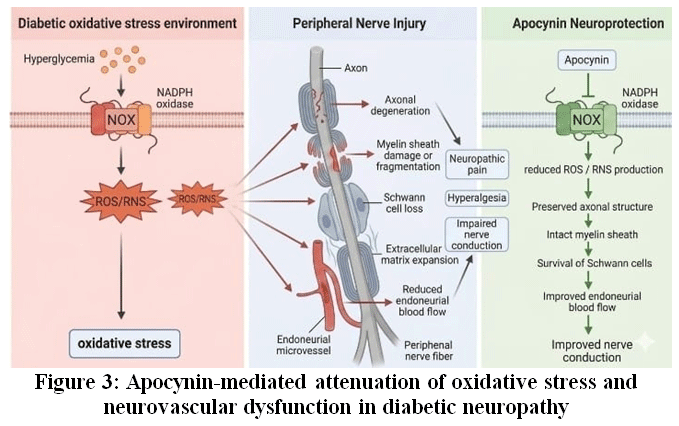 |
Figure 3: Apocynin-mediated attenuation of oxidative stress and neurovascular dysfunction in diabetic neuropathy.
|
Apocynin in Diabetic Cardiomyopathy
Diabetic cardiomyopathy (DCM) is a distinct cardiac complication of DM characterized by structural and functional abnormalities of the myocardium that occur independently of coronary artery disease, arterial hypertension, or valvular disorders. Clinically, DCM typically progresses from early diastolic dysfunction to overt systolic impairment and heart failure.56 At the cellular and molecular levels, chronic hyperglycemia promotes OS, low-grade inflammation, cardiomyocyte apoptosis, and myocardial fibrosis, collectively driving adverse cardiac remodeling and contractile dysfunction. Among these processes, dysregulated redox signaling and excessive generation of ROS play a central role by activating stress-responsive pathways that impair cardiomyocyte survival and myocardial metabolism. This mechanistic framework provides a strong rationale for targeting redox-driven pathways in the prevention and management of DCM.57,58
The co-existence of DM and arterial hypertension is a common clinical scenario that accelerates myocardial remodeling and functional decline. Evidence from hypertensive–diabetic animal models has provided insight into the cardioprotective potential of apocynin. Rosa et al.59 demonstrated that apocynin significantly reduced OS and attenuated pathological cardiac remodeling in spontaneously hypertensive rats with DM. Treatment was associated with reduced myocardial hypertrophy, decreased interstitial fibrosis, and improved left ventricular function, indicating protection against diabetes-associated cardiac alterations. In experimental models of DCM, apocynin also limits myocardial injury by reducing ROS accumulation and suppressing redox-sensitive signaling pathways. In particular, inhibition of the apoptosis signal-regulating kinase-1 (ASK1)–p38/c-Jun N-terminal kinase (JNK) pathway contributes to reduced cardiomyocyte apoptosis, attenuation of inflammatory signaling, and limitation of pathological remodeling.60 Apocynin further restores endogenous antioxidant defenses, including superoxide dismutase, catalase, and glutathione-dependent enzymes, thereby re-establishing redox balance within the diabetic heart. These effects are accompanied by reduced inflammatory cytokine expression, preservation of mitochondrial integrity, and decreased extracellular matrix deposition. In addition, apocynin improves myocardial insulin sensitivity and glucose utilization, supporting enhanced cardiac metabolic efficiency.60-63 These findings suggest that apocynin protects against DCM by attenuating OS–mediated myocardial injury and modulating inflammatory, apoptotic, and metabolic pathways involved in disease progression (figure 4).
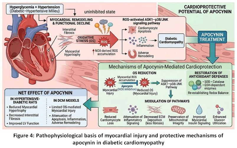 |
Figure 4: Pathophysiological basis of myocardial injury and protective mechanisms of apocynin in diabetic cardiomyopathy
|
Apocynin in Diabetic Retinopathy
Diabetic retinopathy (DR) is a progressive microvascular complication of DM characterized by retinal vascular dysfunction, neuroglial injury, and chronic inflammation. Sustained hyperglycaemia promotes OS, inflammatory signaling, and breakdown of the blood–retinal barrier, ultimately leading to retinal ischemia, neuronal apoptosis, and visual impairment. In addition, hyperglycaemia-induced microvascular damage, increased vascular permeability, and dysregulated angiogenic signaling, including vascular endothelial growth factor activation, contribute to capillary degeneration and pathological neovascularization. Among these pathogenic factors, OS mediated inflammatory pathways play a central role in the initiation and progression of DR, making them important therapeutic targets.64,65
Experimental evidence suggests that apocynin has protective effects in DR, primarily by modulating the inflammatory and redox-sensitive signaling pathways that are influenced by OS, as illustrated in Figure 5. In streptozotocin-induced diabetic rats, apocynin improves retinal structural and vascular abnormalities by inhibitingthe Toll-like receptor 4 (TLR4)/NF-κB pathway. This inhibition leads to decreased nuclear translocation of NF-κB and a downregulation of pro-inflammatory mediators, such as tumour necrosis factor-α, interleukin-1β, and interleukin-6.66 Apocynin also modulates OS markers by reducing malondialdehyde levels and enhancing antioxidant defences, such as superoxide dismutase activity and glutathione levels. Additionally, apocynin regulates apoptotic pathways by decreasing the expression of the pro-apoptotic protein Bax while increasing the levels of the anti-apoptotic protein Bcl-2, which helps limit retinal cell apoptosis.66 Further evidence suggests that the reduction of excessive ROS generation aids in preserving the activity of peroxisome proliferator-activated receptor-γ (PPARγ), an important transcriptional regulator for maintaining retinal vascular stability and anti-inflammatory signaling.67 This preservation is crucial for limiting retinal dysfunction associated with inflammation. The findings by Ahmad et al.68 indicate that apocynin provides protection to glial cells, which play a vital role in neurovascular homeostasis. This protection arises from apocynin’s capacity to diminish oxidative damage and promote cell survival in hypoxic retinal Müller cells and diabetic retinal tissue. The findings indicate that apocynin provides retinal protection by reducing inflammation driven by OS, preserving signaling pathways sensitive to redox changes, and maintaining the integrity of retinal cells. This ultimately helps to slow the progression of DR.
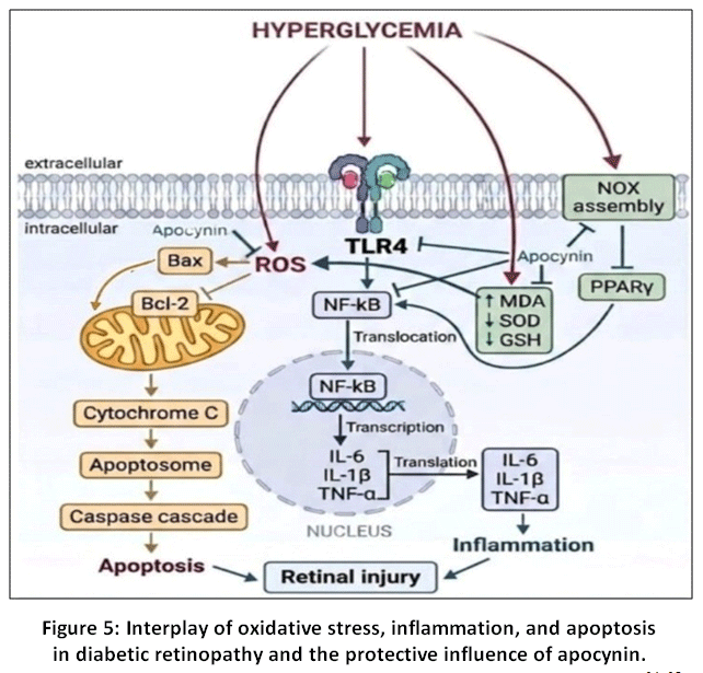 |
Figure 5: Interplay of oxidative stress, inflammation, and apoptosis in diabetic retinopathy and the protective influence of apocynin.
|
Limitations and Translational Challenges of Apocynin
A key limitation in the current evidence base is the heavy reliance on animal models, particularly streptozotocin induced and high fat diet induced models.32,34,40,54,62,66 While these models provide important mechanistic insights, they do not fully reflect the complexity and progressive nature of human diabetes.69 STZ models primarily mimic acute insulin deficiency rather than the gradual development of type 2 DM,70 whereas HFD models show variable metabolic phenotypes across strains.71,72In addition, interspecies differences in NOX expression, redox regulation, and inflammatory signaling limit direct extrapolation to human biology.73 Moreover, doses used in preclinical studies often exceed levels that may be achievable in clinical settings, and study durations are typically short, limiting their ability to model chronic disease progression.40,54,62,66
The absence of human clinical trial data represents the most critical evidentiary gap limiting the translation of apocynin to clinical practice. To date, no studies have evaluated apocynin in patients with DM or its associated complications. Evidence in humans is limited to small exploratory investigations in oxidative stress–related conditions such as chronic obstructive pulmonary disease and osteoarthritis, where modest reductions in oxidative biomarkers have been reported. However, these studies did not progress to advanced trial phases, and no definitive conclusions regarding clinical efficacy can be drawn.74,75 This limited translational progress likely reflects challenges related to pharmacokinetics, bioavailability, and sustained target engagement, which remain insufficiently characterized in humans.
Beyond the absence of clinical data, several pharmacokinetic and mechanistic barriers further complicate the translational development of apocynin. Following oral administration, it undergoes rapid first-pass metabolism, resulting in low bioavailability and uncertainty regarding whether effective concentrations can be maintained in target tissues.76 Its context-dependent mechanism, consistent with the mechanistic considerations outlined above, further complicates its activity, as effective NOX inhibition requires myeloperoxidase-mediated activation, which is limited in many tissues relevant to diabetic complications. Consequently, its effects in these settings may rely predominantly on non-specific antioxidant activity, potentially limiting sustained efficacy.28–31
Table 2: Molecular mechanisms and protective effects of apocynin in major diabetic complications
| Diabetic complications | Major pathological changes | Key molecular targets of apocynin | Therapeutic outcomes |
| Diabetic nephropathy | Glomerular injury; mesangial expansion; podocyte damage; GBM thickening; ECM accumulation; proteinuria; ↓ GFR | ↓ NOX-derived ROS; ↓ p47phox translocation and gp91phox expression; ↓ NLRP3/XIAP signaling; ↓ iNOS, COX-2, MCP-1; activates HO-1/Nrf2 pathway | ↓ OS and inflammation; ↓ proteinuria and fibrosis; preserves glomerular structure; improves renal function; anti-apoptotic effects |
| Diabetic neuropathy | Peripheral nerve degeneration; Schwann cell loss; axonal/myelin damage; neuropathic pain; impaired nerve blood flow | ↓ NOX activity and ROS accumulation; ↓ p47phox-associated signaling; ↓ nitrosative stress; improves microvascular function and endoneurial blood flow | ↓ Hyperalgesia and neuropathic pain; preserves peripheral nerve structure; improves nerve conduction and functional recovery |
| Diabetic cardiomyopathy | Myocardial hypertrophy; interstitial fibrosis; cardiomyocyte apoptosis; mitochondrial dysfunction; | ↓ Myocardial ROS; inhibits ASK1–p38/JNK signaling; restores antioxidant enzymes (SOD, catalase, GSH); improves mitochondrial integrity and metabolic signaling | ↓ Hypertrophy, fibrosis, and apoptosis; improves myocardial contractility, ventricular function, and cardiac metabolic efficiency |
| Diabetic retinopathy | Retinal vascular dysfunction; neuroglial injury; inflammation; OS; retinal apoptosis | Suppresses TLR4/NF-κB signaling; ↓ TNF-α, IL-1β, IL-6, and MDA; ↑ SOD and GSH; modulates Bax/Bcl-2 balance; preserves PPARγ signaling | ↓ Retinal inflammation and oxidative damage; inhibits apoptosis; preserves retinal neurovascular integrity; delays DR progressionretinopathy |
These challenges are further reflected in the considerable variability of dosing protocols across preclinical studies, where inconsistent doses and routes hinder identification of a clear therapeutic window, compounded by the lack of systematic dose–response evaluation.40,54,62,66 Several emerging strategies have been proposed to improve bioavailability, including nanoparticle-based delivery systems and targeted formulations designed to exploit myeloperoxidase rich inflammatory environments for site-specific activation. However, these approaches remain in early stages of development.77 Additional constraints, including formulation instability, limited tissue penetration, and the risk of disrupting physiological redox signaling at higher doses, further complicate the design of safe and effective therapeutic regimens.78
When considered alongside other antioxidant and NOX-targeting strategies investigated in diabetes, apocynin presents a distinct but incompletely validated therapeutic profile. Among NOX-targeting agents, compounds such as setanaxib (GKT137831) have progressed further in clinical evaluation; however, clinical studies have not consistently demonstrated clear therapeutic benefit, highlighting the challenges associated with translating NOX-targeted therapies into effective treatments.79 In comparison, commonly studied antioxidants such as resveratrol and curcumin have shown variable clinical efficacy, largely due to limitations in bioavailability and target engagement.80,81 In this context, the therapeutic relevance of apocynin remains uncertain, and its clinical potential will depend on systematic bioavailability optimization, well-designed dose-finding studies, and ultimately randomized clinical trials in diabetic populations.
Conclusion
Diabetes mellitus is characterized not only by chronic hyperglycemia but also by sustained oxidative stress and redox-sensitive inflammatory signaling that collectively drive the development of multiorgan complications. The present review highlights apocynin as a mechanistically coherent redox-modulating agent with the capacity to target these convergent pathogenic pathways. By inhibiting NOX assembly and limiting excessive reactive oxygen species generation, apocynin attenuates downstream activation of NF-κB and inflammasome signaling, thereby reducing oxidative injury, inflammation, fibrosis, and cellular dysfunction. Experimental evidence consistently demonstrates protective effects of apocynin in diabetic nephropathy, neuropathy, cardiomyopathy, and retinopathy, where modulation of oxidative and inflammatory cascades contributes to preservation of structural integrity and functional outcomes.
Importantly, apocynin acts not merely as a conventional antioxidant but as a regulator of redox-dependent signaling networks influencing metabolic stability, insulin responsiveness, mitochondrial integrity, and cellular survival. Its ability to simultaneously modulate oxidative, inflammatory, and metabolic pathways positions apocynin as a promising adjunct therapeutic candidate in diabetes. However, despite encouraging preclinical findings, translation into clinical application remains limited, underscoring the need for further pharmacological and translational evaluation.
Future research should focus on addressing key translational challenges associated with apocynin, including optimization of formulation strategies to improve bioavailability, detailed pharmacokinetic and pharmacodynamic profiling, and clarification of tissue-specific mechanisms of action. Well-designed clinical studies are required to establish safety, efficacy, and long-term therapeutic benefit in diabetic patients. Additionally, exploration of combination strategies integrating apocynin with standard antidiabetic therapies may offer synergistic benefits by targeting both glycemic control and redox-inflammatory pathways. A deeper understanding of its interaction with emerging molecular targets, including inflammasome signaling and redox-sensitive transcription factors, will further define its therapeutic potential in mitigating diabetic complications.
Acknowledgement
The authors gratefully acknowledge Navsahyadri Institute of Pharmacy, Pune for all the support.
Funding Sources
The author(s) received no financial support for the research, authorship, and/or publication of this article.
Conflict of Interest
The authors do not have any conflict of interest.
Data Availability Statement
This statement does not apply to this article.
Ethics Statement
This research did not involve human participants, animal subjects, or any material that requires ethical approval.
Informed Consent Statement
This study did not involve human participants, and therefore, informed consent was not required.
Clinical Trial Registration
This research does not involve any clinical trials.
Permission to Reproduce Material from other Sources
Not Applicable
Author Contributions
- Ubaidur Rahman Khan: Conceptualization, Visualization, Methodology, Writing.
- Kishor Otari: Visualization, Supervision, Review.
- Ajay Kale: Visualization, Supervision, Review.
References
- Zhao L, Yuan J, Yang Q, et al. Diabetes and its complications: molecular mechanisms, prevention and treatment. Signal Transduct Target Ther. 2026;11(1):22. doi:10.1038/s41392-025-02401-w.
CrossRef - Rehman G, Umar M, Shah N, et al. Green synthesis and characterization of silver nanoparticles using Azadirachta Indicaseeds extract: in vitro and in vivo evaluation of anti-diabetic activity. Pharmaceuticals. 2023; 16(12):1677. doi:10.3390/ph16121677
CrossRef - Umar M, Gul K, Manshoor N, Hadi F, Abdus Samad, Khan A. Green synthesis of silver nanoparticles using Azadirachta indica seeds aqueous extract and evaluation of their anti-diabetic potentials through in vitro and in silico analysis. Indus J Biosci Res. 2025;3(10):28-36. doi:10.70749/ijbr.v3i10.2240
CrossRef - GBD 2021 Diabetes Collaborators. Global, regional, and national burden of diabetes from 1990 to 2021, with projections of prevalence to 2050: a systematic analysis for the global burden of disease study 2021. Lancet. 2023;402(10397):203-234. doi:10.1016/S0140-6736(23)01301-6
CrossRef - Wani ZA, Sharma AK, Muzamil S, et al. Molecular pathways of oxidative stress in diabetes: redox imbalance and insulin pathway dysregulation. Mol Biol Rep. 2025;53(1):222. doi:10.1007/s11033-025-11389-z
CrossRef - Singh A, Kukreti R, Saso L, Kukreti S. Mechanistic insight into oxidative stress-triggered signaling pathways and type 2 diabetes. Molecules. 2022;27(3):950. doi:10.3390/molecules27030950
CrossRef - Chen X, Xie N, Feng L, et al. Oxidative stress in diabetes mellitus and its complications: From pathophysiology to therapeutic strategies. Chin Med J (Engl). 2025;138(1):15-27. doi:10.1097/CM9.0000000000003230
CrossRef - Forrester SJ, Kikuchi DS, Hernandes MS, Xu Q, Griendling KK. Reactive oxygen species in metabolic and inflammatory signaling. Circ Res. 2018;122(6):877-902. doi:10.1161/CIRCRESAHA.117.311401
CrossRef - Caturano A, D’Angelo M, Mormone A, et al. Oxidative stress in type 2 diabetes: impacts from pathogenesis to lifestyle modifications. Curr Issues Mol Biol. 2023; 45(8):6651-6666. doi:10.3390/cimb45080420
CrossRef - Osman AAM, Seres-Bokor A, Ducza E. Diabetes mellitus therapy in the light of oxidative stress and cardiovascular complications. J Diabetes Complications. 2025;39(2):108941. doi:10.1016/j.jdiacomp.2024.108941
CrossRef - Teodoro JS, Nunes S, Rolo AP, Reis F, Palmeira CM. Therapeutic options targeting oxidative stress, mitochondrial dysfunction and inflammation to hinder the progression of vascular complications of diabetes. Front Physiol. 2019;9:1857. doi:10.3389/fphys.2018.01857.
CrossRef - Dong H, Sun Y, Nie L, et al. Metabolic memory: mechanisms and diseases. Signal Transduct Target Ther. 2024;9(1):38. doi:10.1038/s41392-024-01755-x
CrossRef - Tian X, Wang L, Zhang L, et al. New discoveries in therapeutic targets and drug development pathways for type 2 diabetes mellitus under the guidance of precision medicine. Eur J Med Res. 2025;30(1):450. doi:10.1186/s40001-025-02682-5
CrossRef - Akpoveso OO, Ubah EE, Obasanmi G. Antioxidant phytochemicals as potential therapy for diabetic complications. Antioxidants. 2023;12(1):123. doi:10.3390/antiox12010123.
CrossRef - Boshtam M, Kouhpayeh S, Amini F, et al. Anti-inflammatory effects of apocynin: a narrative review of the evidence. All life. 2021;14(1):997-1010. doi:10.1080/26895293.2021.1990136
CrossRef - Nesovic Ostojic J, Kovacevic S, De Luka SR, Ivanov M, Nenadovic A, Vukovic A. Therapeutic potential of apocynin: a promising antioxidant strategy for acute kidney injury. Antioxidants. 2025;14(8):1025. doi:10.3390/antiox14081025
CrossRef - Stefanska J, Pawliczak R. Apocynin: molecular aptitudes. Mediators Inflamm. 2008;2008(1):106507. doi:10.1155/2008/106507
CrossRef - Almeleebia TM, Alsayari A, Wahab S. Pharmacological and clinical efficacy of picrorhiza kurroaand its secondary metabolites: a comprehensive review. Molecules. 2022;27(23):8316. doi:10.3390/molecules27238316
CrossRef - Bhatia A, Thakur S, Kohal R, Brar S, Gupta GD, Verma SK. A comprehensive update on phytochemistry and medicinal developments of apocynin. Fitoterapia. 2025;183:106558. doi:10.1016/j.fitote.2025.106558
CrossRef - Xie W, Zhang X, Wang T, Hu J. Botany, traditional uses, phytochemistry and pharmacology of Apocynum venetum L. (Luobuma): a review. J Ethnopharmacol. 2012;141(1):1-8. doi:10.1016/j.jep.2012.02.003
CrossRef - Yousefsani BS, Boozari M, Shirani K, Jamshidi A, Dadmehr M. A review on phytochemical and therapeutic potential of Iris germanica. J Pharm Pharmacol. 2021;73(5):611-25.doi:10.1093/jpp/rgab008.
CrossRef - Simonyi A, Serfozo P, Lehmidi TM, et al. The neuroprotective effects of apocynin. Front Biosci. 2012;4(6):2183-2193. doi:10.2741/535
CrossRef - Cosme P, Rodríguez AB, Espino J, Garrido M. Plant phenolics: bioavailability as a key determinant of their potential health-promoting applications. Antioxidants. 2020;9(12):1263. doi:10.3390/antiox9121263
CrossRef - Zeb A. Concept, mechanism, and applications of phenolic antioxidants in foods. J Food Biochem. 2020;44(9):13394. doi:10.1111/jfbc.13394
CrossRef - Touyz RM. Apocynin, NADPH oxidase, and vascular cells: a complex matter. Hypertension. 2008;51(2):172-174. doi:10.1161/HYPERTENSIONAHA.107.103200
CrossRef - Xia N, Tenzer S, Lunov O, et al. Regulation of NADPH oxidase-mediated superoxide production by acetylation and deacetylation. Front Physiol. 2021;12:693702. doi:10.3389/fphys.2021.693702
CrossRef - Volpe CMO, Villar-Delfino PH, Dos Anjos PMF, Nogueira-Machado JA. Cellular death, reactive oxygen species (ROS) and diabetic complications. Cell Death Dis. 2018;9(2):119. doi:10.1038/s41419-017-0135-z
CrossRef - Savla SR, Laddha AP, Kulkarni YA. Pharmacology of apocynin: a natural acetophenone. Drug Metab Rev. 2021;53(4):542-562. doi:10.1080/03602532.2021.1895203
CrossRef - Alessandra, Gualberto R, Maris S, et al. Coffee beverage reduces ROS production and does not affect the organism s response against Candida albicans. Rev Cienc Farm Basica Apl. 2020;41. doi:10.4322/2179-443x.0684
CrossRef - Petrônio MS, Zeraik ML, Fonseca LM, Ximenes VF. Apocynin: chemical and biophysical properties of a NADPH oxidase inhibitor. Molecules. 2013;18(3):2821-2839. doi:10.3390/molecules18032821
CrossRef - Heumüller S, Wind S, Barbosa-Sicard E, et al. Apocynin is not an inhibitor of vascular NADPH oxidases but an antioxidant. Hypertension. 2008;51(2):211-217. doi:10.1161/HYPERTENSIONAHA.107.100214
CrossRef - Gimenes R, Gimenes C, Rosa CM, et al. Influence of apocynin on cardiac remodeling in rats with streptozotocin-induced diabetes mellitus. Cardiovasc Diabetol. 2018;17(1):15. doi:10.1186/s12933-017-0657-9
CrossRef - Trevelin SC, Dos Santos CX, Ferreira RG, et al. Apocynin and Nox2 regulate NF-κB by modifying thioredoxin-1 redox-state. Sci Rep. 2016;6:34581. doi:10.1038/srep34581
CrossRef - Meng R, Zhu DL, Bi Y, Yang DH, Wang YP. Apocynin improves insulin resistance through suppressing inflammation in high-fat diet-induced obese mice. Mediators Inflamm. 2010;2010:858735. doi:10.1155/2010/858735
CrossRef - Barutta F, Bellini S, Gruden G. Mechanisms of podocyte injury and implications for diabetic nephropathy. Clin Sci. 2022;136(7):493-520. doi:10.1042/CS20210625
CrossRef - Nishad R, Tahaseen V, Kavvuri R, et al. Advanced-glycation end-products induce podocyte injury and contribute to proteinuria. Front Med. 2021;8:685447. doi:10.3389/fmed.2021.685447.
CrossRef - Zhang L, Jiang L, Xu R, Zhang X, Zhang B, Yue R. Epidemiological research on diabetic nephropathy at global, regional, and national levels from 1990 to 2021: an analysis derived from the global burden of disease 2021 study. Front Endocrinol. 2025;16:1647064. doi:10.3389/fendo.2025.1647064.
CrossRef - Dou L, Jourde-Chiche N. Endothelial toxicity of high glucose and its by-products in diabetic kidney disease. Toxins (Basel). 2019;11(10):578. doi:10.3390/toxins11100578
CrossRef - Wu T, Ding L, Andoh V, Zhang J, Chen L. The mechanism of hyperglycemia-induced renal cell injury in diabetic nephropathy disease: an update. Life (Basel). 2023;13(2):539. doi:10.3390/life13020539
CrossRef - Fernandes SM, Cordeiro PM, Watanabe M, Fonseca CD, Vattimo MF. The role of oxidative stress in streptozotocin-induced diabetic nephropathy in rats. Arch Endocrinol Metab. 2016;60(5):443-449. doi:10.1590/2359-3997000000188
CrossRef - Jin Q, Liu T, Qiao Y, et al. Oxidative stress and inflammation in diabetic nephropathy: role of polyphenols. Front Immunol. 2023;14:1185317. doi:10.3389/fimmu.2023.1185317
CrossRef - Asaba K, Tojo A, Onozato ML, et al. Effects of NADPH oxidase inhibitor in diabetic nephropathy. Kidney int. 2005;67(5):1890-8. doi:10.1111/j.1523-1755.2005.00287.x.
CrossRef - Chen J, Liu Q, He J, Li Y. Immune responses in diabetic nephropathy: Pathogenic mechanisms and therapeutic target. Front Immunol. 2022;13:958790. doi:10.3389/fimmu.2022.958790
CrossRef - Islamuddin M, Qin X. Renal macrophages and NLRP3 inflammasomes in kidney diseases and therapeutics. Cell Death Discov. 2024;10(1):229. doi:10.1038/s41420-024-01996-3
CrossRef - Gao J, Cui JZ, Wang A, Chen HHR, Fong A, Matsubara JA. The reduction of XIAP is associated with inflammasome activation in RPE: implications for AMD pathogenesis. J Neuroinflammation. 2019;16(1):171. doi:10.1186/s12974-019-1558-5
CrossRef - Xin R, Sun X, Wang Z, et al. Apocynin inhibited NLRP3/XIAP signalling to alleviate renal fibrotic injury in rat diabetic nephropathy. Biomed Pharmacother. 2018;1106:1325-31. doi:10.1016/j.biopha.2018.07.036
CrossRef - Ageena SA, Bakr AG, Mokhlis HA, Abd-Ellah MF. Renoprotective effects of apocynin and/or umbelliferone against acrylamide-induced acute kidney injury in rats: role of the NLRP3 inflammasome and Nrf-2/HO-1 signaling pathways. Naunyn Schmiedebergs Arch Pharmacol. 2025;398(1):569-580. doi:10.1007/s00210-024-03271-9
CrossRef - Bhatt NP, Park JY, Lee HJ, Kim SS, Kwon YS, Chun W. Apocynin protects mesangial cells from lipopolysaccharide-induced inflammation by exerting heme oxygenase 1-mediated monocyte chemoattractant protein-1 suppression. Int J Mol Med. 2017;40(4):1294-301. doi: 10.3892/ijmm.2017.3090
CrossRef - Nesovic Ostojic J, Kovacevic S, De Luka SR, Ivanov M, Nenadovic A, Vukovic A. Therapeutic potential of apocynin: a promising antioxidant strategy for acute kidney injury. Antioxidants. 2025;14(8):1025. doi:10.3390/antiox14081025
CrossRef - Ahmad A, Mondello S, Di Paola R, et al. Protective effect of apocynin, a NADPH-oxidase inhibitor, against contrast-induced nephropathy in the diabetic rats: a comparison with n-acetylcysteine. Eur J Pharmacol. 2012;674(2-3):397-406. doi:10.1016/j.ejphar.2011.10.041
CrossRef - Feldman EL, Callaghan BC, Pop-Busui R, et al. Diabetic neuropathy. Nat Rev Dis Primers. 2019;5(1):41. doi:10.1038/s41572-019-0092-1
CrossRef - Lin Q, Li K, Chen Y, et al. Oxidative stress in diabetic peripheral neuropathy: pathway and mechanism-based treatment. Mol Neurobiol. 2023;60(8):4574-94. doi:10.1007/s12035-023-03342-7.
CrossRef - Silva ÁJC, de Lavor MSL. Nitroxidative stress, cell-signaling pathways, and manganese porphyrins: therapeutic potential in neuropathic pain. Int J Mol Sci. 2025;26(5):2050. doi:10.3390/ijms26052050
CrossRef - Olukman M, Önal A, Çelenk FG, et al. Treatment with NADPH oxidase inhibitor apocynin alleviates diabetic neuropathic pain in rats. Neural regen res. 2018;13(9):1657-64. doi:10.4103/1673-5374.232530
CrossRef - Cotter MA, Cameron NE. Effect of the NAD(P)H oxidase inhibitor, apocynin, on peripheral nerve perfusion and function in diabetic rats. Life Sci. 2003;73(14):1813-24. doi:10.1016/s0024-3205(03)00508-3
CrossRef - Miki T, Yuda S, Kouzu H, Miura T. Diabetic cardiomyopathy: pathophysiology and clinical features. Heart Fail Rev. 2013;18(2):149-66. doi:10.1007/s10741-012-9313-3
CrossRef - Peng ML, Fu Y, Wu CW, Zhang Y, Ren H, Zhou SS. Signaling pathways related to oxidative stress in diabetic cardiomyopathy. Front Endocrinol. 2022;13:907757. doi:10.3389/fendo.2022.907757
CrossRef - Ye Z, Wang S, Wan Z, Huang B, Guo J. Targeting NADPH oxidase-driven oxidative stress in diabetic cardiomyopathy: mechanisms and therapeutic perspectives. Front Pharmacol. 2025;16:1610429. doi:10.3389/fphar.2025.1610429
CrossRef - Rosa CM, Gimenes R, Campos DH, et al. Apocynin influence on oxidative stress and cardiac remodeling of spontaneously hypertensive rats with diabetes mellitus. Cardiovasc Diabetol. 2016;15(1):126. doi:10.1186/s12933-016-0442-1
CrossRef - Ding W, Feng H, Li WJ, et al. Apocynin attenuates diabetic cardiomyopathy by suppressing ASK1-p38/JNK signaling. Eur J Pharmacol. 2021;909:174402. doi:10.1016/j.ejphar.2021.174402
CrossRef - Bravo-Sánchez E, Peña-Montes D, Sánchez-Duarte S, et al. Effects of apocynin on heart muscle oxidative stress of rats with experimental diabetes: Implications for mitochondria. Antioxidants. 2021;10(3):335. doi:10.3390/antiox10030335.
CrossRef - Gimenes R, Gimenes C, Rosa CM, et al. Influence of apocynin on cardiac remodeling in rats with streptozotocin-induced diabetes mellitus. Cardiovasc Diabetol. 2018;17(1):15. doi:10.1186/s12933-017-0657-9
CrossRef - Roe ND, Thomas DP, Ren J. Inhibition of NADPH oxidase alleviates experimental diabetes‐induced myocardial contractile dysfunction. DiabetesObes Metab. 2011;13(5):465-73. doi:10.1111/j.1463-1326.2011.01369.x
CrossRef - He W, Tang P, Lv H. Targeting oxidative stress in diabetic retinopathy: Mechanisms, pathology, and novel treatment approaches. Front Immunol. 2025;16:1571576. doi:10.3389/fimmu.2025.1571576.
CrossRef - Mimura T, Noma H. Oxidative stress in diabetic retinopathy: A comprehensive review of mechanisms, biomarkers, and therapeutic perspectives. Antioxidants. 2025;14(10):1204. doi:10.3390/antiox14101204.
CrossRef - Wang Y, Tao J, Jiang M, Yao Y. Apocynin ameliorates diabetic retinopathy in rats: Involvement of TLR4/NF-κB signaling pathway. Int Immunopharmacol. 2019;73:49-56. doi:10.1016/j.intimp.2019.04.062.
CrossRef - Moran E, Ding L, Wang Z, et al. Protective and antioxidant effects of PPARα in the ischemic retina. Invest Ophthalmol Vis Sci. 2014;55(7):4568-4576. doi:10.1167/iovs.13-13127
CrossRef - Ahmad A, Nawaz MI, Siddiquei MM, Abu El-Asrar AM. Apocynin ameliorates NADPH oxidase 4 (NOX4) induced oxidative damage in the hypoxic human retinal Müller cells and diabetic rat retina. Mol Cell Biochem. 2021;476(5):2099-109. doi: 10.1007/s11010-021-04071-y.
CrossRef - Peng X, Rao G, Li X, Tong N, Tian Y, Fu X. Preclinical models for type 1 diabetes mellitus – a practical approach for research. Int J Med Sci. 2023;20(12):1644-1661. doi:10.7150/ijms.86566
CrossRef - Attrill EH, Scharapow O, Perera S, et al. Controlled induction of type 2 diabetes in mice using high fat diet and osmotic-mini pump infused streptozotocin. Sci Rep. 2025;15(1):8812. doi:10.1038/s41598-025-89162-2
CrossRef - Stott NL, Marino JS. High fat rodent models of type 2 diabetes: from rodent to human. Nutrients. 2020;12(12):3650. Published 2020 Nov 27. doi:10.3390/nu12123650
CrossRef - Elshareif N, Gornick E, Gavini CK, Aubert G, Mansuy-Aubert V. Comparison of western diet-induced obesity and streptozotocin mouse models: insights into energy balance, somatosensory dysfunction, and cardiac autonomic neuropathy. Front Physiol. 2023;14:1238120. doi:10.3389/fphys.2023.1238120
CrossRef - Vermot A, Petit-Härtlein I, Smith SME, Fieschi F. NADPH Oxidases (NOX): an overview from discovery, molecular mechanisms to physiology and pathology. Antioxidants. 2021;10(6):890. doi:10.3390/antiox10060890
CrossRef - Bihlet AR, Byrjalsen I, Andersen JR, et al. The efficacy and safety of a fixed-dose combination of apocynin and paeonol, APPA, in symptomatic knee OA: a double-blind, randomized, placebo-controlled, clinical trial. Osteoarthritis Cartilage. 2024;32(7):952-962. doi:10.1016/j.joca.2024.02.948
CrossRef - Stefanska J, Sarniak A, Wlodarczyk A, et al. Apocynin reduces reactive oxygen species concentrations in exhaled breath condensate in asthmatics. Exp Lung Res. 2012;38(2):90-99. doi:10.3109/01902148.2011.649823
CrossRef - Liu F, Fan LM, Michael N, Li JM. In vivo and in silico characterization of apocynin in reducing organ oxidative stress: A pharmacokinetic and pharmacodynamic study. Pharmacol Res Perspect. 2020;8(4):00635. doi:10.1002/prp2.63
CrossRef - Maghrabia AE, Boughdady MF, Khater SM, Hashim IIA, Meshali MM. Innovative nanostructured lipid-particles of apocynin and clove oil tagged with Chitin oligosaccharide for amelioration of tacrolimus-induced nephrotoxicity. Sci Rep. 2025;15(1):29011. doi:10.1038/s41598-025-13978-1
CrossRef - Wang K, Li L, Song Y, et al. Improvement of pharmacokinetics behavior of apocynin by nitrone derivatization: comparative pharmacokinetics of nitrone-apocynin and its parent apocynin in rats. PLoS ONE. 2013;8(7):70189. doi:10.1371/journal.pone.0070189
CrossRef - Reutens AT, Jandeleit-Dahm K, Thomas M, et al. A physician-initiated double-blind, randomised, placebo-controlled, phase 2 study evaluating the efficacy and safety of inhibition of NADPH oxidase with the first-in-class Nox-1/4 inhibitor, GKT137831, in adults with type 1 diabetes and persistently elevated urinary albumin excretion: protocol and statistical considerations. Contemp Clin Trials. 2020;90:105892. doi:10.1016/j.cct.2019.105892
CrossRef - Zhu P, Jin Y, Sun J, Zhou X. The efficacy of resveratrol supplementation on inflammation and oxidative stress in type-2 diabetes mellitus patients: randomized double-blind placebo meta-analysis. Front Endocrinol (Lausanne). 2025;15:1463027. doi:10.3389/fendo.2024.1463027
CrossRef - Marton LT, Pescinini-E-Salzedas LM, Camargo MEC, et al. The effects of curcumin on diabetes mellitus: a systematic review. Front Endocrinol (Lausanne). 2021;12:669448. doi:10.3389/fendo.2021.669448
CrossRef
Abbreviations
DM: Diabetes mellitus
ROS: Reactive oxygen species
OS: Oxidative stress
NOX: NADPH oxidase
NF-κB: Nuclear factor-κB
DN: Diabetic nephropathy
H2O2: Hydrogen peroxide
NLRP3: Nod-like receptor pyrin domain-containing 3
XIAP: X-linked inhibitor of apoptosis protein
iNOS: inducible nitric oxide synthase
MCP-1: Monocyte chemoattractant protein-1
HO-1: Heme oxygenase-1
Nrf2: Nuclear factor erythroid 2–related factor 2
Dn: Diabetic neuropathy
DCM: Diabetic cardiomyopathy
ASK-1: Apoptosis signal-regulating kinase-1
JNK: c-Jun N-terminal kinase
DR: Diabetic retinopathy
TLR4: Toll-like receptor 4
PPARγ: Proliferator–activated receptor-γ
Accepted on: 26-04-2026
Second Review by: Dr. Karuna Priyachitra
Final Approval by: Dr. Mohammad Fareed


![]()
![]()
