
Comparative diffusion Kinetics of Lawsonia and Rubia in ‘Shatadhuata Ghrita’ and Aloe vera based Formulations
 , Sulochana Sonya Gavit and Bhagyashree Nanasaheb Borde
 Department of Pharmacognosy, Mahatma Gandhi Vidyamandir’s Pharmacy College, Nashik, India.
Correspondence Author Email: daksha511@gmail.com
DOI : http://dx.doi.org/10.13005/bbra/3502
Download this article as:
![]()
Foot cracks are a common dermatological condition that require intensive and long-term care for effective management. Ayurvedic Shatadhauta Ghrita, a lipid-rich base, is known to enhance skin permeation and provide sustained soothing effects. The present study focuses on the use of Shatadhauta Ghrita as a base incorporated with herbal extracts of Lawsonia inermis and Rubia cordifolia, and compares its transdermal diffusion performance with Aloe Vera gel-based formulations. Diffusion studies were carried out using both synthetic and biological membranes over a period of 300 minutes employing Franz diffusion cells. Simultaneous analysis of the metabolites was determined using dual wavelength UV spectroscopy. The results demonstrated that Lawsone and Rubia metabolites exhibited significantly higher cumulative drug release, flux, and permeability from the Ghrita-based formulations, particularly through the biological membrane. Kinetic analysis suggested a controlled, erosion-assisted diffusion mechanism for Lawsone, while Rubia showed sustained Fickian diffusion from the lipid-rich base. An increased lag time was observed for Lawsone, whereas Rubia exhibited moderate lag characteristics in the Ghrita formulation through the biological membrane. Overall, Shatadhauta Ghrita demonstrated superior enhancement of transdermal diffusion for both hydrophilic Lawsone and lipophilic Rubia constituents, supporting its potential as an effective base for herbal foot crack management.
KEYWORDS:Franz diffusion study; Lawsone; Permeation release study; Rubia; Shatadhauta Ghrita; Transdermal diffusion
Introduction
Cracked feet are common problem often caused by lack of moisture, harsh weather, standing for long period, medical condition like diabetes, which can cause bleeding, pain, discomfort and secondary infections.1 Although conventional topical formulations provide symptomatic relief, their long-term application is often limited by the presence of synthetic excipients, which may provoke irritation or hypersensitivity reactions. This has led to increased interest in natural and herbal alternatives that are both safe and effective in promoting sustained skin repair.2
In this context, a novel foot crack cream was developed using the Shatadhauta Ghrita, a traditional Ayurvedic preparation made by washing ghee hundred times with water.3 It’s moisturizing, calming and wound healing qualities as well as skin penetrating properties make it excellent base for treating cracked heels.4 For further enhancement of this base ethanolic extract of Henna and Manjistha were added to make it more potent and effective to repair cracked heels.
Henna is a plant known as Lawsonia inermis Linn. belonging to Lythraceae. It contains active compounds like lawsone (2-hydroxy-1,4-naphthoquinone), alkaloids, proteins, terpenoids, quinines, coumarins, flavonoids, and saponins.5,6 It has been traditionally used for various skin conditions like fungal infections, seborrheic dermatitis, burns and to aid in wound healing. In Asian countries henna paste is applied to the hands, hair and feet. Its cooling and soothing properties help calm irritated skin, while its ability to stimulate epithelial regeneration supports tissue repair.7,8
Manjistha, known as Rubia cordifolia Linn. belonging to Rubiaceae. It is a well -known herb in Ayurveda, traditionally used for blood purifying, and detoxifying properties.9 Traditional Ayurveda uses it as a natural dye for fabrics and herbal cosmetics. Its major bioactive constituents are rubiadin, purpurin, munjistin, including xanthopurpurin, triterpenoids, flavonoids, and tannins. These compounds contribute to its wide range of pharmacological actions, such as anti-inflammatory, anti-microbial, antioxidant, and anti-bacterial effects. It helps to treat skin disorders, remove toxins, and promote wound healing and tissue regeneration, making it effective in formulations for repairing cracked or damaged skin.10-12
To investigate the dermal permeation potential of both plants bioactive like Lawsone and rubiadin, a Franz diffusion study were performed comparing Shatadhauta Ghrita based and Aloe Vera gel based formulations by using synthetic and biological membranes.
In this research work, simultaneous analysis of the diffusion profiles of Lawsonia and Rubia extracts were performed using UV spectroscopy at dual wavelength mode. The cumulative released metabolites were further analyzed using various kinetic models. This comparative approach provided insight into the permeation efficiency of the herbal actives and also highlighted the functional superiority of Shatadhauta Ghrita as a transdermal delivery enhancer in herbal foot care.
Materials and Methods
Crude plant powder of Henna (L. inermis) and Manjistha (R. cordifolia) collected from local store (Dagduteli Kashtashaudhi), Nashik. Cow’s ghee , Aloe Vera gel, Glycerin, Tea Tree oil, Sodium Benzoate, and Ascorbyl palmitate, Disodium hydrogen phosphate, Potassium dihydrogen phosphate, Sodium chloride, Ethanol 95 %, Distilled water.
Standard- Lawsone, Yucca Enterprises (Batch no. 83-72-7)
Membrane: Dialysis membrane -70 (LA 393) (Dolphin, Mumbai), Goat ear skin (Slaughter House)
Instruments: Soxhlet apparatus, UV-Visible Spectroscopy (LABINDIA Analytical, UV 3200), Franz Diffusion Cell (with receptor compartment volume: 30 mL, Diffusion area: 3.14 cm²), Make: Dolphin, Mumbai.
Sample preparation
Preparation of Plant Extracts
The powdered plant materials of Henna and Manjistha were subjected to Soxhlet extraction using ethanol, individually. The extracts were filtered, concentrated and designated as LIE for L. inermis extract and RCE for R. cordifolia extract.
Preparation of Shatadhauta Ghrita13
Shatadhauta Ghrita was prepared following traditional Ayurvedic methods by washing cow’s ghee 100 times with purified water. The process involved ten thousand manual rotations in a copper vessel, with the water being decanted after every 100 rotations. This washing and decanting cycle was repeated until all 100 washes were completed. The mixture was triturated repeatedly until a smooth, cream like consistency was achieved.
Foot crack cream containing Shatadhauta Ghrita (Designated as FCSG ) Master Formula: L. inermis extract (LIE) 2%, R. cordifolia extract (RCE) 2%, Aloe Vera gel 15 %, Glycerine 2%, Tea tree oil 1%, Ascorbyl palmetate 0.2 %, Sodium benzoate 0.1 %, Shatadhauta Ghrita Quantity sufficient (Q.S).
Non-Shatadhauta Ghrita Aloe Vera gel (Designated as NSG-AV) containing:
L. inermis extract (LIE) 2%, R. cordifolia extract (RCE) 2%, Glycerine 2%, Tea tree oil 1%, Ascorbyl palmetate 0.2 %, Sodium benzoate 0.1 %, Aloe Vera gel quantity sufficient (Q.S).
Preparation of Buffer Solution (Phosphate Buffer pH 6.4) as per I.P 2022
Phosphate buffer of pH 6.4 was prepared by dissolving 1.79 g of disodium hydrogen phosphate, 1.36 g of potassium dihydrogen phosphate and 8.0 g of sodium chloride in distilled water and the volume made to 1000 mL in a volumetric flask. The pH was checked and adjusted, if necessary, using dilute NaOH or HCl.. This buffer solution was used as the solvent for the preparation of standard and sample solutions, as the blank for UV analysis and for Franz diffusion study.
Determination of λ max and Calibration curve for L. inermis extract using Standard Lawsone
To determine the maximum absorbance wavelength (λ max) of Lawsone, a standard solution of 100 µg/mL was prepared in phosphate buffer (pH 6.4). The solution was scanned in the UV-Spectroscopy over a range of 200–700 nm using phosphate buffer as the blank. The λ max was found to be 452 nm, which was used for all further UV absorbance measurements.
From this stock solution, serial dilutions were prepared to obtain concentrations ranging from 5 to 25 µg/mL the absorbance of each solution was measured at 452 nm using a UV-Visible spectrophotometer. A calibration curve of absorbance versus concentration was plotted, and the linear regression equation was derived to quantify Lawsone content in the test samples of diffusion study.
Determination of λ max and calibration curve for Rubiadin in R. cordifolia extract
To determine the maximum absorbance wavelength (λ max) of Rubiadin in R. cordifolia extract, a standard solution of 100 μg/mL was prepared in phosphate buffer (pH 6.4) and scanned in the UV- Spectroscopy in the wavelength range of 200–700 nm using the buffer as blank. The maximum absorbance (λ max) was observed at 322 nm, which was used for all further analysis.
From this stock solution, serial dilutions were prepared to obtain concentrations ranging from 60 to 100 µg/mL the absorbance of each solution was measured at 322 nm using a UV-Visible spectrophotometer. A calibration curve of absorbance versus concentration was plotted, and the linear regression equation was derived to quantify R. cordifolia extract content in the test samples of diffusion study.
Drug diffusion Study Using Franz Diffusion Cell apparatus 14
Preparation of Membranes
Dialysis Membrane: Commercial dialysis membrane was cut in round shape of Diameter 30 mm and soaked in phosphate buffer (pH6.8) 24 hr.
Franz Diffusion Cell Setup
Franz diffusion cells have an effective diffusion area of 3.14 cm2. The receptor compartment was filled with phosphate buffer pH 6.4 and maintained at 37 ± 0.5°C. Continuous stirring was done with magnetic stirrer of Dolphin cell.
Mounting of Membranes
Dialysis Membrane: Clamped between donor and receptor compartments with no air bubbles.
Goat Ear Skin: Mounted with the stratum corneum facing the donor compartment.
Four sets of cells are mounted as follows: Cell 1 and 3: Dolphin dialysis membrane; Cell 2 and 4: Goat ear skin.
Sample Application
500 mg Foot Crack Cream Shatadhauta Ghrita based (FCSG) and 500 mg Non-Shatadhauta Ghrita Aloe Vera gel (NSG-AV) were applied separately to donor compartments as follows.
Cell 1: Dialysis membrane – FCSG is applied,
Cell 2: Goat ear skin – FCSG is applied,
Cell 3: Dialysis membrane -NSG- AV is applied,
Cell 4: Goat ear skin – NSG- AV is applied.
Sampling
At 0, 1, 2, 3, 4, and 5 hours, 1 mL of sample was withdrawn from the receptor compartment and replaced with 1mL of fresh phosphate buffer pH (6.4). Withdrawn 1mL sample diluted with fresh Phosphate buffer pH (6.4) up to 3mL in glass stoppered test tube.
Simultaneous Analysis and kinetic modeling 15-17
Samples were analyzed simultaneously at two λ max, 452 nm for Lawsone of L. inermis extract (LIE) and the 322 nm for R. cordifolia extract (RCE) in UV spectroscopy by setting this two λ max using dual wavelength mode. Absorbance noted for each time samples and converted to concentration using corresponding Y equation of linearity of standard Lawsone and R. cordifolia extract.
These concentrations enabled the calculation of cumulative drug release and permeation per cm² as Shown in Table 1.
Additionally, flux (J) and permeability coefficient (Kp) were calculated and shown in Table 2.
Flux (J) is the rate of drug passage through a membrane per unit area, calculated as: J = Slope / Area (mg/cm²/hr).
The permeability coefficient (Kp), indicating membrane permeability, was derived from Kp = J / C₀ (cm/hr), where C₀ is the initial drug concentration. A graph of X axis-Time vs. Y axis- cumulative drug permeated per cm² was plotted for each analysis set for LIE and RCE, with comparative values for flux (J) and Kp. The highest flux and kp are selected for optimal diffusion.
Graphs for drug diffusion kinetic studies were plotted for the following kinetic models: Zero-order kinetic (% cumulative drug released against the Time), First–order (log cumulative drug remaining vs. Time), Korsmeyer-Peppas model (Log% cumulative drug released Vs. Log time), Higuchi model (% cumulative drug released vs. Square root of time), and Hixson-Crowell model (Cube root of % drug remaining vs. Time.15-19
The linear regression coefficient (R2) was used to determine the best fit kinetic model; the model was closest to one is the best fit.
Results
The percentage cumulative drug release (% CDR) of Lawsone from L. inermis extract (LIE) and of R. cordifolia extract (RCE) from two different formulations Shatadhauta Ghrita based (FCSG) and Non- Shatadhauta Ghrita Aloe Vera gel based (NSG-AV) formulation using two types of membranes synthetic dialysis and biological are as shown in Table 1, and graph plotted as % CDR on Y axis and time on X axis for comparison of diffusion between membrane and formulation as shown in Fig. 1.
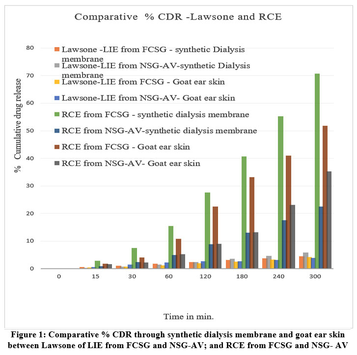 |
Figure 1: Comparative % CDR through synthetic dialysis membrane and goat ear skin between Lawsone of LIE from FCSG and NSG-AV; and RCE from FCSG and NSG- AV
|
Table 1: Comparative % CDR through synthetic dialysis membrane and goat ear skin membrane between *FCSG, and *NSG-AV analyzed at λ max 452 nm (for Lawsone from *LIE) and at max 322 nm (for *RCE).
| % CDR for Lawsone from LIE | ||||
| Time | Synthetic Dialysis membrane | Goat ear skin | ||
| % CDR –FCSG (Cell-1) | % CDR -NSG-AV (Cell-3) | % CDR –FCSG (Cell-2) | % CDR -NSG-AV (Cell-4) | |
| 15 | 0.5353 | 0.3432 | 0.375996 | 0.504 |
| 30 | 1.084774 | 0.756 | 0.7848 | 1.5288 |
| 60 | 1.74951 | 1.4568 | 1.205604 | 2.2764 |
| 120 | 2.344774 | 2.3964 | 1.852404 | 2.6184 |
| 180 | 3.102669 | 3.5304 | 2.474004 | 2.7336 |
| 240 | 3.767405 | 4.596 | 3.2532 | 3.0564 |
| 300 | 4.50951 | 5.8368 | 4.217604 | 3.8136 |
|
% CDR for RCE |
||||
| 15 | 2.90143 | 0.92402 | 1.84 | 1.57 |
| 30 | 7.417144 | 2.36215 | 4.07 | 2.152857 |
| 60 | 15.42143 | 4.91128 | 10.80143 | 5.19 |
| 120 | 27.61143 | 8.79345 | 22.48143 | 8.944286 |
| 180 | 40.75143 | 12.97816 | 33.21429 | 13.21857 |
| 240 | 55.31429 | 17.61602 | 40.99 | 23.18 |
| 300 | 70.79 | 2.54459 | 51.87 |
35.32 |
*FCSG (Foot Crack Cream Shatadhauta Ghrita), *NSG-AV (Non Shatadhauta Ghrita Aloe vera gel), *LIE (L. inermis extract),*RCE (R. cordifolia extract
As per Fig. 1 regarding % CDR for Lawsone of LIE, the highest release is observed from Non- Shatadhauta Ghrita Aloe Vera gel (NSG-AV) using the synthetic dialysis membrane (~6% at 300 min) as compare to Foot Crack Cream Shatadhauta Ghrita (FCSG) shows slightly less release (~4.5%). Both formulations in goat ear skin membranes shows lower % CDR compared to synthetic membranes. However, FCSG shows slightly better release through goat ear skin compared to NSG-AV and is more consistent and linear in release
As per Fig. 1 regarding % CDR of RCE (R. cordifolia extract) highest cumulative release (~70%) is seen from Foot Crack Cream Shatadhauta Ghrita (FCSG) with synthetic dialysis membrane. FCSG with goat skin also shows better release (~50%) than all Non- Shatadhauta Ghrita Aloe Vera gel (NSG-AV) groups. NSG-AV shows the least release, especially through goat skin (~30%) and synthetic membrane (~22%).
Flux, permeability coefficient (Kp), and lag time of Lawsone (from L. inermis extract, LIE) and Rubiadin (from R. cordifolia extract, RCE) were evaluated from FCSG and NSG-AV across synthetic dialysis and biological membranes as shown Table 2.
The dialysis membrane, Lawsone showed higher flux with the Aloe Vera-based formulation and slightly lower flux with the Shatadhauta Ghrita based formulation. However, when considering both membrane types overall, the highest flux was observed from the Ghrita based formulation. Similarly, the permeability coefficient (Kp) for Lawsone was highest through the goat skin membrane, although it exhibited a longer lag time likely due to its hydrophilic nature.
Secondly, in the dialysis membrane, RCE showed the highest flux and permeability Kp with the Shatadhauta Ghrita based formulation, along with a quick release and comparatively low lag time. While in biological membrane its flux, permeability and lag time are moderate average through Ghrita formulation.
Table 2: Permeation parameters- Lawsone of *LIE and *RCE from *FCSG and *NSG- AV through Dialysis and Goat ear skin membrane
| Membrane Type, λ max and molecule | Drug Formulation | Graph
X axis= Time Vs Cumulative drug amount in mg |
Flux (J)=
Slope/Area (mg/cm²/hr) |
Permeability Coefficient (Kp) = J / C₀ (cm/hr) | Lag Time min |
| Dialysis, 452 nm, Lawsone | FCSG | y = 0.0221x + 0.0322 | 0.00701
|
0.0007 | 10- 15 |
| NSG-AV | y = 0.0349x + 0.0088 | 0.01083 | 0.00110 | 10 | |
| Goat ear skin, 452 nm, Lawsone | FCSG | y = 0.0742x + 0.0373 | 0.0236
|
0.0035 | 20-25 |
| NSG-AV | y = 0.0351x + 0.1846 | 0.0111 | 0.00111 | 20-25 | |
| Dialysis, 322nm, RCE | FCSG | y = 0.186x + 0.687 | 0.40892 | 0.0409 | 10-15 |
| NSG-AV | y = 1.384x + 0.044 | 0.1866 | 0.0187 | 20-25 | |
| Goat ear skin,322nm, RCE | FCSG | y = 0.320x + 0.053 | 0.10191 | 0.0102 | 15- 20 |
| NSG-AV | y = 0.237x – 0.164 | 0.00755 | 0.0076 | 25- 30 |
*LIE (L.inermis extract), *RCE (R.cordifolia extract), *FCSG (Foot crack cream Shatadhauta Ghrita), *NSG-AV (Non Shatadhauta Ghrita-Aloe vera), J *- Flux, Kp- Permeability coefficient, C₀ – initial drug concentration (10mg), Area=3.14 cm2.
The in vitro release kinetics of Lawsone and RCE (R.cordifolia extract) from two different formulations, were evaluated using various mathematical models, including Zero-order, First-order, Higuchi, Korsmeyer–Peppas, and Hixson–Crowell. The corresponding linear regression coefficient values (R²) were calculated to determine the best-fitting release model for both formulations, as shown in Table 3. Fig. 2 (a) to (e) and Fig. 3 (a) to (f) shows selected graphs for best fitting models through Synthetic dialysis membrane and goat ear skin membrane respectively for Lawsone and RCE.
Table 3: Comparative kinetic modeling and best fit analysis – Lawsone of LIE and RCE release through Dialysis membrane and goat ear skin membrane
| Metabolite | Zero order | First Order | Higuchi | Peppas | Hixson–Crowell | Best fitting model | ||||
| Dialysis Membrane – Values of R2 | ||||||||||
| Lawsone from FCSG | 0.981 | 0.967 | 0.969 | 0.962 | 0.982 | Hixson | ||||
| Lawsone from NSG-AV | 0.997 | 0.997 | 0.929 | 0.969 | 0.997 | Zero order, First Order, Hixson | ||||
| RCE from FCSG | 0.860 | 0.991 | 0.995 | 0.993 | 0.904 | Higuchi | ||||
| RCE from NSG-AV | 0.998 | 0.959 | 0.974 | 0.996 | 0.979 | Zero order | ||||
| Goat Ear skin Membrane– Values of R2 | ||||||||||
| Lawsone from FCSG | 0.989 | 0.989 | 0.939 | 0.983 | 0.989 | Zero order,
First order, Hixson |
||||
| Lawsone from NSG-AV | 0.940 | 0.909 | 0.772 | 0.825 | 0.920 | Zero Order | ||||
| RCE from FCSG | 0.997 | 0.993 | 0.928 | 0.997 | 0.997 | Zero order, Peppas
Hixson |
||||
| RCE from NSG-AV | 0.940 | 0.909 | 0.772 | 0.952 | 0.9205 | Peppas | ||||
*FCSG (Shatadhauta Ghrita based foot crack cream), *NSG-AV (Non Shatadhauta Ghrita– Aloe Vera gel formulation), *LIE (L.inermis extract), *RCE (R.cordifolia extract).
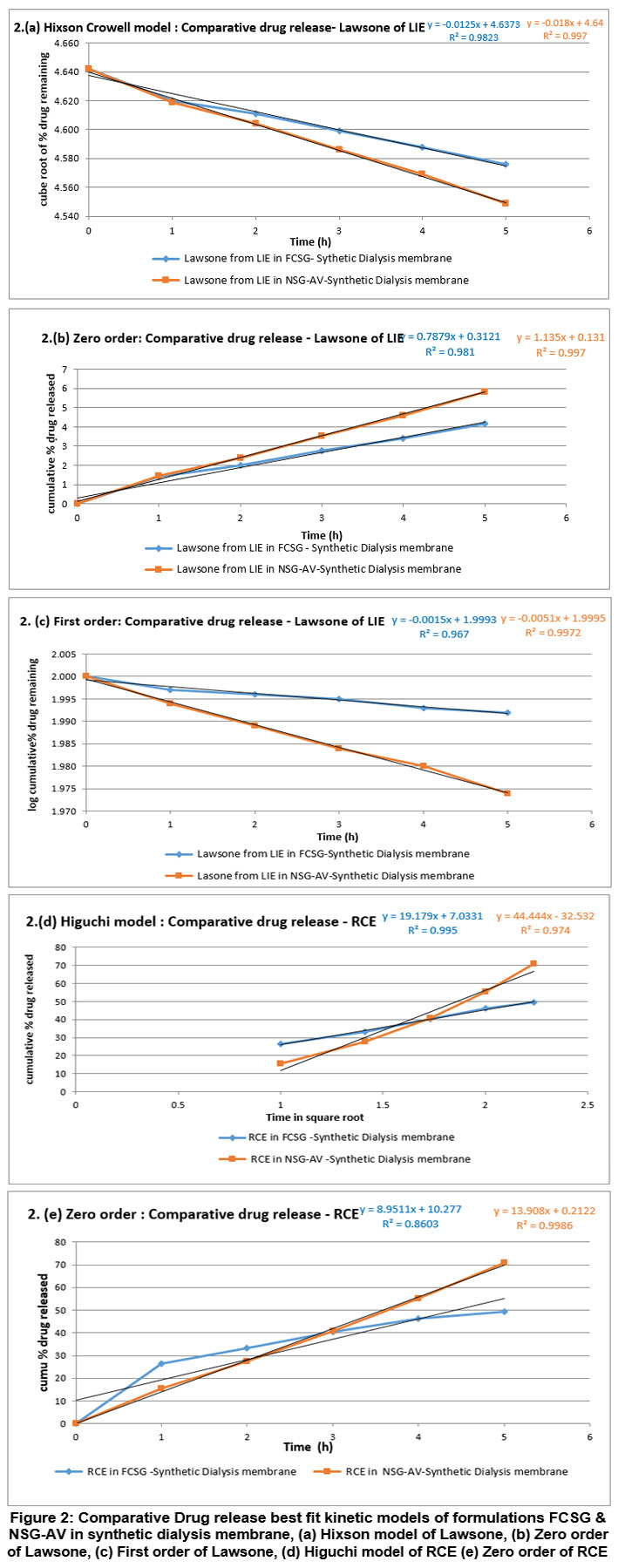 |
Figure 2: Comparative Drug release best fit kinetic models of formulations FCSG & NSG-AV in synthetic dialysis membrane, (a) Hixson model of Lawsone, (b) Zero order of Lawsone, (c) First order of Lawsone, (d) Higuchi model of RCE (e) Zero order of RCE.
|
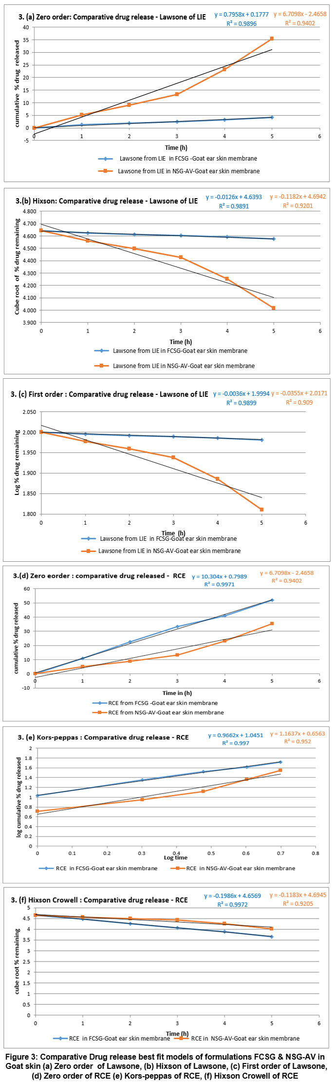 |
Figure 3: Comparative Drug release best fit models of formulations FCSG & NSG-AV in Goat skin (a) Zero order of Lawsone, (b) Hixson of Lawsone, (c) First order of Lawsone, (d) Zero order of RCE (e) Kors-peppas of RCE, (f) Hixson Crowell of RCE.
|
Discussion
Synthetic membranes allow higher permeation overall. Aloe Vera gel based formulation promotes better Lawsone diffusion through synthetic membrane, likely due to its hydrophilic nature. However Shatadhauta Ghrita based formulation (FCSG), enhances goat ear skin permeation of Lawsone compared to aloe vera base.
Shatadhauta Ghrita based formulation (FCSG) significantly enhances the release and permeation of RCE, especially for hydrophobic bioactives like Rubiadin. Goat ear skin shows a real skin barrier, so the enhanced performance of FCSG here confirms better transdermal potential. Aloe vera based formulation NSG-AV fails to deliver RCE effectively due to possibly poor lipid compatibility or insufficient permeation enhancer.
Therefore overall, a Diffusion insight shows that, Synthetic membranes offer less resistance and hence higher CDR values. Goat ear skin, being a biological barrier, offers a realistic model of human skin permeability. Shatadhauta Ghrita (FCSG) formulation improves diffusion across both synthetic and biological membranes, especially for lipophilic compounds like Rubiadin from RCE. Aloe vera may offer better delivery for hydrophilic drugs like Lawsone but is inferior for lipophilic compounds.
Due to Ghrita’s better lipid solubility and transdermal effectiveness, Shatadhauta Ghrita formulation significantly increases RCE penetration into biological membrane as compared to NSG-AV, indicating enhanced transdermal delivery prospective.
However, Shatadhauta Ghrita based formulation showed consistently increased diffusion flux, permeability and moderate lag time, particularly across biological membranes making it more efficient for transdermal delivery of both bioactives Lawsone and Rubiadin. Aloe vera based formulation showed a faster onset in some cases but lacked sustained and efficient permeation.
Shatadhauta Ghrita based formulation (FCSG) maintained effective diffusion even across this barrier, demonstrating its therapeutic promise in contemporary skin diseases.
Kinetic modeling and best fit models:
As per Table 3, the release of lawsone from the Shatadhauta Ghrita based formulation (FCSG) through the dialysis membrane predominantly followed the Hixson–Crowell model, indicating that changes in particle size and surface area due to matrix erosion play a major role in controlling drug release (Fig. 2.(a) ). In contrast, release through the biological membrane (goat ear skin) exhibited higher correlation values for zero-order, first-order, and Hixson–Crowell models ( Fig. 3.(a) to 3.(c) ). This suggests that erosion acts as an initial release mechanism, followed by diffusion and a relatively constant drug release profile.
Although lawsone is hydrophilic in nature, its release from the aloe vera–based formulation (NSG-AV) through the dialysis membrane followed zero-order, first-order, and Hixson–Crowell kinetics (Fig.2.(b) and Fig.2.(c) ), indicating a complex release mechanism involving continuous, concentration-dependent, and erosion-controlled processes. However, in goat ear skin, lawsone release from NSG-AV showed a higher correlation with the zero-order model ( Fig.3.(a) ), suggesting a more uniform and sustained release across the biological membrane.
For RCE, the Higuchi model provided the best fit for the Shatadhauta Ghrita–based formulation using the synthetic dialysis membrane, indicating that drug release was primarily governed by diffusion ( Fig 2.(d) ). In contrast, release through goat ear skin followed both zero-order and Korsmeyer–Peppas models ( Fig.3.(d) and Fig.3.(e) ), suggesting a sustained release governed predominantly by Fickian diffusion through the skin barrier.
RCE release from the aloe vera–based formulation closely followed zero-order kinetics in the dialysis membrane ( Fig.2.(e) ), indicating a consistent drug release rate independent of concentration. In the goat ear skin membrane, the Korsmeyer–Peppas and Hixson–Crowell models ( Fig.3.(e) and Fig.3.(f) ) showed moderate correlation, suggesting that although diffusion and erosion contribute to the overall release process, their contribution is secondary to the dominant zero-order release mechanism.18-25
Conclusion
This research demonstrates, the Shatadhauta Ghrita based formulation provides considerably better topical delivery of bioactive compounds such as Lawsone and Rubiadin than the Non-Shatadhauta Ghrita Aloe Vera gel based formulation. The Shatadhauta Ghrita based formulation showed higher flux with better permeability and sustained release across both membranes Synthetic and goat ear skin. The kinetic modeling indicates that Lawsone follows a controlled release mechanism involving both diffusion and matrix erosion, while Rubiadin exhibits a sustained, Fickian diffusion driven release. The lipid rich Shatadhauta Ghrita base effectively supports the dual release behavior, enhancing the delivery of both phytoconstituents. These results showed that the traditional use of Shatadhauta Ghrita as a scientifically reliable and effective carrier for herbal bioactives, making it a potential natural alternative for the topical treatment of cracked heels and other skin conditions. The result demonstrates that the Shatadhauta Ghrita enhances the dermal drug delivery, provides a scientifically verified base for natural therapeutic foot care application.
Acknowledgement
The authors are thankful to Mahatma Gandhi Vidyamandir’s Pharmacy College, Nashik, for providing laboratory facilities
Funding Sources
The author(s) received no financial support for the research, authorship, and/or publication of this article.
Conflict of Interest
The authors do not have any conflict of interest.
Data Availability Statement
This statement does not apply to this article.
Ethics Statement
This research did not involve human participants, animal subjects, or any material that requires ethical approval.
Informed Consent Statement
This study did not involve human participants, and therefore, informed consent was not required. Clinical Trial.
Clinical Trial Registration
This research does not involve any clinical trials.
Permission to reproduce material from other sources
Not Applicable.
Author Contributions
Daksha Lalit Attarde : Conceptualization, Methodology, Data Collection, Investigation,Writing, Review ,Editing and Supervision,,
Sulochana Sonya Gavit : Conceptualization, Literature review, Methodology, Formal analysis, Investigation, Data Collection, Writing,
Bhagyashree Nanasaheb Borde : Literature review, Data Collection, Writing ,Review and Editing,
References
- Sanap TD, Pawar JR. Review on heel fissure and their management. Emerg. Technol. Innov. Res. 2024;11(11). doi:10.6084/m9.jetir.jetir2411441.
CrossRef - Hashmi F, Wright C, Nester C, Lam S. The reliability of non-invasive biophysical outcome measures for evaluating normal and hyperkeratotic foot skin. J Foot Ankle Res. 2015;8(1):28. doi: 10.1186/s13047-015-0083-8.
CrossRef - Wayal SR, Gurav SS. Bhallatakadi Ghrita: Development and evaluation with reference to Murcchana and Shata-Dhauta process. J Ayurveda Integr Med. 2020;11(3):261-269. org/10.1016/j.jaim.2020.05.005.
CrossRef - Chhangani JA, Kathale PS, Agnihotri AK, Murkute AA, Katre SG. A review on utilizing traditional knowledge: incorporating cow ghee and shata dhauta ghrita in modern moisturizing creams. World J Pharm Res. 2025;14(1):649–660. doi:10.20959/wjpr20251-35152
CrossRef - Nadkarni KM. Indian Materia Medica. 3rd ed. Mumbai: Popular Prakashan. 2002; vol 1:714-716,1062-1065.
- Batiha GE, Teibo JO, Shaheen HM, et al. Therapeutic potential of Lawsonia inermis Linn: a comprehensive overview. Naunyn Schmiedebergs Arch Pharmacol. 2024;397(6):3525-3540. doi: 10.1007/s00210-023-02735-8.
CrossRef - Khare CP. Pharmacopoeal and Related Drugs of Biological Origin. New Delhi: Business Horizons. 2005;293-295, 367-369.
- The Wealth of India. New Delhi: Council of Scientific and Industrial Research. 1972;vol 9:288-292.
- Indian Pharmacopoeia. Ghaziabad: Indian Pharmacopoeia Commission. 2022;vol I:1064, vol.III:1354.
- Kakad S, Bajpai N. Manjistha (Rubia cordifolia): A herbal treasure of India. Int J Multidiscip Res. 2024;6(4):1-9. doi.org/gt3ngf.
CrossRef - Singh B, Dadhich OP, Deepa. A review study of medicinal uses of Manjishtha (Rubia cordifolia). Int J Adv Res. 2017; 5(8):1394-1401. doi:10.21474/IJAR01/5196.
CrossRef - Evans WC. Trease and Evans Pharmacognosy. 16th ed. Edinburgh: Saunders Elsevier. 2009;545-547.
- Deshmukh MD, Patil MP, Ahire ED, Gosavi SB. Shatdhauta ghrita: A promising agent in the development of herbal creams. J. Pharm. Negat. Results. 2022;13(S1):1332–1343. doi:10.47750/pnr.2022.13.S01.159.
- Kumar M., Sharma A., Mahmood S., Thakur A., Mirza MA., Bhatia, A. Franz diffusion cell and its implication in skin permeation studies. Journal of Dispersion Science and Technology, 2024;45(5): 943–956. doi.org/10.1080/01932691.2023.2188923
CrossRef - Askarizadeh M, Esfandiari N, Honarvar B, Sajadian SA, Azdarpour A. Kinetic modeling to explain the release of medicine from drug delivery systems. Chem Bio Eng Rev. 2023;10(6):1006–1049. doi.org/10.1002/cben.202300027
CrossRef - Siepmann J, Siepmann F. Mathematical modeling of drug delivery. Int J Pharm. 2008;364(2):328–343. doi.org/10.1016/j.ijpharm.2008.09.004.
CrossRef - Earle RR, Bandaru KK, Lakshmi UA. Formulation and characterization of sustained release coated matrix granules of metformin hydrochloride. Asian J Pharm Clin Res. 2018;11(7):387-392. doi.org/10.22159/ajpcr.2018.v11i7.24996.
CrossRef - Jahromi PL, Ghazali M, Ashrafi H, Azadi A. A comparison of models for the analysis of the kinetics of drug release from PLGA-based nanoparticles. Heliyon. 2020;6(2):e03451. doi.org/10.1016/j.heliyon.2020.e03451.
CrossRef - Mahshid A, Nadia E, Bizhan H, Seyed Ali S, Amin A. Kinetic modeling to explain the release of medicine from drug delivery systems. Chem Bio Eng Rev. 2023;10(6):1006-1049. doi.org/10.1002/cben.202300027.
CrossRef - Lachman L, Lieberman HA, Kanig JL. The Theory and Practice of Industrial Pharmacy. 3rd ed. Mumbai: Varghese Publishing House. 1991.
- Simona A, Amaro MI, Healy AM, Cabral LM, Sousa VP. Comparative evaluation of rivastigmine permeation from a transdermal system in the Franz cell using synthetic membranes and pig ear skin with in vivo–in vitro correlation. Int J Pharm. 2016;512(1):234-241. doi.org/10.1016/j.ijpharm.2016.08.052.
CrossRef - Kumar M, Sharma A, Mahmood S, Thakur A, Mirza MA, Bhatia A. Franz diffusion cell and its implication in skin permeation studies. J Dispersion Sci Technol. 2023;45(5):1-14. doi.org/10.1080/01932691.2023.
CrossRef - Torlak C, Guleli M, Kizilok S, Kartop RA, Caliskan C, Baysak FK. Comparison of permeability of topical cream drug through polymer synthetic membranes of different structures using Franz Cell diffusion test. J Dispersion Sci Technol. 2024;45(14):1-9. org/10.1080/01932691.2024.2390970.
CrossRef - Kim KM. A Study of the Transdermal Permeation of Lotion Formulations containing Angelica gigas Nakai Extracts in Franz Diffusion Cells. J Life Sci. 2021; 31(11): 1004-9. doi.org/10.5352/JLS.2021.31.11.1004
- Liebenberg W, Engelbrecht E, Wessels A, Devarakonda B, Yang W, De VM. A comparative study of the release of active ingredients from semisolid cosmeceuticals measured with Franz, enhancer or flow-through cell diffusion apparatus. J Food Drug Anal. 2004;12(1):10 . org/10.38212/2224-6614.2669.
Abbreviations : Cm- centimeter, CDR –Cumulative drug release, FCSG- Foot crack cream containing Shatadhauta Ghrita, g- gram, hr-hour, J– Flux, Kp– permeability coefficient, LIE– Lawsonia inermis extract, min- minute, mL—milliliter, µg– microgram, nm- nanometer, NSG-AV– Non-Shatadhauta Ghrita Aloe Vera gel, Q.S– Quantity sufficient, RCE– Rubia cordifolia extract , R2 – linear regression coefficient, UV– Ultraviolet, λ max- lambda máxima, °C– degree centigrade , % – percentage
Accepted on: 20-01-2026
Second Review by: Dr. Ramya Sri
Final Approval by: Dr. Eugene A. Silow


![]()
![]()
