

Manuscript accepted on : 12 June 2017
Published online on: --
Plagiarism Check: Yes
Nadiah Mutluq Alkammash1,2
1Department of Botany and Microbiology, College of Science, King Saud University, P.O. Box 2455 Riyadh, 11451, Saudi Arabia.
2Department of Botany and Microbiology, King Saud University, 11451 Riyadh, Saudi Arabia.
Corresponding Author E-mail: N.alkammash@hotmail.com
DOI : http://dx.doi.org/10.13005/bbra/2474

ABSTRACT: The synthesis, characterization and application of biologically synthesized nanomaterials have become important research areas in nanotechnology, and the green synthesis of nanoparticles using plants is being increasingly studied largely because this approach is considered to lack the problems associated with conventional synthesis. Here we report the synthesis and characterization (using a scanning electron microscope) of silver nanoparticles (AgNPs) obtained using extracts of leaves of the medicinal plants, Artemisia sieberi and Calotropis procera. Scanning electron microscopy (SEM) studies revealed the characteristics of the synthesized nanoparticles which were confirmed by analyzing the excitation of surface plasmon resonance (SPR) using UV–vis spectrophotometer at 482 nm. SEM analysis of the synthesized Ag NPs clearly showed that the particles were predominantly spherical in shape, mostly aggregated and having a size around 8–20 nm. Finally, we consider that the nanoparticles synthesized in this study have potential for wide application in nanotechnology and nanomedicine.
KEYWORDS: Artemisia sieberi; Calotropis procera; Green synthesis; Silver nanoparticles; SEM analysis
Download this article as:| Copy the following to cite this article: Alkammash N. M. Synthesis of Silver Nanoparticles from Artemisia Sieberi and Calotropis Procera Medical Plant Extracts and Their Characterization using SEM Analysis. Biosci Biotech Res Asia 2017;14(2). |
| Copy the following to cite this URL: Alkammash N. M. Synthesis of Silver Nanoparticles from Artemisia Sieberi and Calotropis Procera Medical Plant Extracts and Their Characterization using SEM Analysis. Biosci Biotech Res Asia 2017;14(2). Available from: https://www.biotech-asia.org/?p=25958 |
Introduction
Nanoparticles are extremely small synthesized structures which have an extremely wide applicability1 largely due to their unique thermal,2 electrical3 and optical4 properties. They can be used products ranging from photovoltaics5 to biological and chemical sensors6 while nanosilver particles are extensively used as an anti-bacterial agent in the health industry7 in food storage,8 as textile coatings9 as well as in a range of environmental applications.10 Currently nanosilver is prepared using methods which include electrolysis, and physical, chemical, and biological methods.11,12 The bioreduction of a precursor salt of silver by a bioactive compounds present in plants is a so-called biomimetic method in which; natural products are used as reducing agents to produce nanoparticles.13
Silver nanoparticles play an increasingly important role in biology and medicine due to their attractive physiochemical properties The most important application of silver and silver nanoparticles is in medical industry, where they are used for example, in topical ointments to prevent infection against burn and open wounds.14 Silver products have long been known to have strong inhibitory and bactericidal effects, as well as a broad spectrum antimicrobial activities, which has been used for centuries to prevent and treat various diseases, particularly bacterial infections15; silver nanoparticles also exhibit antifungal, anti-inflammatory, antiviral, antiangiogenesis and antiplatelet activity.16
Nanoparticles can be synthesized using a variety of chemical, physical, and biological approaches. Although chemical synthesis requires short time periods for the synthesis of large quantities of nanoparticles, this method the need requires capping agents for size stabilization of the nanoparticles. The chemicals used in this approach to nanoparticle-synthesis and stabilization are also toxic and the process leads to non-eco-friendly by-products. For environmentally nontoxic nanoparticle synthesis has led to the development of a number of biological approaches, which largely avoid the use or production of toxic chemicals. As a result, there is an increasing demand for “green nanotechnology.” A number of biological approaches for both extracellular and intracellular nanoparticles synthesis have been reported to date using microorganisms including bacteria,17 fungi18,19 and plants.20,21,22 Plants generally provide a better platform for nanoparticle synthesis as they are free from toxic chemicals and contain natural capping agents. Moreover, the use of plant extracts reduces the cost of the isolation and culture of micro-organisms thereby increasing the cost effectiveness of nanoparticle synthesis.23
Calotropis procera (family: Asclepiadaceae) (C. procera) is a cultivable wild xerophytic shrub which is found across Africa, Asia and South America.24 It produces milky white latex which has a range of curative properties.25,26,27 Latex is found in special branching tubes called latex tubes28,29 and has been the subject of interest due to its biological activities including, antibacterial,30 antifungal,31 antiviral,32 anticandidal33 and anticarcinogenic propertoes.34,35 More than 80% of the dry mass of the crude latex corresponds to rubber and the rest 20% covers soluble fractions rich in protein including antioxidant enzymes, cysteine protease with free thiol group and tryptophan.36,37
The genus Artemisia has always been of considerable pharmaceutical interest and is used in traditional medicines to treat a variety of diseases.38,39,40 The genus includes small herbs found in Northern temperate regions and belonging to the important family Compositae (Asteraceae), which comprises about 1,000 genera and over 20,000 species. Within this family, Artemisia is included into the family Anthemideae, which is itself, made up of over 400 species and is found in Europe and North America, and Asia.41,42,43 Among the Asian Artemisia flora, 150 species have been recorded for China, 50 species have been reported in Japan, and 34 species in Iran, of which the following are probably endemic: A. melanolepis Boiss and A. kermanensis Pold,44 A. absinthium45, A. annua,46 A. dracunculus,47 A. aucheri,48 A. haussknechtii Boiss, 49A. scoparia, A. sieberi50 and A. sieberi Besser.51 Here, I described a simple one step method for the synthesis of silver nanoparticles using leaf extracts of Artemisia sieberi and Calotropis procera; the nanoparticles were then characterized by SEM analysis.
Materials and Methods
Plant Extract
Fresh leaves of Artemisia sieberi and Calotropis procera were collected from Saudi Arabia, Riyadh and washed three times with tap water and then distilled water and then dried at room temperature. In order to obtain leaf extracts, five grams of air dried leaves were cut into fine pieces and boiled for 10 min in microwave oven. The extract was then cooled at room temperature and filtered by using Whatman filter paper No. 1; the filtered plant extract was then used to synthesize silver nanoparticles.
Silver Nitrate Solution
Silver nitrate (Merck) solution of 4 mM was prepared by dissolving appropriate amount of silver salt in distilled water and storing in an amber coloured bottle.
Synthesis of Silver Nanoparticles
For the synthesis of silver nanoparticles, 100 ml of leaf extract was mixed with 100 ml of silver nitrate aqueous solution in an Erlenmeyer flask and stored at room temperature. A change in colour was observed after mixing plant extract and silver solution. The reduction of silver in the colloidal solution was monitored by UV–Visible spectroscopy analysis. The mixture was then centrifuged at 15,000 rpm for 5 min and the supernatant was subjected to characterization by the use of a UV-visible spectrophotometer and by scanning electron microscopy. A further two flasks were used as controls where one flask contained only an aqueous plant extract, while the second contained a silver solution.
Characterization of Silver Nanoparticles
The synthesized silver nanoparticles were characterized using the following techniques:
UV–Visible Spectroscopy
The reduction of silver ions in the colloidal solution was confirmed by UV–Visible spectroscopy. A small aliquot of sample was taken in a quartz cuvette and observed for wavelength scanning between 300-700 nm with control samples as a reference. PerkinElmer Lambda 950 UV/Vis spectrometer was used for UV Visible spectroscopy.
Scanning Electron Microscopy
Surface morphology of silver nanoparticles was characterized by scanning electron microscopy. The sample was prepared by centrifuging colloidal solution after 6 h of reaction at 14,000 rpm for 4 min. The pellet was the re-dispersed in deionized water and again centrifuged. The process was repeated three times and the material was finally washed with acetone. The purified silver nanoparticles were sonicated for 10 min for making the suspension and then a drop from the suspension was placed on the carbon coated copper grid. The sample was kept under lamp until completely dry. The prepared sample was subjected to SEM analysis by using Jeol JSM-6490A Analytical Scanning Electron Microscope at king Saud University, Riyadh.
Results and Discussion
In this study, when the leaf extract was mixed with silver nitrate solution its colour started to change which is important since silver nanoparticles in aqueous solution exhibit dark brown colour. A change in colour occurred because of excitation in surface Plasmon resonance which indicates the formation of silver nanoparticles. These findings are similar to those reprted previously where the colour of fresh suspension of Vitex negundo and a silver nitrate solution was also seen to be dark brown52 and also reported that silver nanoparticles exhibit a yellow-brown colour in aqueous solution due to the excitation of surface plasmon vibrations in silver nanoparticles.53 Confirmation of the presence of silver nonoparticles in our work as done using UV–Visible spectroscopy. A small aliquot from the reaction mixture was taken in a quartz cuvette and observed for absorption spectrum. It was noted that a colloidal solution after 6 h of reaction showed absorption peak at 482 nm, confirming the presence of silver nanoparticles in the solution. The result of the UV–Visible spectrum was well-correlated with previous work where the absorption peak was observed at 475 nm for a silver solution of leaf extracts of Memecylon edule54 and also similarly reported that the absorption spectra of silver nanoparticles formed in the reaction media have an absorbance peak at 430–440 nm.55
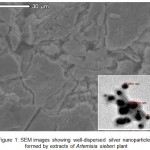 |
Figure 1: SEM images showing well-dispersed silver nanoparticles formed by extracts of Artemisia sieberi plant Click here to View figure |
The synthesized silver nanoparticles were further characterized by SEM analysis where it was noted that the particles were predominantly spherical in shape, although other than the spherical shaped particles were also present. The particle size ranged from 8 nm to 20 nm (Fig. 1 and 2). The different sizes of particles may be correlated with the variable shapes. Our results are similar to those reported by Elavazhagan and Arunachalam (2011).54 Further, Savithramma et al., (2011)56 who observed the formation of relatively spherical shaped silver nanoparticles formed with diameter ranging from 30 to 40 nm in Boswellia ovalifoliolata and 40 nm in Shorea tumbuggaia.
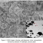 |
Figure 2: SEM images showing well-dispersed silver nanoparticles formed by extracts of Calotropis procera plant Click here to View figure |
In the present study, the biosynthesis of silver nanoparticles was successfully obtained using a green method of preparation. This method used showed that select plants can be used as an effective stabilizing reducing agent for the synthesis of AgNPs. The methodology employed here is very simple, easy to perform, inexpensive, eco-friendly and provide an improved alternative to chemical synthesis. The formed-AgNPs are highly stable spherical shaped particles, (when observed under the SEM). It is hoped that novel and improved silver nanoparticles will be produced in further studies.
Conclusion
In conclusion, leaves of Artemisia sieberi and Calotropis procera were found to be excellent sources for synthesis of silver nanoparticle. Silver nanoparticles were synthesized by applying an environmentally safe method which minimizes the addition of hazardous wastes in the environment. The synthesized nanoparticles were spherical, 8 – 20 nm in size, crystal in nature and showed absorption spectrum at 482 nm characterized by using different techniques. Important outcomes of the study are likely to be in the further development of value added products from the medicinal plants, Artemisia sieberi and Calotropis procera for use in biomedical and nanotechnology based industries.
References
- Duncan T. V. Applications of nanotechnology in food packaging and food safety: barrier materials, antimicrobials and sensors. J. Coll. Interface Sci. 2011;363:1–24.
CrossRef - Moon K. S., Dong H., Maric R., Pothukuchi S., Hunt A., Li Y and Wong C. P. Thermal behavior of silver nanoparticles for low temperature interconnect applications. J. Electron. Mater. 2005;34:168–175.
CrossRef - Chen D., Qiao X., Qiu X and Chen J. Synthesis and electrical properties of uniform silver nanoparticles for electronic applications. J.Mater Sci. 2009;44:1076–1081.
CrossRef - Kelly K. L., Coronado E., Zhao L. L and Schatz G. C. The optical properties of metal nanoparticles: the influence of size, shape, and dielectric environment. J. Phys. Chem. B. 2003;107:668–677.
CrossRef - Yoon W. J., Jung K. Y., Liu J., Duraisamy T., Revur R., Teixeira F. L and Berger P. R. Plasmon-enhanced optical absorption and photocurrent in organic bulk heterojunction photovoltaic devices using self-assembled layer of silver nanoparticles. Sol Energy. Mater.Sol.Cells. 2010;94:128–132.
CrossRef - McFarland A. D and Van Duyne R. P. Single silver nanoparticles as real-time optical sensors with zeptomole sensitivity. Nano. Lett. 2003;3:1057–1062.
CrossRef - Jain P and Pradeep T. Potential of silver nanoparticle-coated polyurethane foam as antibacterial water filter. Biotechnol. Bioeng. 2005;90:59–63.
CrossRef - Costa C., Conte A., Buonocore G. G., and Del Nobile M. A. Antimicrobial silver-montmorillonite nanoparticles to prolong the shelf life of fresh fruit salad. Int. J. Food. Microbiol. 2011;148:164–167.
- Perelshtein I., Applerot G., Perkas N., Guibert G., Mikhailov S and Gedanken A. Sonochemical coating of silver nanoparticles on textile fabrics (nylon, polyester and cotton) and their antibacterial activity. Nanotechnology. 2008;24:245705.
CrossRef - Li Q., Mahendra S., Lyon D. Y., Brunet L., Liga M. V., Li D and Alvarez P.J. Antimicrobial nanomaterials for water disinfection and microbial control: potential applications and implications. Water Res. 2008;42:4591–4602.
CrossRef - Dubey S. P., Lahtinen M and Sillanpaa M. Green synthesis and characterizations of silver and gold nanoparticles using leaf extract of Rosa rugos. Process Biochem. 2010;45:1065–1071.
CrossRef - Rani P. U and Reddy R. Green synthesis of silver-protein (coreshell) nanoparticles using Piper betle L. leaf extract and its ecotoxicological studies on Daphnia magna. Coll. Surf. A. Physicochem. Eng. Asp. 2011;389:188–194.
CrossRef - Bhainsa K. C and D’Souza S. F. Extracellular biosynthesis of silver nanoparticles using the fungus Aspergillus fumigates. Coll. Surf. B. Biointerfaces. 2006;47:160–164.
CrossRef - Ip M., Lui S. L., V. K. M., Poon V. K. M., Lung I and Burd A. Antimicrobial activities of silver dressings an in vitro comparison. J. Med. Microbiol. 2006;55(1):59–63.
CrossRef - Shankar S. S., Rai A., Ahmad A and Sastry M. Controlling the optical properties of lemongrass extract synthesized gold nanotriangles and potential application in infrared-absorbing optical coatings. Chem. Mat. 2005;17(3)566– 572.
CrossRef - Wiley B. J., Im S. H., Li Z. Y., McLellan J., Siekkinen A and Xia Y. Maneuvering the surface plasmon resonance of silver nanostructures through shape-controlled synthesis.J. of Physical Chemistry B. 2006;110(32):15666–15675.
CrossRef - Nair B and Pradeep T. Coalescence of nanoclusters and the formation of sub-micron crystallites assisted by Lactobacillus strains. Crystal Growth and Design. 2002;2(4):293–298.
CrossRef - Mukherjee P., Ahmad A., Mandal D. Fungus-mediated synthesis of silver nanoparticles and their immobilization in themycelialmatrix a novel biological approach to nano particle synthesis. Nano. Letters. 2001;1(10):515–519.
CrossRef - Duran N., Marcato P. D., Alves O. L., De Souza G. I. H and Esposito E. Mechanistic aspects of biosynthesis of silver nanoparticles by several Fusarium oxysporum strains. J. Nanobiotechnol. 2005;3(8):1-7.
- Chandran S. P., Chaudhary M., Pasricha R., Ahmad A and Sastry M. Synthesis of gold nanotriangles and silver nanoparticles using Aloevera plant extract. Biotechnol. Progr. 2006;22(2):577–583.
CrossRef - Li S., Shen Y and Xie A. Green synthesis of silver nanoparticles using Capsicum annuum L. extract. Green Chemist. 2007;9:852–858.
CrossRef - Huang J., Li Q and Sun D. Biosynthesis of silver and gold nanoparticles by novel sundried Cinnamomum camphora leaf. Nanotechnology. 18(10) Article ID 105104.
CrossRef - Garima S., Riju B., Kunal K., Ashish R. S and Rajendra P. S. Biosynthesis of silver nano particles using Ocimum sanctum (Tulsi) leaf extract and screening its antimicrobial activity. J. Nanopart. Res. 2011;13:2981–2988.
CrossRef - Mascolo N., Sharma R., Jain S. C and Capasso F. Ethnopharmacology of Calotropis procera flowers. J Ethnopharmacol. 1988;22(2):211-221.
CrossRef - Iqbal Z., Lateef M., Jabbar A., Muhammad G and Khan M. N. Anthelmintic activity of Calotropis procera (Ait.) Ait. F. flowers in sheep. J. Ethnopharmacol. 2005;102(2):256-261.
CrossRef - Saadabi A. M. A., Ali N. M., Mohammed H. I., Alsafi F. N and Mustafa H. B. An in vitro antimicrobial activity of Calotropis procera (Ait.) R.Br. extracts on certain groups of pathogenic microorganisms. Res. J. Med. Sci. 2012;6(1):13-17.
CrossRef - Ramos M. V., Aguiar V. C., Melo V. M., Mesquita R. O., Silvestre P. P and Oliveira J. S. Immunological and allergenic responses induced by latex fractions of Calotropis procera (Ait.) R.Br. J. Ethnopharmacol . 2007;111(1):115-122.
CrossRef - Mahajan R. T and Badgujar S. B. Phytochemical investigations of some laticiferous plants belonging to Khandesh region of Maharashtra. Ethnobot. Leaflets. 2008;12:1145-1152.
- Pandey B. P. Plant Anatomy. New Delhi: S. Chand Limited. 2001;57-58.
- Ishnava K. B., Chauhan J. B., Garg A. A and Thakkar A. M. Antibacterial and phytochemical studies on Calotropis gigantia (L.) R.Br. latex against selected cariogenic bacteria. Saudi J. of Biologi. Sci. 2012;19(1):87-91.
CrossRef - De Freitas C. D. T., Nogueira F. C. S., Vasconcelos I. M., Oliveira J. T. A and Domont G. B., Ramos M. V. Osmotin purified from the latex of Calotropis procera biochemical characterization, biological activity and role in plant defense. Plant. Physiol. Biochem. 2011;49(7):738-743.
CrossRef - Oliveira J. S., Costa-Lotufo L. V., Bezerra D. P., Alencar N. M and Marinho-Filho J. D., Figueiredo I. S. In vivo growth inhibition of sarcoma 180 by latex proteins from Calotropis procera. Naunyn Schmiedebergs Arch. Pharmacol. 2010;382(2):139-149.
CrossRef - Sehgal R., Arya S and Kumar V. L. Inhibitory effect of extracts of latex of Calotropis procera against Candida albicans a prilimary study. Indian J. Pharmacol. 2005;37(5):334-335.
CrossRef - Silva M. C. C., Da Silva A. B., Teixeira F. M., Sousa P. C. P. D., Rondon R. M. M and Júnior J. E. R. H. Therapeutic and biological activities of Calotropis procera (Ait). R. Br. Asian Pac. J. Trop. Med. 2010;3(4):332-336.
CrossRef - Samy R. P., Rajendran P., Li F., Anandi N. M., Stiles B. G and Ignacimuthu S. Identification of a novel Calotropis procera protein that can suppress tumor growth in breast cancer through the suppression of NF-κB pathway. PLoS One. 2012;7(12):e48514.
CrossRef - Pal G and Sinha N. K. Isolation, crystallization, and properties of calotropins DI and DII from Calotropis gigantean. Arch. Biochem. Biophys. 1980;202(2):321-329.
CrossRef - Freitas C. D., Oliveira J. S., Miranda M. R., Macedo N. M., Sales M. P and Villas-Boas L. A. Enzymatic activities and protein profile of latex from Calotropis procera. Plant. Physiol. Biochem. 2007;45(10-11):781-789.
CrossRef - Willoughby Sr J. A., Sundar S. N., Cheung M., Tin A. S., Modiano J and Firestone G. L. Artemisin in blocks prostate cancer growth and cell cycle progression by disrupting Sp1 interactions with the cyclin-dependent kinase-4 (CDK4) promoter and inhibiting CDK4 gene expression. J. Biol. Chem. 2009;284(4):2203–2213.
CrossRef - Arab H. A., Rahbari S., Rassouli A., Moslemi M. H and Khosravirad F. Determination of artemisinin in Artemisia sieberi and anticoccidial effects of the plant extract in broiler chickens. Trop. Animal Hlth. Prod. 2006;38(6):497–503.
CrossRef - Romero M. R., Serrano M. S., Vallejo M., Efferth T., Alvarez M and Marin J. J. Antiviral effect of artemisinin from Artemisia annua against a model member of the Flaviviridae family the bovine viral diarrhoea virus (BVDV). Planta. Medica. 2006;72(13):1169–1174.
CrossRef - Heywood V. H and Humphries J. C. Anthemideae systematic review, in The Biology and Chemistry of the Compositae V. H., Heywood J. B., Harbord and Turner B. L., Eds., chapter 31.Academic Press, London, UK. 1977;852–888.
- White N. J. Qinghaosu (artemisinin): the price of success. Science. 2008;320(5874):330–334.
CrossRef - Liu C., Zhao Y and Wang Y. Artemisinin: current state and perspectives for biotechnological production of an antimalarial drug. Appl Microbiol. Biotechnol. 2006;72(1):11–20.
CrossRef - Rechinger K. H. Artemisia in flora iranica,” in Compositae K. H., Rechinger and I. C., Hedge Eds. Akademische Druck and Verlagsanatalt, Graz, Austria. 1986;158:214.
- Rezaeinodehi, A., and Khangholi, S. “Chemical composition of the essential oil of Artemisia absinthium growing wild in Iran,” Pakistan J of Biol. Sci., 2008; 11(6): 946– 949.
CrossRef - Khangholil S and Rezaeinodehi A. Effect of drying temperature on essential oil content and composition of sweet wormwood (Artemisia annua) growing wild in Iran,” Pakistan J. Biol. Sci. 2008;11(6):934–937.
CrossRef - Maleki A and Zarasvand M. A. Heavy metals in selected edible vegetables and estimation of their daily intake in Sanandaj, Iran,” Southeast Asian J. Trop. Med. Pub. Hlth. 2008;39(2):335–340.
- Asgary S., Dinani N., Madani H and Mahzouni P. Ethanolic extract of Artemisia aucheri induces regression of aorta wall fatty streaks in hypercholesterolemic rabbits. Pharmazie. 2008;63(5):394–397.
- Heravi M. J and Sereshti H. Determination of essential oil components of Artemisia haussknechtii Boiss. Using simultaneous hydrodistillation-static headspace liquid phase microextraction-gas chromatography mass spectrometry. J. Chromat. 2007;1160(1-2):81–89.
CrossRef - Farzaneh M., Ahmadzadeh M., Hadian J and Tehrani A. S. Chemical composition and antifungal activity of the essential oils of three species of Artemisia on some soil-borne phytopathogens. Comm. Ag. Appl. Biol. Sci. 2006; 71(3):1327–1333.
- Bagheri R., Chaichi M. R., Mohseni-Saravi M., Amin G. R and Zahedi G. “Grazing affects essential oil compositions of Artemisia sieberi Besser. Pakistan J. Biol. Sci. 2007;10(5):810–813.
CrossRef - Zargar M., Hamid A. A., Bakar F. A., Shamsudin M. N., Shameli, K., Jahanshiri, F., and Farahani, F., 2011. Green synthesis and antibacterial effect of silver nanoparticles using Vitex negundo L. Molecul. 2011;16:6667–6676.
CrossRef - Shankar S.S., Ahmad A., Pasricha R and Sastry M. Bioreduction of chloroaurate ions by geranium leaves and its endophytic fungus yields gold nano particles of different shapes. J. Mat. Chem. 2003;13(7):1822–1826.
CrossRef - Elavazhagan T and Arunachalam K. D. Memecylon edule leaf extract mediated green synthesis of silver and gold nanoparticles. Int. J. Nanomed. 2011;6:1265–1278.
CrossRef - Prasad T. N. V and Elumalai E. K. Biofabrication of Ag nanoparticles using Moringa oleifera leaf extract and their antimicrobial activity. Asian Pacific J. Trop. Biomed. 2011;1(6):439–442.
CrossRef - Savithramma N.,Rao M. L and Devi P. S. Evaluation of antibacterial efficacy of biologically synthesized silver Nanoparticles using stem barks of Boswellia ovalifoliolata Bal and Henry and Shorea tumbuggaia Roxb. J. Biol. Sci. 2011;11(1)39–45.
CrossRef

This work is licensed under a Creative Commons Attribution 4.0 International License.


