

Manuscript accepted on : 30 May 2017
Published online on: --
Plagiarism Check: Yes
Kulkarni C. P.1 and Maurya C. B.2
1Department of Chemistry, Kirti M. Doongursee College of Arts, Science and Commerce, Dadar (W), Mumbai - 400028, India.
2Department of Chemistry, G.N. Khalsa College of Arts, Science and Commerce, Matunga (E), Mumbai - 400019, India.
Corresponding Author E-mail: sabhutada13@gmail.com
DOI : http://dx.doi.org/10.13005/bbra/2495

ABSTRACT: Cellulase enzyme plays an important role in converting cellulosic biomass in high value products and therefore finds various applications in a number of industries such as pulp and paper, textile, laundry, biofuel production, food and feed industry, brewing and agriculture. Although there are various sources available for obtaining enzymes of industrial applications such as bacteria, fungi, actinomycetes, microalgae, animals and plants; microorganisms represent the most common source of enzymes because of their broad biochemical diversity, feasibility of mass culture and ease of genetic manipulation. Among the microorganisms, actinomycetes are increasingly becoming an important resource for the production of therapeutic molecules and industrially important enzymes. Considering this, the present investigation was undertaken to isolate a novel actinomycete strain from a sea sediment sample and explore its ability to produce the enzyme cellulase. After morphological and biochemical studies, this actinomycete strain was further characterized using its 16S rRNA gene sequence and a phylogenetic tree was constructed. The activity and stability of the extracted enzyme from this actinomycete was analyzed. Finally, the applications of this extracted enzyme were studied.
KEYWORDS: Actinomycetes; Cellulase; Enzyme applications; Phylogenetic analysis; 16S rRNA; Polymerase Chain Reaction
Download this article as:| Copy the following to cite this article: Kulkarni C. P, Maurya C. B. Characterization of the Cellulase Enzyme Produced by Actinomycetes Isolated from the Mangrove Coastal Areas. Biosci Biotech Res Asia 2017;14(2). |
| Copy the following to cite this URL: Kulkarni C. P, Maurya C. B. Characterization of the Cellulase Enzyme Produced by Actinomycetes Isolated from the Mangrove Coastal Areas. Biosci Biotech Res Asia 2017;14(2). Available from: https://www.biotech-asia.org/?p=25918 |
Introduction
Enzymes are organic catalysts produced by living cells. Many industrial processes are simplified due to the use of enzymes. Enzymes are having applications in several industries such as biofuel, paper, animal feed, biomedicine and food. There are various sources for obtaining enzymes having industrial applications. These sources include bacteria, fungi, actinomycetes, microalgae, animals and plants. Among all these sources, microorganisms represent the most common source of enzymes because of their broad biochemical diversity, feasibility of mass culture and ease of genetic manipulation (Niehaus et al., 1999). Actinomycetes are the most economically and biotechnologically valuable prokaryotes. They are virtually unlimited sources of novel compounds with many therapeutic applications and hold a prominent position due to their diversity and proven ability to produce novel bioactive compounds. There are more than 22,000 known microbial secondary metabolites, 70% of which are produced by actinomycetes, 20% from fungi, 7% from Bacillus spp. and 1–2% by other bacteria (Subramani and Aalbersberg, 2012).
Cellulase is a complex enzyme which carries out the conversion of cellulose to a simpler monomeric glucose. The enzyme holds significant importance as it has industrial applications in the animal feed, food, textiles and detergents and in the paper industry (Sadhu and Maiti, 2013). In the present investigation, microorganisms with special emphasis on actinomycetes were used for the exploration of cellulase enzyme having importance in fruit and vegetable processing industries.
Materials and Methods
Isolation of Actinomycetes
Sediment samples from the mangrove coastal areas of Maharashtra-Goa border were collected in sterile zip lock bags, brought to the laboratory, stored at room temperature till processing within 24 hours.
Isolation of the microorganisms from these samples was carried out by crushing 1 g of each sample and suspending in 10 ml of sterile seawater by vortexing. A 10-3 serial dilution of the suspension was prepared. This dilution of the sample was spread onto Actinomycete isolation agar, GYM Streptomyces medium and Glycerol Yeast extract (GYE) agar using the standard spread plate technique. All the media were inoculated with two kinds of antibiotics viz. Rifampicin and cycloheximide. After incubation for 2-3 weeks, actinomycetes were selected based on their morphology, size and colour appearance on the agar plates. The selected colonies were then sub-cultured onto separate agar plates to ensure their purity.
Identification of Actinomycetes Isolates
The Actinomycetes isolates were largely characterized on the basis of their morphological characters. The macroscopic and macroscopic studies of an actinomycetes growing on agar can provide useful and rapid clues for identification of their respective genus. Macroscopic characters include colony characteristics such as size, shape, color, consistency on different media, the absence or presence of aerial mycelium and extent of spore formation. Indole test, Methyl-red and Voges-Proskauer tests, Citrate utilization test and starch hydrolysis test were carried out using the standard protocols to identify the presence of actinomycetes by biochemical means.
Screening of Actinomycetes Cultures for Cellulase Enzyme Production Ability by Plate Assay
Screening Media
The twenty actinomycetes isolates were spot inoculated on Carboxymethyl cellulose agar (CMC) plates. The plates were stained with 0.1% Congo red staining solution. After 10- 15 minutes the congo red solution was drained out and the plates were flooded with 1M NaCl de-staining solution for 5 minutes to remove the excess unbound congo red. Diameter of clear yellow zones on red background were measured.
Enzyme Activity and Stability Studies
Submerged fermentation of the cultures was carried out on the in Vogel’s minimal media supplemented with 1% Rice straw. After microfiltration, activity if the enzyme was studies using CMC agar cup assay and DNS assay. The cellulase enzyme was further concentrated using microfiltration and TFF. Stability with respect to the temperature and pH was analyzed for the extracted enzyme. Optimum temperature and pH were also recorded following standard protocols.
Identification of Actinomycetes Isolates using 16S Rrna Gene Sequencing
DNA Extraction was carried out using Uniflex DNA Isolation Kit (GeNei, 612117000051730). DNA concentration was determined using Quant-iT TM dsDNA BR Assay Kit of Invitrogen.
PCR Amplification
The DNA isolated from actinomycetes isolates was subjected to polymerase chain reaction (PCR) amplification using Biometra thermal cycler (T-Personal 48). The PCR amplification cycle consisted of a cycle of 5 min at 94°C; 35 cycles of 1min at 94°C, 1 min at 60˚C, 2 min at 72°C; and additionally 1 cycle of 7 min at 72°C. The PCR reagents used were procured from GeNei and the primers were synthesized by Bioserve Biotechnologies (INDIA) Pvt. Ltd (Table 1).
Table 1: Primers used for partial 16S rRNA amplification
| Primers | Primer Sequence (5’-3’) |
| 235F | CGCGGCCTATCAGCTTGTTG |
| 878R | CCGTACTCCCCAGGCGGGG |
DNA Sequencing
The PCR product was purified using AxyPrep PCR Clean up kit (Axygen, AP-PCR-50). It was further sequenced using Applied Biosystems 3730xl DNA Analyzer USA and chromatogram was obtained. The sequencing primer used was 235F- 5’ CGCGGCCTATCAGCTTGTTG 3’.
Bioinformatics Analysis
The DNA sequences were analyzed using online BLASTn (nucleotide Basic Local Alignment Search Tool) facility of National Center for Biotechnology Information (NCBI). The BLAST results were used to find out evolutionary relationship of isolates. Altogether eleven sequences, including sample were used to generate phylogenetic tree.
Application o the Extracted Cellulases
Paper Degradation
Strips of A4 paper of equal size were cut and dipped in to the enzyme reaction mix as the substrate. The reaction mixture contained 0.1M phosphate buffer of pH 6 and the TFF concentrated enzyme in the ratio 4:1. The tubes were incubated at 50°C for 8 hours.
Vegetable Degradation
0.3g of vegetable mix were mixed in 5ml of distilled water and kept in water bath at 90°C for 30mins. These were cooled and the water extracted out. 2ml of water was added again and the flasks sterilized by autoclaving. 0.5ml of TFF concentrated enzyme and 0.5ml of 1: 100 diluted sigma cellulase was added in laminar air flow. The flasks were incubated at 50°C for 48 hrs.
Potato Peel Degradation
0.2g of potato peels were cut and mixed in 5ml of distilled water and kept in water bath at 90°C for 30mins. These were cooled and the water extracted out. 2ml of water was added again and the flasks sterilized by autoclaving. 0.5ml of TFF concentrated enzyme and 0.5ml of 1: 100 diluted sigma cellulase was added in laminar air flow. The flasks were incubated at 50°C for 48 hrs.
Seed Coat Degradation
Tur seeds were peeled and the seed coat used for enzymatic degradation assay. 0.2 g of the seed coats were mixed in 5ml of distilled water and kept in water bath at 90°C for 30mins. These were cooled and the water extracted out. 2ml of water was added again and the flasks sterilized by autoclaving. 0.5ml of TFF concentrated enzyme and 0.5ml of 1: 100 diluted sigma cellulase was added in laminar air flow. The flasks were incubated at 50°C for 48 hrs.
Rice Straw Degradation by Cellulase
0.5g of finely cut rice straw was suspended in 10ml of pH 5 phosphate buffer. The flasks were heated at 90°C for 15min. The enzyme samples were filter sterilized and 1ml inoculated in the substrate mix. The flasks were incubated at 50°C for 4 days. The amount of glucose released from 0.5g of substrate by 1ml of enzyme was estimated by DNSA method.
Clarification of Fruit Juice
Fruit juice was obtained by centrifugation and filtration of the local Pairi variety of mango pulp through 0.3µ glass fibre filter. 500µl of the juice was added to 8ml of buffer pH 5 to obtain enough turbidity. Buffer pH 5 was used for negative control and cellulase from Sigma was used as the positive control. 500µl of enzyme was added and the mixture incubated at 50°C. The tubes were checked for clarity after 4 days. Absorbance was taken at 500nm for the three samples.
Results
From the samples collected, a total twenty actinomycetes isolates were obtained and named as Ac1 to Ac20. Observation under oil immersion lens and through the phase contrast microscope clearly indicated that the isolates were actinomycetes. All the isolates were also found to be gram positive.
Identification of Actinomycetes Isolates using Biochemical Tests
Negative results for the Indole, Methyl-red and Voges-Proskauer tests whereas positive results for the Citrate utilization and starch hydrolysis test indicated the presence of actinomycetes by biochemical means.
CMCase Assay for Cellulase Production
Out of 20 isolates screened for cellulase production 14 were found to be cellulase producers. Isolates Ac1, Ac6, Ac13, Ac15 and Ac20 showed zone of clearance of 30mm and above. Ac3 showed the smallest zone of 10mm. Ac6 showed maximum zone of 34 mm whereas Ac1 showed a zone of 32 mm hence these two isolated were selected for further studies.
Table 2 shows the zones of clearances recorded for the crude cellulase enzyme extracted from actinomycete isolates Ac1 and Ac6.
Table 2: Zone of Clearance for the crude cellulase enzyme by Ac1 and Ac6
| Sr. No | Enzyme sample | Dilution | Zone in cm |
| 1. | Ac1 | Undiluted | 3.0 |
| 2. | Ac1 | 1:2 | 1.5 |
| 3. | Ac6 | Undiluted | 2.0 |
| 4. | Ac6 | 1:2 | 0.7 |
Table 3: Enzyme activity for the selected actinomycetes for cellulases
| Culture code | Reaction Conditions | Activity in U/ml |
| Ac1 | 40˚C/pH 7 | 0.19 |
| Ac6 | 40˚C/pH 7 | 0.05 |
The culture showing higher activity that is Ac1 was selected for further studies (Table 3).
Enzyme Kinetics
The Vmax and Km was found to be 160.2 and 0.8542 for AC1 cellulase respectively.
Stabilities Studies of Enzyme at Different Ph and Temperature
The extracted cellulase protein was found to be highly stable in a broad range of buffers for the test incubation period (1 h). More than 70% of initial activity was retained after 1 h incubation at pH 11.0. The cellulase protein was also found to be a highly thermostable. After 1 h incubation, the enzyme showed 60% and more than 95% residual activity at 80oC and 90oC, respectively. Interestingly, there is approximately, 50% activity retained after 30 min incubation at 100oC.
The optimum temperature for the enzyme and the control was found to be 55°C. The optimum pH at which there was maximum activity was obtained at pH 5 for the sample and the control enzyme.
Identification of Actinomycetes Isolates using 16S Rrna Gene Sequencing
On PCR amplification of the 16S rRNA gene, the isolate Ac1 produced an amplification product of around 600 bp which when sequenced yielded a sequence length of 403 bp (Fig. 1).
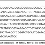 |
Figure 1: Sequence of the amplified 16S rRNA gene of the actinomycetes isolate Ac1
|
This sequence when analyzed by BLASTn tool, indicated that the isolate was from the Genus Streptomyces as indicated 98% sequence similarity with many existing entries of Streptomyces sps.
Application of the Extracted Cellulases
Paper Degradation
The paper dipped in the test and the positive control showed degradation to fine fibers. The difference between the two was very large. The test enzyme showed very little visible degradation with fibers released and reduction in the size of the paper while the positive control showed complete degradation of the part of paper dipped in the liquid. The negative control was however intact.
Vegetable Degradation
Fine fibres released from the vegetable matter were observed in the test as well as the positive control. Pigment was also released in case of the test sample.
Potato Peel Degradation
No visible degradation of potato peels observed in the test as well as the control.
Seed Coat Degradation
There was no visual degradation seen in the test enzyme after incubation though the positive control showed turbidity after incubation. There was however darkening of seed coat colour or release of pigments in the test which was not observed in case of the either controls.
Rice Straw Degradation by Cellulase
Rice straw degradation was observed in case of cellulase extracted from Ac1. The amount of glucose released from 0.5g of substrate by 1ml of enzyme was estimated at 3.4 mg compared to 10.7 mg by standard commercial enzyme by Sigma.
Clarification of Fruit Juice
Optical density of the tested samples showed the effect of the extracted enzyme on fruit juice clearly. The percent clarification obtained for the test was approx. 48.24% and that for the positive control was 97.7%.
Discussion
Programmes to select new microbes for enzyme production are increasing now. Enzymes from bacterial and fungal sources are most commonly used for industrial applications today. Possibility of using actinomycetes for the enzyme production is now being explored on a large scale. They can be easily grown in submerged fermentations and down-streaming of products obtained from them is convenient. In the past few years many actinomycetes has been proved to be a valuable source of enzymes (Jang and Chen, 2003; El-Sersey et al., 2010; Gulve and Deshmukh, 2011; Jani et al., 2012; Magare et al., 2015). It is suggested that microorganisms such as actinomycetes from unexplored or underexploited habitats such as mangroves could have a better potential to produce bioactive compounds and enzymes as they are better adapted to survive in harsh conditions such as the salinity (Sharma, 2014). Results obtained in the present study clearly support this rationale as the cellulase enzyme extracted from the Ac1 strain was found to be both thermo and pH stable indicating its usefulness in harsh industrial processes.
Parulekar (2016) and Abraham (2016) have recently carried out isolation of microorganisms from the mangrove habitat for the extraction of enzymes. Parulekar reported production of manganese peroxidase enzyme from the fungi isolated from the degrading wood whereas Abraham reported production of lignin peroxidase enzyme from the bacteria isolated from the degrading wood. These studies clearly indicate that neglected habitats such as mangroves of Konkan region of Maharashtra and Goa states should be thoroughly screened as it could act as a hot spot of novel microbes with bioactive compounds and secondary metabolites such as enzymes having industrial applications. Identification done by only morphological and biochemical studies often leads to error so the use of molecular marker such as 16S rRNA gene sequence is highly recommended. In the present study, sequence similarity of the amplified gene clearly indicated that the isolate was from the genus Streptomyces vindicating the fact that this molecular tool can be effectively used for reliable species identification up to genus level (Isik et al., 2014; Mabrouk and Saleh, 2014; Sari et al., 2014)
Conclussion
Enzymes from bacterial and fungal sources are most commonly used for industrial applications today. Among the microorganisms, actinomycetes are increasingly becoming an important resource for the production of therapeutic molecules and industrially important enzymes. In the present investigation, a novel actinomycete strain from a sea sediment sample having an ability to produce the enzyme cellulase. The study suggested that, the cellulase enzyme extracted from the Ac1 strain was found to be both thermo and pH stable indicating its usefulness in harsh industrial processes
Acknowledgements
The authors are very much thankful to Dr. R. C. Patil for providing all the facilities and support for this research work and Thankful to Dr Sarita Bhutada, Director, Vihaan Life Science and Research Centre, Kopargaon for help in the preparation of manuscript.
References
- Abraham K. G. Extraction of Lignin Peroxidase Enzyme from Bacteria Isolated from the Mangrove Wood. Paripex- Indian Journal of Research. 2016;5(6):7-9.
- El-Sersy N. A., Abd-Elnaby H., Abou-Elela G. M., Ibrahim H. A. H and El-Toukhy N. M. K. Optimization, economization and characterization of cellulase produced by marine Streptomyces ruber. Afr J Biotechnol. 2010;9(38):6355-6364.
- Gulve R. M and Deshmukh A. M. Enzymatic activity of actinomycetes isolated from marine sediments. Recent Research in Science and Technology. 2011;3(5):80-83.
- Isik K., Gencbay T., Özdemir- Kocak F and Cil E. Molecular identification of different actinomycetes isolated from East Black Sea region plateau soil by 16S rDNA gene sequencing. African Journal of Microbiology Research. 2014;8(9):878-887.
CrossRef - Jang H. D and Chen K. S. Production and characterization of thermostable cellulases from Streptomyces transformant T3-1.World J Microbiol Biotechnol. 2003;19:263-268.
CrossRef - Jani S. A., Chudasama C. J., Patel D. B., Bhatt P. S and Patel H. N. Optimization of extracellular protease production from alkali thermo tolerant actinomycetes: Saccharomonospora viridis SJ-21. Bull. Environ. Pharmacol. Life Sci. 2012;1:84-92.
- Mabrouk M. I and Saleh N. M. Molecular Identification and Characterization of Antimicrobial Active Actinomycetes Strains from Some Egyptian Soils. American-Eurasian J. Agric. & Environ. Sci. 2014;14(10):954-963.
- Magare V. N., Kulkarni C. P and Maurya C. B. Extraction of cellulase enzyme from isolated actinomycetes. Paripex- Indian Journal of Research. 2015;4(5):3-5.
- Niehaus F., Bertoldo C., Kahler M and Antranikian G. Extremophiles as a source of novel enzymes for industrial application. Appl. Microbiol. Biotechnol. 1999;51:711-729.
CrossRef - Parulekar G. T. Extraction of manganese peroxidase from mangrove wood degrading fungi. Paripex- Indian Journal of Research. 2016;5(7):269-270.
- Sadhu S and Maiti T. K. Cellulase production by bacteria: A review. British Microbiology Research Journal. 2013;3(3):235-258.
CrossRef - Sari W. E., Solihin D. D and Lestari Y. Identification of endophytic actinomycetes from Indonesian rice plant based on 16S rRNA and nifH genes analyses. Adv Environ Biol. 2014;8:2357-65.
- Sharma C. Characterization of amylolytic and cellulolytic actinomycetes of salterns of Sambhar salt lake, Rajasthan. Ph. D. Thesis submitted to the IIS University, Jaipur, India. 2014.
- Subramani R and Aalbersberg W. Marine actinomycetes an ongoing source of novel bioactive metabolites. Res. 2012;167(10):571-580.
CrossRef

This work is licensed under a Creative Commons Attribution 4.0 International License.


