Invitro Antidiabetic Evaluation of Biogenic Iron oxide Nanoparticles derived from Punica granatum Peel Extract
 and Spriha Bairagi
Department of Biotechnology, School of Life Sciences, Vels Institute of Science, Technology, And Advanced Studies (VISTAS), Chennai, India
Corresponding author’s E-mail: vardhana88@ymail.com
Download this article as:
ABSTRACT:The Diabetes mellitus is characterized by persistent hyperglycemia and associated metabolic complications. Pomegranate (Punica granatum) peel is rich in polyphenolic and flavonoid compounds and has reported biological activity. The present study investigated the green synthesis and characterization of iron-containing nanoparticles using aqueous pomegranate peel extract and evaluated their in vitro α-amylase inhibitory activity. Aqueous pomegranate peel extract was reacted with ferric chloride, followed by characterization using UV–Vis spectroscopy, FTIR, XRD, SEM, EDS, and DLS. A change in color from pale yellow to black and an absorption maximum at approximately 268 nm were observed after synthesis. FTIR indicated hydroxyl, carbonyl and Fe–O-associated bands. SEM showed predominantly spherical particles with primary particle diameters of 17.18–27.76 nm, whereas DLS showed hydrodynamic size distributions of approximately 250–600 nm, consistent with aggregation and/or a phytochemical-associated hydration layer. The aqueous peel extract and the synthesized FeONPs showed concentration-dependent α-amylase inhibitory activity. At the highest tested sample volume, the extract showed 89.61% inhibition and the FeONPs showed 80.34% inhibition. These findings demonstrate in vitro α-amylase inhibitory activity of both the pomegranate peel extract and the synthesized iron-containing material; however, therapeutic antidiabetic efficacy, safety, mechanism, and in vivo effectiveness remain to be established.
KEYWORDS:Amylase Inhibition; Antidiabetic Activity; Diabetes Mellitus; Green Synthesis; Iron Oxide Nanoparticles; Punica granatum
Introduction
The Pomegranate (Punica granatum L.), an ancient medicinal fruit belonging to the family Punicaceae (Lythraceae), has been extensively used in traditional medicines on account of its nutritional and pharmacological potential.1,2 Indigenous to Iran and Afghanistan, this fruit is currently cultivated in diverse geographical locations across the globe, including India, where fruits like ‘Ganesh’ variety are considered to be of high yield and good quality.3 The fruit comprises vitamins, minerals, fiber, and several other phytochemicals, which render medicinal attributes to it. Several studies have revealed that the peel of the fruit has been the subject of great scientific interest as it contains a high level of active phytoconstituents. However, it may be noted here that the peel of the fruit accounts for about 50–60% of its weight and is normally thrown away as farm waste material despite being a good source of polyphenols, flavonoids, tannins, anthocyanins, punicalagin, and ellagic acid.4,5
Several biological activities of pomegranate peel and its extracts have already been reported. These include antioxidant, antimicrobial, anti-inflammatory, anticancer, cardioprotective, and antidiabetic activities.6,7,2 Moreover, studies have also suggested that pomegranate peel has a stronger antioxidant capacity than other antioxidant foodstuffs.8 Therefore, pomegranate peel has been of great significance in developing a value-added product for its possible application in the biomedical field.
The rapid expansion of nanotechnology over recent years has resulted in its application in medicine, biotechnology, agriculture, and environment among other areas of research. The distinctive features of nanoparticles in terms of physiochemical properties, which arise from the nanoscale sizes and large surface area to volume ratios, contribute greatly to reactivity and biological interaction.9 Iron oxide nanoparticles (FeONPs), among other nanomaterials, have received much focus owing to their unique features such as magnetism, biocompatibility, stability, and application in biosensors and drug delivery systems, among others.10,11 However, the conventional methods employed in preparing nanoparticles involve the use of hazardous chemicals and high energy requirements. Green synthesis through the use of biological agents offers a solution to these limitations.9
Plant-mediated fabrication of nanoparticles is especially useful due to the fact that plant extracts consist of different secondary metabolites which can act as both reducers and stabilizers. Phytochemicals found in the extracts from pomegranate peel enable the transformation of metal ions to stable nanoparticles without using any toxic chemicals.12 There were successful experiments on the synthesis of nanoparticles from the extract from pomegranate peels with improved antimicrobial, antioxidative, and anticancer properties.13,5 This means that pomegranate peels can be an efficient and eco-friendly source of nanoparticles.
Diabetes mellitus is an acute metabolic disease, accompanied by constant hyperglycemia as a consequence of insufficient insulin secretion, resistance, or both.14 Diabetes mellitus cases around the globe are constantly growing, which leads to finding better ways to fight the disease. One of the well-known ways of fighting postprandial hyperglycemia is inhibiting α-amylase activity since α-amylase is an enzyme accountable for the degradation of starch into glucose.6 However, there are plenty of adverse side effects associated with α-amylase inhibitors used for therapy; therefore, natural products became especially popular as antidiabetic drugs. It has been proven that pomegranate peel extracts exhibit high α-amylase inhibiting activity because of the presence of ellagitannins, punicalagin, and other polyphenolic compounds.3,15 Moreover, it was proven in several previous experiments that pomegranate peel extract possesses hypoglycemic properties as well as the ability to regenerate pancreatic β-cells in an experimental diabetic model.16
Despite the proven biological activities of the pomegranate peel extracts and increased interest towards green synthesis of nanoparticles, there is a lack of scientific knowledge about the antidiabetic activity of FeONPs synthesized using pomegranate peel extract. In view of this, it was assumed that the phytochemicals present in pomegranate peel extract could be used for the synthesis of FeONPs and improve their α-amylase inhibiting abilities. Thus, the current experiment aimed to synthesis iron oxide nanoparticles via aqueous pomegranate peel extract, characterization of the synthesized nanoparticles, and evaluation of their antidiabetic activity by means of α-amylase inhibiting assay.
Materials aand Methods
Preparation of Extract
The fresh and ripe fruits of Punica granatum were harvested and cleaned carefully with water in order to wash off any foreign materials that might be present on the fruits. The peel was then removed by hand, dried in the shade, powdered using a laboratory mill, and sieved in order to ensure that a consistent particle size was attained.
In aqueous extraction, 10 g of powdered pomegranate peel was suspended in 100 mL of distilled water in a 250 mL flask and shaken for 24 h at room temperature. This extract was then filtered using Whatman No. 1 filter paper and stored at 4°C for subsequent preparation of nanoparticles.
Preparation of Ferric Chloride Solution
An aqueous ferric chloride (FeCl₃) solution was prepared as the iron precursor. The required quantity (0.1mM) of anhydrous FeCl₃ was dissolved in distilled water to obtain the desired concentration. The solution was freshly prepared and used for the synthesis of iron oxide nanoparticles.
Synthesis of Iron Oxide Nanoparticles (FeONPs)
Iron oxide nanoparticles were synthesized using aqueous pomegranate peel extract as a reducing and stabilizing agent. Firstly, 10 mL of pomegranate peel extract solution was slowly poured into a solution of ferric chloride containing 100 mL and continuously stirred using magnetic stirring in a controlled room temperature environment. Gradual conversion from a pale yellow color to a black color in the mixture showed that ferric ions had been reduced to form iron oxide nanoparticles.
Centrifugation of the mixture was then done at 10,000 revolutions per minute for 15 minutes, and the resultant pellet obtained was rinsed several times with distilled water before being dried at 60°C to produce powder-form FeONPs.
Characterization of Iron Oxide Nanoparticles
The synthesized iron oxide nanoparticles (FeONPs) were characterized through several characterization methods. The presence of the nanoparticles was first determined by UV-Visible spectroscopy (JAZ) in the wavelength range of 200-800 nm. The identification of functional groups responsible for the reduction and stabilization of nanoparticles using pomegranate peel extract was done using Fourier Transform Infrared (FTIR) spectroscopy (Spectrum Two FT-IR/Sp10 Software). The crystallinity and phases of the synthesized FeONPs were studied using X-ray Diffraction (XRD (SmartLab). On the other hand, the surface morphology and size of the particles were measured using Scanning Electron Microscopy (SEM) whereas the elemental analysis was performed using Energy Dispersive X-ray Spectroscopy (EDS) (FESEM: Quattro S). Hydrodynamic particle size distribution and stability of the colloids were studied using Dynamic Light Scattering (DLS) (Nanotrac Wave II).
In Vitro α-Amylase Inhibitory Assay
The antidiabetic activity of the synthesized FeONPs was assessed using the α-amylase inhibition assay. Different concentrations of FeONPs (100-400 µg/mL) were prepared. A reaction mixture containing 0.25 mL of α-amylase (diastase) solution (0.1 mg/mL in phosphate-buffered saline, pH 6.8) and 0.25 mL of the test sample was incubated at 37°C for 5 min.
Succeeding incubation, 0.5 mL of 1% starch solution was added to initiate the reaction, and the mixture was further incubated at 37°C for 3 min. The reaction was completed by adding 0.5 mL of DNS reagent and heating the tubes in a boiling water bath at 100°C for 5 min. After cooling to room temperature, absorbance was measured at 540 nm using a UV–Visible spectrophotometer.
The percentage inhibition of α-amylase activity was calculated using the following equation:
% Inhibition = (Acontrol − Asample) / Acontrol × 100
where A control is the absorbance of the control reaction and Asample is the absorbance of the reaction containing FeONPs.
Results
Preparation of Pomegranate Peel Extract
Aqueous extract of Punica granatum peel was effectively extracted from shade-dried peel powder. About 10 g of dried powder was stirred with 100 mL of deionized water, after which it was sieved using Whatman filter paper. The deposit was collected, and it was brown in color, and was used in the synthesis of iron oxide nanoparticles.
Synthesis of Iron Oxide Nanoparticles
Synthesis of iron oxide nanoparticles (FeONPs) was done by stirring 15 mL of extract with 100 mL of 0.1 Mm Ferric Chloride (FeCl₃) solution. There was an observable change in colour from pale yellow to deep black, suggesting the synthesis of iron oxide nanoparticles.
Characterization of Iron Oxide Nanoparticles
UV–Visible Spectral Analysis
The UV spectrum of the synthesized FeONPs showed an absorption maximum at around 268 nm (Fig 1). The occurrence of the maximum was evidence for the successful synthesis of iron oxide nanoparticles via the reduction of the Fe precursor ions by phytochemicals found in the pomegranate peel extract. This absorption feature provides preliminary evidence of nanoparticle formation; however, UV–Visible spectroscopy alone is not sufficient to confirm the specific iron oxide phase. Therefore, the UV–Visible results were interpreted together with the FTIR, XRD, SEM, and EDS findings for characterization of the synthesized material.
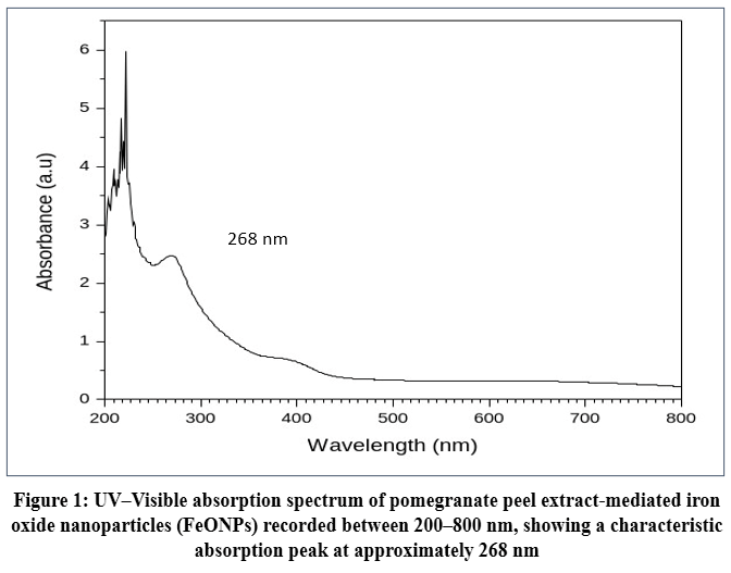 |
Figure 1: UV–Visible absorption spectrum of pomegranate peel extract-mediated iron oxide nanoparticles (FeONPs) recorded between 200–800 nm, showing a characteristic absorption peak at approximately 268 nm |
FTIR Spectral Analysis
FTIR analysis was performed in order to study the functional groups associated with nanoparticle formation and stability. The strong and wide-ranging absorption band at around 3330 cm⁻¹ indicated the presence of O-H stretching vibrations for phenolic compounds like flavonoids and tannins. The sharp absorption band at around 1635 cm⁻¹ suggested carbonyl (C=O) stretching or C=C vibrations of aromatic compounds. The absorption bands at less than 700 cm⁻¹ indicated Fe-O stretching vibrations, supporting the presence of iron–oxygen bonds in the synthesized material (Fig 2).
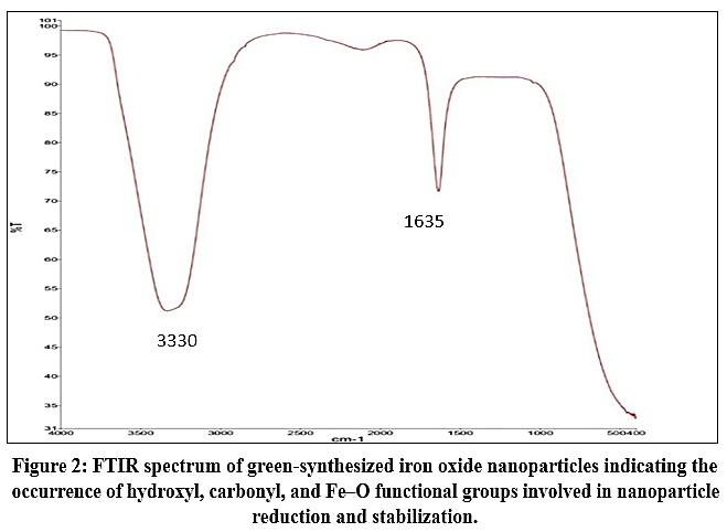 |
Figure 2: FTIR spectrum of green-synthesized iron oxide nanoparticles indicating the occurrence of hydroxyl, carbonyl, and Fe–O functional groups involved in nanoparticle reduction and stabilization. |
X-Ray Diffraction Analysis
The crystalline characteristics of the synthesized FeONPs were analyzed using X-ray diffraction (XRD). The complete diffraction pattern is presented in Fig. 3. The XRD pattern showed broad diffraction features with major reflections observed at approximately 30.24° and 35.61° (2θ). The broad nature of the diffraction features indicates a relatively low degree of crystallinity and/or nanoscale crystallite size. The observed reflections were compared with reported diffraction patterns for iron oxide materials; however, the available peaks were insufficient for definitive assignment of a specific iron oxide phase. Therefore, the material is described cautiously as a poorly crystalline iron-containing nanostructured material. The broad diffraction features may also be influenced by the phytochemical/organic matrix associated with the pomegranate peel extract.
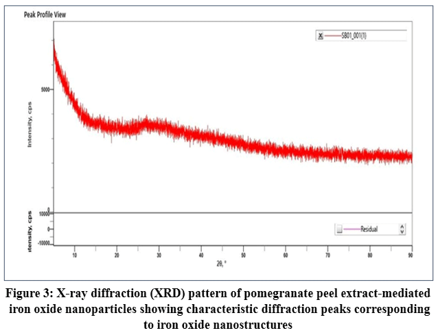 |
Figure 3: X-ray diffraction (XRD) pattern of pomegranate peel extract-mediated iron oxide nanoparticles showing characteristic diffraction peaks corresponding to iron oxide nanostructures |
Scanning Electron Microscopy Analysis
The results obtained from SEM analysis showed that the synthesized FeONPs had spherical shapes with diameters of between 17.18 nm and 27.76 nm (Fig. 4). The size of the nanoparticles was within the nano range and their shapes were relatively uniform. The observed spherical morphology and nanoscale dimensions support the formation of nanoscale particles. However, SEM morphology alone cannot establish the chemical composition or specific iron oxide phase of the material.
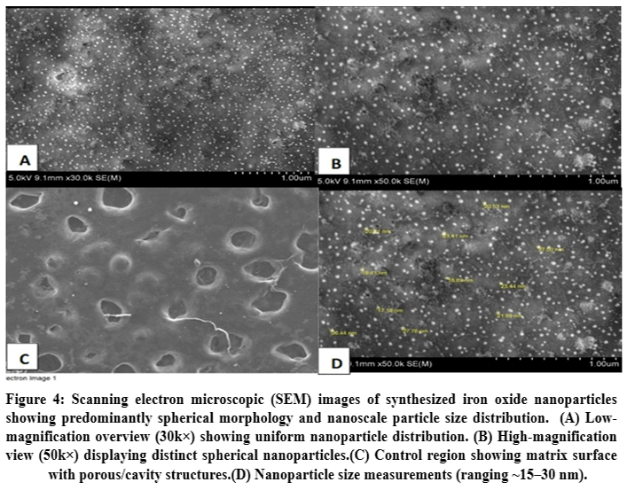 |
Figure 4: Scanning electron microscopic (SEM) images of synthesized iron oxide nanoparticles showing predominantly spherical morphology and nanoscale particle size distribution. |
Energy Dispersive X-ray Spectroscopy Analysis
The EDS spectrum showed the presence of iron and oxygen, supporting the formation of an iron-containing oxygen-rich material (Fig. 5). The analysis confirmed the presence of iron and oxygen, showing that the iron oxide nanoparticles had been successfully synthesized. The percentage compositions of iron and oxygen were found to be 0.94 wt% and 30.87 wt% respectively, whereas that of iron atoms was found to be 0.22 at%. The relatively high carbon content (66.86 wt%) may be attributed to phytochemicals associated with the pomegranate peel extract.
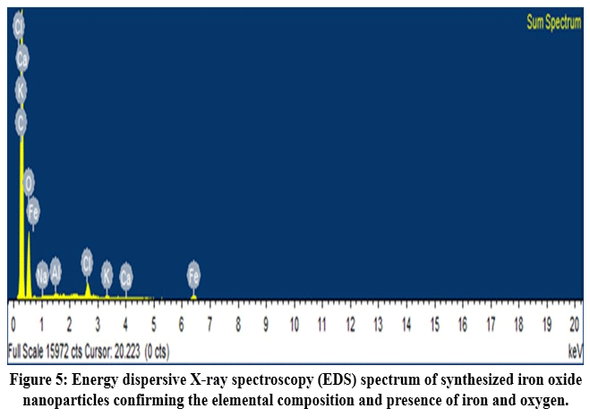 |
Figure 5: Energy dispersive X-ray spectroscopy (EDS) spectrum of synthesized iron oxide nanoparticles confirming the elemental composition and presence of iron and oxygen. |
Dynamic Light Scattering Analysis
The hydrodynamic size distribution of the synthesized FeONPs was determined using DLS. The measured hydrodynamic size ranged from approximately 250–600 nm, which was considerably larger than the primary particle diameter of 17.18–27.76 nm observed by SEM (Fig. 6). This difference indicates that the particles may undergo aggregation in suspension and/or possess a substantial hydration and phytochemical-associated surface layer. Therefore, the SEM measurements represent the primary particle diameter, whereas the DLS measurements represent the hydrodynamic diameter of dispersed particles and possible aggregates.
As per the data obtained from the three runs, the nanoparticles had a peak diameter of about 550 nm, 280 nm, and 380 nm. The larger hydrodynamic sizes observed by DLS compared with the SEM measurements may be attributed to the hydration layer, surface-associated phytochemicals, and possible aggregation of nanoparticles in suspension. The observed sub-micron hydrodynamic size distribution therefore suggests that the particles form aggregates or phytochemical-associated assemblies in the dispersed state. DLS measurements should be interpreted as hydrodynamic particle size rather than primary particle size.
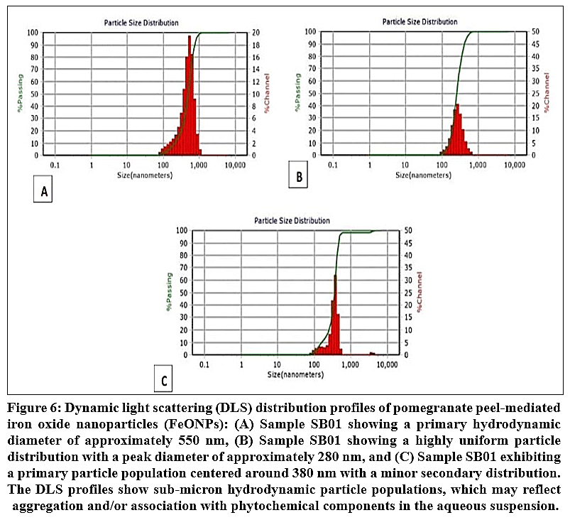 |
Figure 6: Dynamic light scattering (DLS) distribution profiles of pomegranate peel-mediated iron oxide nanoparticles (FeONPs): Click here to View Figure |
In Vitro Antidiabetic Activity
α-Amylase Inhibition Assay
Alpha Amylase inhibitory activities were displayed by aqueous pomegranate peel extract and synthesized FeONPs in a concentration-dependent manner (Table 1). The aqueous extract presented high inhibitory power, showing an increment from 73.88% at 100 µL to 89.61% at 400 µL. In the case of FeONPs, it presented a concentration-dependent manner of activity, where the inhibitory activity went from 28.79% at 100 µL to 80.34% at 400 µL. Maximum inhibitory activity was observed in the case of 400 µL concentration for both aqueous extract and FeONPs. Greater inhibition was shown by aqueous extract compared to FeONPs in all concentrations. FeONPs also showed an appreciable increment in activity with an increase in concentration, where it showed over 80% inhibitory power at maximum concentration tested.
Table 1: α-Amylase inhibitory activity of aqueous pomegranate peel extract and synthesized iron oxide nanoparticles (FeONPs) at different concentrations, expressed as optical density (OD) and percentage inhibition.
|
Sample Volume |
Phase | OD (Trial A) | OD (Trial B) | Average OD | Inhibition (%) |
| 100 µl | Plant Extract | 0.510 | 0.493 | 0.502 |
73.88% |
| FeONP | 1.326 | 1.303 | 1.315 | 28.79% | |
| 200 µl | Plant Extract | 0.328 | 0.305 | 0.317 |
83.51% |
| FeONP | 1.026 | 1.015 | 1.021 | 44.72% | |
| 300 µl | Plant Extract | 0.269 | 0.258 | 0.264 |
86.27% |
| FeONP | 0.720 | 0.721 | 0.721 | 60.97% | |
| 400 µl | Plant Extract | 0.198 | 0.201 | 0.200 |
89.61% |
| FeONP | 0.369 | 0.357 | 0.363 | 80.34% | |
| Control | Plant/NP | 1.920 | 1.846 | 1.883 |
– |
Discussion
Green synthesis of iron-containing nanostructured material using Punica granatum peel extract was carried out in the present study. The visible colour change from pale yellow to deep black following the reaction between the pomegranate peel extract and FeCl₃ provided a preliminary indication of nanoparticle formation. The UV–Visible spectrum showed an absorption maximum at approximately 268 nm (Fig. 1). However, UV–Visible spectroscopy alone is not sufficient to establish the formation or specific phase of iron oxide; therefore, the results were interpreted together with FTIR, XRD, SEM, and EDS analyses.
FTIR spectral analysis of the obtained nanoparticles indicated the existence of hydroxyl and carbonyl group bands below 700 cm⁻¹ associated with iron-oxide (Fe–O) bonds, suggesting that phytochemicals in pomegranate peel extract played role of reducing and stabilizing agentoxide nanoparticles.9,17,11
The presence of iron and oxygen in the EDS spectrum further supported the formation of an iron-containing oxygen-rich material (Fig. 5). The relatively high carbon signal may be associated with residual phytochemical components from the pomegranate peel extract that remained attached to or associated with the nanoparticle surface.5 The XRD pattern showed broad diffraction features with major reflections around 30.24° and 35.61° (2θ), indicating a relatively low degree of crystallinity and/or nanoscale crystallite size (Fig. 3). The observed reflections have been reported for iron oxide materials.5
However, the available diffraction data were insufficient to conclusively assign a specific iron oxide phase. Therefore, the XRD findings are interpreted cautiously as supporting the formation of a poorly crystalline/nanocrystalline iron-containing material rather than confirming a specific iron oxide phase. The broad diffraction features may also be influenced by the organic/phytochemical matrix associated with the pomegranate peel extract.
SEM analysis revealed predominantly spherical particles with primary particle diameters ranging from 17.18 to 27.76 nm (Fig. 4). In contrast, DLS analysis showed considerably larger hydrodynamic sizes ranging from approximately 250 to 600 nm (Fig. 6). This difference can be explained by the fact that SEM measures the primary particle diameter in the dried state, whereas DLS measures hydrodynamic diameter in suspension. The larger DLS values may therefore be attributed to particle aggregation and/or the presence of hydration and phytochemical-associated layers surrounding the particles. The peak sizes observed in the three DLS measurements were approximately 550, 280, and 380 nm. Thus, the DLS values represent hydrodynamic particle or aggregate sizes rather than the primary nanoparticle diameter.9,17
For α-amylase inhibition, both the aqueous pomegranate peel extract and synthesized FeONPs exhibited inhibitory activity. However, the crude aqueous extract showed greater inhibition than the FeONPs at all tested sample volumes, with maximum inhibition of 89.61% compared with 80.34% for FeONPs. The higher activity of the crude extract may be attributed to the greater availability of soluble bioactive phytochemicals, including punicalagin, ellagic acid, flavonoids, and tannins, which have been reported to possess enzyme inhibitory properties and may contribute to the inhibition of carbohydrate-digesting enzymes.2-4 Similar antidiabetic activities have also been reported for pomegranate peel extracts.6,16,18
The green synthesis approach using pomegranate peel extract provides a potentially simple and environmentally friendly route for producing iron-containing nanostructured materials, with phytochemicals serving important roles during nanoparticle formation and stabilization.5,17 However, the present findings should be interpreted as preliminary evidence of in-vitro α-amylase inhibitory activity and not as proof of therapeutic antidiabetic efficacy. The study did not include a positive reference inhibitor such as acarbose or separate sample blanks to evaluate possible absorbance interference from the coloured plant extract and FeONPs in the DNS assay. In addition, the assay was performed using duplicate trial readings, which provide consistency of absorbance measurements but do not constitute independent biological replicates. Therefore, robust statistical comparison and strong comparative claims are limited.
The study findings confirm the hypothesized ability of pomegranate peel bioactive substances to aid in the production of stable FeONPs via a green nanotechnological approach that is environmentally and economically friendly.5 More research on other enzyme activities such as α-glucosidase as well as toxicity tests and diabetic animal models should be done to further explore the medicinal role played by the synthesized nanoparticles.
Conclusion
Iron oxide nanoparticles (FeONPs) were successfully synthesized via green technique using an aqueous extract of pomegranate (Punica granatum) peel. Characterization techniques, such as UV-Vis, FTIR, XRD, SEM, EDS, and DLS analysis, proved that phytochemical-capped iron oxide nanoparticles have been successfully synthesized and had a nanoscale size. FeONPs exhibited substantial α-amylase inhibition, showing promising in vitro antidiabetic properties; at the same time, it revealed the importance of pomegranate peel extract in producing environmentally friendly nanoparticles. This work proves the possibility of employing pomegranate peel-based nanoparticles in nanobiotechnology and antidiabetic researches while contributing to utilizing agricultural waste materials. Nevertheless, future research is needed to reveal whether FeONPs increase the bioavailability of phytochemicals from pomegranate, understand the mechanisms of antidiabetic properties of phytochemicals, and prove the effectiveness of these nanoparticles in vivo.
Acknowledgement
The authors would like to Thank Vels Institute of Science ,Technology, and Advanced Studies, for providing necessary support to carry out the research work.
Funding Sources
The author(s) received no financial support for the research, authorship, and/or publication of this article.
Conflict of Interest
The authors do not have any conflict of interest.
Data Availability Statement
This statement does not apply to this article.
Ethics Statement
This research did not involve human participants, animal subjects, or any material that requires ethical approval.
Informed Consent Statement
This study did not involve human participants, and therefore, informed consent was not required.
Clinical Trial Registration
This research does not involve any clinical trials.
Permission to reproduce material from other sources
Not Applicable.
Author Contributions:
- Spriha Bairagi: Methodology, Writing – Original Draft
- Vardhana Janakiraman: Conceptualization, Supervision, Project Administration, Writing – Review & Editing.
References
- Das S, Barman S. Antidiabetic and antihyperlipidemic effects of ethanolic extract of leaves of Punica granatum in alloxan-induced non-insulin-dependent diabetes mellitus albino rats. Indian J Pharmacol. 2012;44(2):219-224.
CrossRef - Bacha WA, Hamza A, Khan AA, et al. Scrutinizing the antidiabetic, antidiarrheal, and anti-inflammatory activities of methanolic extract of pomegranate peel via different approaches. Ital J Food Sci. 2024;36(1).
CrossRef - More RK, Pingale PL, Upasani CD, Amrutkar SV. In vitro evaluation of Punica granatum fruit peel extract for its potential antidiabetic effects. J Appl Pharm Res. 2024;12(6):137-143.
CrossRef - El-Hadary AE, Ramadan MF. Phenolic profiles, antihyperglycemic, antihyperlipidemic, and antioxidant properties of pomegranate (Punica granatum) peel extract. J Food Biochem. 2019;43:e12803.
CrossRef - Monika P, Chandraprabha MN, Hari Krishna R, et al. Recent advances in pomegranate peel extract-mediated nanoparticles for clinical and biomedical applications. Biotechnol Genet Eng Rev. 2024;40(4):3379-3407.
CrossRef - Mahalakshmi R, Aswini M, Banu AT. Anti diabetic activity of pomegranate peels. In: Rethinapandy A, ed. Research Journal of SRNMC. 2015:76-80.
- Eghbali S, Askari SF, Avan R, Sahebkar A. Therapeutic effects of Punica granatum (pomegranate): An updated review of clinical trials. J Nutr Metab. 2021;2021:5297162.
CrossRef - Saad EA, Hassanien MM, El-Hagrasy MA, Radwan KH. Antidiabetic, hypolipidemic and antioxidant activities and protective effects of Punica granatum peels powder against pancreatic and hepatic tissue injuries in streptozotocin-induced IDDM in rats. Int J Pharm Pharm Sci. 2015;7(7):397-402.
- Kumar B, Smita K, Galeas S, et al., Characterization and application of biosynthesized iron oxide nanoparticles using Citrus paradisi peel: A sustainable approach. Inorg Chem Commun. 2020;119:108116.
CrossRef - Abdullah JAA, Lagos SNP, Sanchez EJE, et al., Innovative agrowaste banana peel extract-based magnetic iron oxide nanoparticles for eco-friendly oxidative shield and freshness fortification. Food Bioprocess Technol. 2024;17(12):5083-5096.
CrossRef - Ikhuoria EU, Uwidia IE, Okojie RO, et al. Advancing green nanotechnology: Harnessing the bio-reducing properties of Musa paradisiaca peel extract for sustainable synthesis of iron oxide nanoparticles. J Multidiscip Appl Nat Sci. 2024;4(1):108-119.
CrossRef - Ifeanyichukwu UL, Fayemi OE, Ateba CN. Green synthesis of zinc oxide nanoparticles from Punica granatum extracts and characterization of their antibacterial activity. 2020;25(19):4521.
CrossRef - Faddladdeen K, Ojaimi A. Protective effect of pomegranate (Punica granatum) extract against diabetic changes in adult male rat liver: Histological study. J Microsc Ultrastruct. 2019;7(4):165-170.
CrossRef - Amri Z, Ben Khedher MR, Zaibi MS, et al., Anti-diabetic effects of pomegranate extracts in long-term high fructose-fat fed rats. Clin Phytosci. 2020;6:55.
CrossRef - Shi R, Zhou N, Zhang H, Gong M, Han L. Bioaffinity ultrafiltration coupled with HPLC-ESI-MS/MS for screening potential α-glucosidase inhibitors from pomegranate peel. Front Nutr. 2022;9:1014862.
CrossRef - Salwe KJ, Sachdev DO, Bahurupi Y, Kumarappan M. Evaluation of antidiabetic, hypolipidemic and antioxidant activity of hydroalcoholic extract of leaves and fruit peel of Punica granatum in male Wistar albino rats. J Nat Sci Biol Med. 2015;6(1):56-62.
CrossRef - Aida MS, Alonizan N, Zarrad B, Hjiri M. Green synthesis of iron oxide nanoparticles using Hibiscus plant extract. J Taibah Univ Sci. 2023;17(1).
CrossRef - Thapa S, Pandey D, Biradar MS, et al. Phytochemical profile and α-amylase inhibitory activity of pomegranate peel extract: An in silico and in vitro investigation. Food Sci Nutr. 2025;13(11):e71191.
CrossRef
Abbreviations
DM Diabetes Mellitus
FeONP s Iron Oxide Nanoparticles
FeCl₃ Ferric Chloride
UV–Vis Ultraviolet–Visible Spectroscopy
FTIR Fourier Transform Infrared Spectroscopy
XRD X-ray Diffraction
SEM Scanning Electron Microscopy
EDS Energy Dispersive X-ray Spectroscopy
DLS Dynamic Light Scattering
DNS 3,5-Dinitrosalicylic Acid
O–H Hydroxyl Group
C=O Carbonyl Group
C=C Carbon–Carbon Double Bond
Fe–O Iron–Oxygen Bond
wt% Weight Percentage
at% Atomic Percentage
nm Nanometer
µg/mL Micrograms per Milliliter
µL Microliter
mL Milliliter
mM Millimolar
rpm Revolutions Per Minute
°C Degree Celsius
h Hour
min Minute
pH Potential of Hydrogen
Accepted on: 07-09-2026
Second Review by: Dr. Rajendra Jangde and Dr. Rajesh Damodharan
Final Approval by: Dr. Wagih Ghannam