Evaluation of Donkey Milk Whey Extract (Equus asinus) for Hemostatic and Metabolic Applications
 , Rajesh Medar and Bhagyalakshmi Manjappa*
 Department of Studies and Research in Biochemistry, Tumkur University, Tumkur, Karnataka, India
Corresponding Author’s E-mail:bhagyaayanur@gmail.com
DOI : http://dx.doi.org/10.13005/bbra/3565
Download this article as:

The protein and fat structures of the DMWE (Donkey milk whey extract) are very close to that of the human breast milk when compared to that of the bovine milk, and therefore has a significant value in the functional food science. This study uses in vitro and in vivo models to examine DMWE's antithrombotic, antidiabetic, and antioxidant activities. DMWE was acquired after acid induced whey separation and subjected to biological tests. DMWE demonstrated a strong dose-dependent procoagulant effect that reduced plasma recalcification time in 100-500 µg concentrations, reducing plasma recalcification time to 36 s (P < 0.0001) at 100-500 µg.The mouse tail bleeding model significantly reduced the bleeding duration from 217 s to 40 s (P < 0.01).DMWE also experienced a substantial reduction in the APTT (activated partial thromboplastin time) and PT (prothrombin time) that witnessed the activation of intrinsic and extrinsic pathways. DMWE had a procoagulant activity but it was also found to have antiplatelet and thrombolytic potential in decreasing order of lysis of the clot under observation after 90 min at 37ºC. DMWE inhibited alpha-amylase (28-67%) and alpha-glucosidase (35-54%) with an IC50 of 39 µg/mL and this is similar to acarbose. DPP-IV (Dipeptidyl Peptidase-IV) inhibition reached 63% with an IC₅₀ of 395 ± 1.856 µg/mL, compared to sitagliptin (57%, IC₅₀ 3.5 µg/mL). The antioxidant activity demonstrated 79% DPPH (2,2-Diphenyl-1-picrylhydrazyl) scavenging, with IC50 values of 185 µg/mL (DMWE) and 160 µg/mL (ascorbic acid). The biological benefits are linked to bioactive proteins and peptides such as lactoferrin and lysozyme, which have antioxidant, antibacterial, and metabolic activities. DMWE has tremendous opportunities as a functional nutraceutical to be implemented in the metabolic regulation and specialized nutrition.
KEYWORDS:Alpha-amylase inhibition; Alpha-glucosidase inhibition; Antiplatelet aggregation; Bioactive peptides; Lysozyme Milk whey extract; Thrombolytic activity
Introduction
Proteomic and lipidomic properties of donkey milk (Equus asinus) have made it a valuable biological fluid in the functional foods’ context. The fact that the content of the protein of donkey milk is mainly composed of whey as compared to the casein-dominant bovine milkmakes the similarity of the protein content of the donkey milk and the human breast milk much more comparable than the casein-dominant bovine milk.1,2 The whey fraction is a complex matrix of bioactive peptides and strongly encrypted peptides with strong medicinal properties, such as antidiabetic, antibacterial and antioxidant properties.3,4The findings are in line with the rest of the dairy studies that have shown that whey proteins and peptides have multifunctional therapeutic effects, such as antihypertensive, immunomodulatory, and metabolic regulatory effects.5–7The recent efforts of proteomic and peptidomic techniques have shown that donkey milk whey is a source of a wide range of multifunctional, bioactive peptides with multifunctional therapeutic effects. The comparative proteomic studies have revealed that there are specific similarities between the donkey milk whey and the human milk proteins, indicating it has a higher nutritional and therapeutic value.8–11 Due to their activities which include reactive oxygen species scavenging, enzyme inhibition, and control of cellular signalling pathways, these peptides have antioxidant, antimicrobial, anti-inflammatory and antidiabetic properties.12–14 This has been extensively reported in the case of milk peptides where whey peptides have been proven to have the ACE inhibitory, antioxidant and antimicrobial activities.15–17
Antioxidants and Decreased Oxidative Stress
The high concentration of sulfur-containing amino acids and particular bioactive peptides generated during gastrointestinal digestion is the fundamental source of their antioxidant action.18The action of these compounds is that they chelate the pro-oxidant transition metals, and neutralize reactive oxygen species (ROS) and prevent the peroxidation of cellular membranes by lipid peroxidation. In addition, recent studies also confirm that the peptides that are present in milk cause the defense system of antioxidants to be activated and to help maintain the systemic redox balance.12,13 It is claimed that endogenous activity of antioxidant enzymes of the water-soluble fraction of the donkey milk is dramatically increased and it is a crucial factor in the maintenance of systemic redox homeostasis.19
Enzyme Inhibition and Antidiabetic Properties
Donkey milk whey has the most promising medicinal properties, which include the antidiabetic properties.Biochemical tests show that the whey-derived peptides are natural blockers of α-glucosidase, an important enzyme that breaks down complex carbohydrates into glucose.20 The whey components of donkey milk may be used to manage the postprandial hyperglycemia, which is effectively reduced by slowing down the glucose absorption into the blood. Recent research also indicates that milk peptides have a high inhibitory effect on DPP-IV, α-amylase, and α-glucosidase enzymes.13,21,22
Immunomodulatory and Antimicrobial Properties
The antibacterial profile of donkey milk whey is well developed due to high concentrations of both lysozyme and lactoferrin and leads to its therapeutic range. Lysozyme breaks the β (1→4) glycosidic linkages in bacterial peptidoglycan to lyse pathogenic microorganisms.23Furthermore, these proteins regulate immunological responses by modulating cytokine synthesis and enhancing the activity of macrophages and natural killer cells. Donkey milk has been well reported for its antibacterial action against the majority of infections.20Besides, lactoferrin and α-lactalbumin are whey proteins that are important in host defense and immune regulation. Lactoferrin is a large whey glycoprotein that has been widely documented to exert antimicrobial, antiviral, anti-inflammatory, and anticancer effects by sequestration of iron and by modulating the immune system.24 These multi-purpose attributes underscore the therapeutic importance of whey in donkey milk and in diverse sources of mammal milk.
Gut Microbiota Modulation
It is beginning to be shown that donkey milk whey may help in the modulation of gut microbiota by supporting the growth of beneficial bacteria and enhancing the health of the intestines.25–27 This further contributes to its functionality as a nutraceutical.
Materials and Methods
Materials
Donkey milk was collected from Okkodigollaratti, Nisarga Badavane, Bheemsandra, Tumkur, Karnataka
Preparation of donkey milk whey extract
Extraction of DMWE is initiated by collecting milk from Nisarga Badavane, Tumkur. After the milk collection, the milk was boiled to 100ºC, and then once it boiled, 6N HCl was added. Upon addition of HCl, the whey and casein get separated. Then filtering the whey by using the Whatman filter paper. The filtered whey was then lyophilized. Then lyophilized whey was stored at 20°C until it was used for further investigation.
Ethical Approval
The ethical guidelines for the study of human and animal research at the institutional and national levels were followed in this investigation. The Institutional Human Ethical Committee of Tumkur University, Tumakuru (IHEC-TUT No: 03/Res/2024-2025) accepted the designated human blood utilized for all the tests, which were carried out in accordance with ethical guidelines.
All animal studies have been authorized by Sharnbasava University’s Institutional Animal Ethics Committee (IAEC) in Kalaburagi (SUK/ZOL/IAEC/17/2024-25). The Department of Zoology at Sharnbasava University has also given its approval. Before handling, all procedures were carried out in accordance with the Committee for the Control and Supervision of Experiments on Animals’ (CCSEA) requirements.
Male Albino Wistar rats weighing between 190 and 210 grams were split up into groups of six and allowed to acclimate for 24 days in polypropylene cages. The experimental diets followed the AIN-93M regimen, while the rats were fed a standard diet of laboratory pellets. The rats were fed lab pellets and provided with tap water and cow feed ad libitum. The animals were kept at 27±2°C, 12-hour light/dark cycle, and humidity of 55-65% and light of 2000 lux.
Antithrombotic activity
The in vitro and in vivo activity of donkey milk whey extract (DMWE) for its anti-thrombotic potential was investigated. In vitro tests used to assess the extract’s anticoagulant properties were plasma recalcification time, thrombin time, prothrombin time (PT), activated partial thromboplastin time (aPTT), and fibrinogen clotting assays. In vivo investigations like tail bleeding time and clot lysis test were used to further examine its ability to prevent blood clots and its safety.
In vitro and in vivo Antithrombotic Studies
Plasma recalcification time
The time needed for plasma recalcification was determined. The DMWE (10-80 µg) doses were first inoculated for 1 min at 37 °C (Isotherm forced convection lab incubator, In Lab Equipments (Madras) Pvt. Ltd.) with 0.2 ml of citrate based human plasma in the presence of 10 µl of pH 7.4 Tris HCl buffer. The pre-incubated mixture received 10 µl of 0.25 M CaCl₂, and the clotting time was recorded.28
APTT (Activated Partial Thromboplastin Time) and PT (Prothrombin Time)
APTT and PT tests were performed following a 1-minute pre-incubation with DMWE (3mg/ml) and 100 µl of normal citrated human plasma. For APTT, add 100 µl of LIQUICELIN-E phospholipid deposition from rabbit brain agitated for 3 minutes at 37 °C (Isotherm convection induced lab incubator, Inlab Equipments (Madras) Pvt. Ltd.). To initiate the clot formation procedure, 100 µl of 0.02 M CaCl₂ was added. The duration of clotting was observed. To begin the PT method, 200 µl of the reagent (UNIPLASTIN-rabbit brain thromboplastin) was injected. The viewable spots’ endurance was timed (in seconds). The data from the oversight plasma heated (Isotherm forced convection lab incubator, In Lab Equipments (Madras) Pvt. Ltd.) using the buffer for a similar length period were utilized to estimate the APTT rate and international normalized ratio (INR) for PT at each point.29
Whole Blood Clot Retraction Assay
A whole blood clot lysis assay was carried out.28 The blood samples were collected from four healthy men aged 25-35, who were free from drug and alcohol addiction, illness, weight etc. and centrifuged in the tubes (500 µl/tube) and are weighed, and incubated in the Isotherm forced convection lab incubator, Inlab Equipments (Madras) Pvt. Ltd. for 45 minutes at 37 °C. A clot formed and serum was removed and the tubes were again weighed. (0 – 75 µg) was added and incubated for 90 min at 37 °C (Isotherm forced convection lab incubator, Inlab Equipments (Madras) Pvt. Ltd.). The fluid was collected after the incubation period then the % of clot lysis was calculated.30
Direct Hemolytic Assay
Direct lysis ability was assessed using properly cleansed human erythrocytes. Essentially, washed RBC and phosphate buffered saline (PBS) (1:9 v/v) was mixed. 0.1 ml of the suspension was subjected to several concentrations of DMWE (0 – 150 micrograms) for 1 hour at 37 °C. The reaction was halted by adding 9ml of ice-cold PBS and separated at 1000 g (Remi, CB-30 BL, cooling centrifuge, 20000 rpm) for 10 minutes at 37℃ (Isotherm forced convection lab incubator, Inlab Equipments (Madras) Pvt. Ltd). The quantity of hemoglobin discharged in the supernatant was detected at 540nm with a Thermo Nicolet iS50 (Thermo Fisher Scientific USA). Activity was reported as a percentage of blood loss relative to 100% rupture of cells by adding water (positive).31
Hemorrhagic Activity
The hemorrhagic activity was assessed31. Mice got DMWE injections on both the left and right sides of their dermis. Three units (375 µg) of crude milk whey and phosphate buffer were injected into the mice’s left and right flanks, correspondingly. The same sample was introduced into mice in two units: 125 µg on the left side and 250 µg on the right. The mice perished with diethyl ether, and their skins were split on both sides to determine their toxic properties. The dorsal cuticle was cut off and the interior of the hemorrhage region was measured compared to control mice shot with 30 µl of saline. The diameter of the hemorrhagic spot on the inner surface of the skin was measured. The minimum hemorrhagic dose (MHD) was the dose that caused a hemorrhage that was 10 mm in diameter.32
Antidiabetic activity
In vitro α-amylase inhibitory activity
A phosphate buffer (500 µl of 0.1 M, pH 6.9) containing an α-amylase (0.5 mg/ml) solution was mixed with 500 µl of DMWP (100-500 µg) and acarbose (10-70 µg/ml), and the mixture was incubated for 10 min at 37 °C.Next, add 500 μL of 0.5% starch solution to each tube in 0.1 M sodium phosphate buffer (pH 6.9). Incubation then DNS were incorporated to the solution, stirred thoroughly, and cooked in a hot water bath for five minutes. The tubes were then cooled and 5 ml of double distilled water was added, the reading was taken at 540 nm in a UV-Vis spectrophotometer.33–35

Inhibition Assay for α-Glucosidase Activity
A series of reaction mixtures of the varying concentrations of DMWE’s sample (100-500 µg) were prepared by adding 490 µL of 0.1 M phosphate buffer (pH 6.8) and 250 µL of 5 mM p-nitrophenyl α-D-glucopyranoside. The preincubation was done at 37 °C for 5 min and 250 μL of α-glucosidase was added and incubated for 15 min at 37 °C (Isotherm forced convection lab incubator, In Lab Equipments (Madras) Pvt. Ltd.). The process was halted by adding 2000 μL of 0.2 M Na₂CO₃. The absorbance was evaluated at 400 nm using a UV-Vis spectrophotometer (Thermo Nicolet iS50-Thermo Fisher Scientific, USA). A blank with 100% enzyme function was created using 200 μl of buffer in place of the enzyme extract. The extract dosage was represented versus % α-glucosidase inhibition, and the IC50 was determined from the graph.35,36

DPP-IV inhibitory activity
The DPP-IV Drug Discovery Kit (DPP-IV Drug Discovery Kit–BML–AK499) is used to perform this experiment. Kit parts are to be defrosted and stored on ice until required. DMSO (inhibitor and substrates) should be kept in the refrigerator andat ambient temperature, ideally in a cool spot. Spin the vials briefly (Remi, CB-30 BL, 20000 rpm, cold centrifuge). Minimize the time required to thaw any kit parts. Dissolve the antagonist (P32/98) by one-tenth in the test buffer. Dilute the substrate (H Gly-Pro-pNA) to 1/50 (50 µL/well) in assay buffer. After brief defrosting period, immediately spin the DPP-IV vial (BML-SE434-9090) to get the tube bottomed. The stock concentration of DPP-IV is 17.3 µU/µL, and needs to be diluted to the appropriate concentration for the assay in the buffer supplied. Add DPP-IV to the “Control,” “Inhibitor,” and “Test Sample” wells, ensuring that the ultimate dosing of DPP-IV in every well is 0.26 mU. Do not put DPP-IV into blanks. Simply add 10 μL of the P32/98 inhibitor to the “Inhibitor” well! The end concentration of inhibitor is 10µM. Add the indicated amount of test sample(s) to the appropriate well(s). Incubate (Isotherm forced convection lab incubator, In lab Equipments (Madaras) Pvt. Ltd. for 10 minutes at reaction temperature for contact between the inhibitor and enzyme. Adjust the substrates to the reaction temperature and start the experiment by adding 50µL of H-Gly-Pro-pNA or 50µL of H-Gly-Pro-AMC. There is enough of each substrate to do 96 experiments at either 5 µM for the fluorogenic substrate or 100 µM for the chromogenic substrate. Continue to read the plate at 405 nm (Thermo Nicolet iS50-Thermo Fisher Scientific, USA) for the pNA substrate. Or, in case of the AMC substrate. For example, record for 10 to 60 minutes, taking 1-minute samples. Analyze data.21,37
Determination of antioxidant activity by DPPH assay
The DPPH radical scavenging activity was measured by following the method.38 The DPPH (0.04 mg/100mL) solution of the radical was prepared in 95% ethanol. The different milk whey extracts were dissolved in water with 150 μL of DPPH radical solution and the final volume was made up to 600 μL using 95% ethanol incubated in the dark at room temperature in Isotherm forced convection lab incubator (In Lab Equipments (Madras) Pvt. Ltd.). The absorbance was taken at 517 nm using a Thermo Nicolet iS50 (Thermo Fisher Scientific, USA). Ethanol was used as blank and ascorbic acid was used as the positive control. The scavenging activity was calculated using the formula given below.
Statistical Analysis
Measurements were given as mean ± S.E.M., and variations were assessed using one-way ANOVA and Duncan’s Multiple Range Test (DMRT). P-values < 0.001 indicated significant differences in data.
Results
Antithrombotic and Coagulant
Effect of DMWE on clotting time (PPP and PRP)
DMWE Effect on clotting time (PPP and PRP) In an experimental study, Donkey Milk Whey Extract (DMWE) had a significant dose-dependent effect on the human hemostatic system. Fig. 1 showed the outcomes of an experiment to study the influence of DMWE on the platelet-rich plasma (PRP) and platelet-poor plasma (PPP) clotting times within the 10-70 µg/mL concentration range. PPP and PRP had clotting times of 205.4 ± 3.2 s and 195.2 ± 2.8 s, respectively, at baseline (control). Both PPP and PRP samples showed a reduction in the clotting time following DMWE therapy and were concentration dependent. Clotting times dropped to 120.6 ± 2.5 s (PPP) and 110.3 ± 2.2 s (PRP) at 10 µg/mL. With values of 110.2 ± 2.1 s (PPP) and 95.4 ± 1.9 s (PRP), more decrease was seen at 20 µg/mL. Clotting time decreased to 98.7 ± 1.8 s (PPP) and 90.6 ± 1.7 s (PRP) at 30 µg/mL. Considerable reduction (p < 0.05) was noted at higher doses. Clotting times were 85.3 ± 1.6 s (PPP) and 78.5 ± 1.5 s (PRP) at 40 µg/mL. Values dropped to 65.8 ± 1.4 s (PPP) and 60.2 ± 1.3 s (PRP) at 50 µg/mL. Clotting periods were 50.4 ± 1.2 s (PPP) and 45.7 ± 1.1 s (PRP) at 60 µg/mL. At 70 µg/mL, the lowest clotting times were 35.6 ± 1.0 s (PPP) and 30.8 ± 0.9 s (PRP). PRP was found to clot more rapidly than PPP at any concentration, which implies that in the presence of platelets, PRP had a better ability to form a clot.
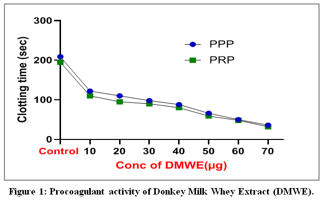 |
Figure 1: Procoagulant activity of Donkey Milk Whey Extract (DMWE).
|
The data illustrates the dose-response relationship between DMWE concentration and plasma clotting duration in both PPP (blue) and PRP (green) models. Significant decreases in bleeding times indicate It is the presence of bioactive peptides in the donkey milk proteome which enable hemostatic activation in specific locations. All values given as mean ± SD (n = 3). One-way ANOVA and Tukey’s post hoc test (p<0.05 vs control) were used to determine statistical significance.
Effect of DMWE on coagulation parameters (APTT and PT)
DMWE was tested on the parameters of coagulation, such as activated partial thromboplastin time (APTT) and prothrombin time (PT), and its findings are shown in Fig. 2. Both APTT and PT had a dose-dependent reduction in clotting time with an increase in the concentration of DMWE (20–100 µg). The control group had an APTT and PT of about 32 s and 13 s, respectively. Upon treatment with DMWE, APTT decreased progressively to about 30 s (20 µg), 24 s (40 µg), 15 s (60 µg), 7 s (80 µg), and 4 s (100 µg). Equally, the PT values decreased to 13 s in the control to about 12 s, 7 s, 4 s, 3 s, and 1-2 s at respective concentrations. The results of statistical analysis showed that the clotting time decreased significantly at greater amounts (***p < 0.001, ****p < 0.0001), and the coagulation activity was improved. These results indicate that DMWE has a strong procoagulant effect because it significantly reduces an intrinsic (APTT) and extrinsic (PT) pathway.
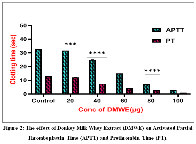 |
Figure 2: The effect of Donkey Milk Whey Extract (DMWE) on Activated Partial Thromboplastin Time (APTT) and Prothrombin Time (PT).
|
The bar chart demonstrates a substantial, dose-dependent reduction in clotting times (in seconds) for both APTT (teal bars) and PT (magenta bars) at dosages ranging from 20 to 100 µg. The noted time modulation of prothrombin is indicative of the presence of bioactive peptides in the DMWE proteome that affect localized hemostatic control. Arrows (***P < 0.001, ****P < 0.0001) denote statistical importance when contrasted with the control group.
The DMWE effect on clotting time in vivo is depicted in Fig. 3.
The clotting time was increased gradually with an increase in dose. The clotting time of the control group was around 180.5 ± 5.2 s. DMWE treatment produced a dosage-dependent decrease of 140.3 ± 4.1 s (low dose), 110.6 ± 3.5 s (moderate dose), 85.2 ± 2.9 s, 60.4 ± 2.3 s, and, at the highest dose tested, 45.7 ± 1.8 s. All subjects treated showed a substantial decrease in bleeding time compared to the control group (p < 0.05), indicating DMWE’s pro-coagulant activity in vivo.
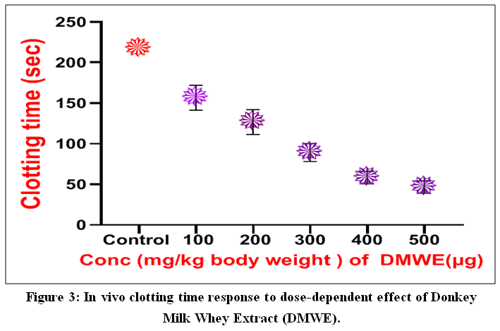 |
Figure 3: In vivo clotting time response to dose-dependent effect of Donkey Milk Whey Extract (DMWE)
|
The graph shows that the clotting time (in seconds) gradually decreases with increase in the concentration of DMWE Between 100-500mg/kg of body weight when compared with the untreated control. This important reduction in bleeding time is an indication of the potential of DMWE to act as an effective local procoagulant, allowing quick hemostatic control. Data are given in terms of mean and SD (n = 3), with statistical significance at *p < 0.05.
Clot formation analysis, both quantitative and qualitative, is indicated in figures 4A and 4B.
Fig. 4A depicted the representative images of the formation of clots in the samples (control and DMWE-treated 50, 100, and 150 µg). It was evident that the samples treated with DMWE had an improved clot density and aggregation, but in the control group, the clot formation was minimal. Clot formation rose from ~20.5 ± 1.6% (control) to 35.4 ± 1.8% (50 µg), 55.6 ± 2.1% (100 µg), and 75.8 ± 2.4% (150 µg), according to quantitative analysis (Fig. 4B). At the highest concentration, the clot started to form at a maximum of about 85.2 ± 2.7. These results validate the concentration-dependent promotion of clot formation by DMWE.
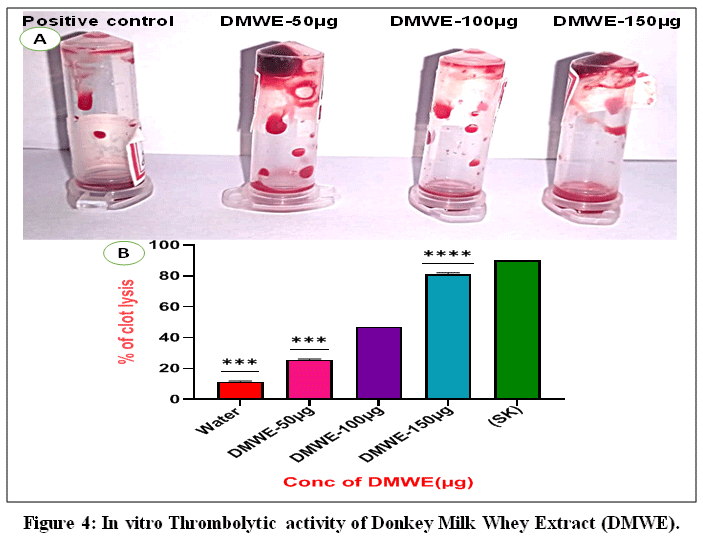 |
Figure 4: In vitro Thrombolytic activity of Donkey Milk Whey Extract (DMWE).
|
(A) Visualization of clot lysis in microcentrifuge tubes in the presence of a positive control and varying concentrations of DMWE (50 -100-150 µg). (B) Quantitative data demonstrating an increase in the proportion of lysis of the clot as a ratio of DMWE concentration. This strong thrombolytic action demonstrates the existence of bioactive peptides in the donkey milk proteome that is capable of preventing pathogenic thrombosis without impairment of the essential repair processes.
The hemocompatibility of DMWE was evaluated in a hemolysis experiment, and the results are presented in Fig. 5.
The hemocompatibility of DMWE was evaluated with the help of a hemolysis experiment, the results of which are presented in Fig. 5. The positive control (distilled water) had a hemolysis of 89.6 ± 3.1, and the negative control (PBS buffer) had a hemolysis of 1.2 ± 0.3%. All cases had low concentrations of DMWE, which had low hemolytic activity. The results for hemolysis were 2.8 ± 0.4% (50 µg), 2.1 ± 0.3% (100 µg), 1.8 ± 0.2% (150 µg), and 1.5 ± 0.2% (200 µg). The excellent biocompatibility was proven by the statistical analysis, which revealed significant differences with the positive control (***p < 0.001; **p < 0.0001). These results show that there does exist special biological duality such that the extract inhibits platelet-mediated thrombosis and promotes extrinsic coagulation.
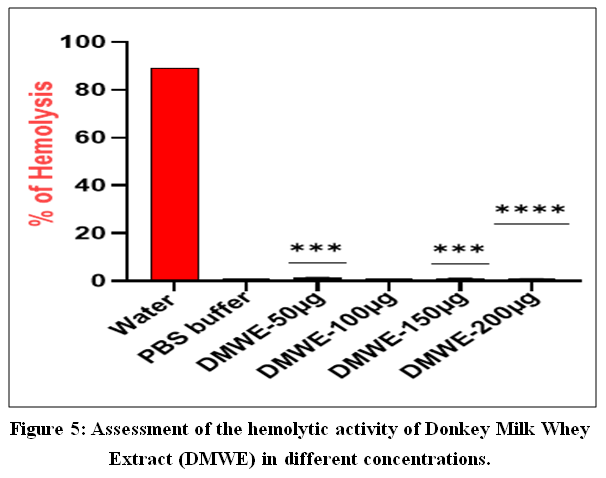 |
Figure 5; Assessment of the hemolytic activity of Donkey Milk Whey Extract (DMWE) in different concentrations.
|
The bar graph shows the percent of hemolysis induced by DMWE (50-200 µg) with respect to water (positive control) and PBS buffer (negative control). The insignificant hemolytic effect of all the dosages tested proves the cytoprotective effect of the extract and its lack of harm to the systemic use. Asterisks (noted as: ***P < 0.001, ****P < 0.0001) above the positive control indicate statistical significance.
Antidiabetic Effect and Enzyme Inhibition
α-amylase
The capability to block the digestion of carbohydrates in the diet, which consists of the most common enzymes in carbohydrate digestion, was one of the characteristics of DMWE that determined its antidiabetic potential. According to Tidona et al,20 it was found that the extract had a strong dose-dependent inhibitory effect on α-amylase and that its inhibitory curve was similar to the clinical standard, acarbose. The findings are presented in Fig. 6, which clearly depicts that the inhibitory activity of both samples, which is dependent on the concentration, increases. Acarbose was discovered to be a stronger α-amylase inhibitor (ranging around 38 to 75% at 10 µg to 65 µg, respectively). However, DMWE was inhibitory to a significant degree with a percentage of about 28% at 10 µg and 67% at 65 µg. The ability of DMWE to be a natural enzyme inhibitor was shown by its consistent increase in inhibitory power with concentration. Acarbose and DMWE differed more at lower doses (10-30 µg); DMWE had a similar but still lower level of inhibition with higher doses (50-65 µg) compared to acarbose. The trend shows that DMWE peptides resemble those of the pharmacological inhibitors that are extremely prevalent in human milk fractions by destabilizing the active sites of the enzyme.2
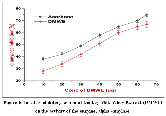 |
Figure 6: In vitro inhibitory action of Donkey Milk Whey Extract (DMWE) on the activity of the enzyme, alpha -amylase
|
The line graph is used to compare the percentage of dose-dependent inhibition of DMWE (red diamonds) with the standard clinical inhibitor, Acarbose (black circles). Both the agents exhibit a progressive rise in the inhibitory activity over a concentration range of 10-65 µg. The fact that DMWE can resemble clinical inhibitors points to the possibility that DMWE can be used as a natural nutraceutical in controlling postprandial hyperglycemia. The data are presented in the form of mean ± SD (n=3).
α-glucosidase
DMWE was tested on the α-glucosidase inhibitory activity at various concenrations and compared with the standard drug acarbose. A clear percentage inhibition concentration-dependent increase was observed in DMWE as shown in Fig. 7.The linear regression analysis (R² = 0.9896) revealed a strong positive connection between concentration and enzyme inhibition at doses of 100 and 500 µg, with inhibition levels ranging from 36 to 54 percent. Comparatively, acarbose showed a relatively higher level of inhibitory activity in all the concentrations tested, with an average level of 42% at 100 µg and 62% at 500 µg. In spite of the fact that acarbose was still stronger, DMWE was found to have a steady and gradual rise in inhibition, hence indicating its potential in being a natural α-glucosidase inhibitor. These results provide a biological rationale to use DMWE to treat postprandial hyperglycemia. At different concentrations.
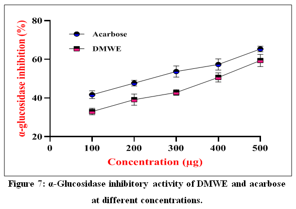 |
Figure 7: α-Glucosidase inhibitory activity of DMWE and acarbose at different concentrations
|
DMWE at different doses (100, 200, 300, 400, and 500 µg) were contrasted to the reference medication, acarbose, for its α-Glucosidase inhibitory action. DMWE and acarbose both had a concentration dependent inhibitory effect on α-glucosidase. The inhibition ranged from 28 ± 2% to 68 ± 2% for DMWE and 35 ± 2% to 75 ± 3% for acarbose. The value represents the average of three (n = 3) determinations with standard deviation.
DPP-IV inhibitory activity by DMWE
DMWE was tested on the DPP-IV inhibitory activity and compared to the standard drug sitagliptin. Fig. 8 illustrates that DMWE has been observed to exhibit an increase in the DPP-IV inhibition in a concentration-related manner. The greatest inhibitory activity was observed with sitagliptin (10 µM), which exhibited an approximate 58% inhibition. In comparison, DMWE did not inhibit it so intensively at 200 µg (around 25), but as the concentration of the inhibitor was raised, there was about 50 and 75 percent inhibition at 400 and 600 µg, respectively. The inhibitory activity was greatly enhanced with successive concentrations of DMWE.Statistical evaluation showed a significant variance in suppression with 200 µg of tetanus toxin (p < 0.001), 400 µg, and 600 µg of tetanus toxin (p < 0.0001) relative to the standard. These results indicate that DMWE has significant DPP-IV inhibitory properties, and it is almost similar to sitagliptin at high concentrations.
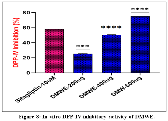 |
Figure 8: In vitro DPP-IV inhibitory activity of DMWE
|
The bar graph is a representation of the percentage inhibitor of Dipeptidyl peptidase-IV (DPP-IV) by different concentrations of Donkey milk whey extract (DMWE) in comparison to the standard drug Sitagliptin (10 µM). The results are reported in the form of Mean ± SD (n=3). When a comparison is done with the standard, statistical significance is indicated as: ****p < 0.0001 and ****p < 0.001.
Antioxidant Activity
The findings of evaluating DMWE’s antioxidant activity at various doses (20–120 µg/mL) are shown in Fig. 9. There was a definite rise in % inhibition that was concentration-dependent. DMWE showed 9.2 ± 0.8% inhibition at the lowest dose (20 µg/mL), which rose to 20.3 ± 1.1% at 40 µg/mL. At 60 µg/mL (31.5 ± 1.4%), a considerable increase in activity was noted. At increasing dosages, antioxidant activity increased significantly (p < 0.05), with inhibition reaching 54.6 ± 1.8% at 80 µg/mL and 68.7 ± 2.0% at 100 µg/mL. At 120 µg/mL (73.9 ± 2.3%), the greatest inhibition was seen. The standard chemical exhibited a relatively higher antioxidant activity at all the doses tested, with a range of inhibition of 10.5 ± 0.9% to 80.2 ± 2.5. However,
The bar graph is a representation of the percentage inhibitor of Dipeptidyl peptidase-IV (DPP-IV) by different concentrations of Donkey milk whey extract (DMWE) in comparison to the standard drug Sitagliptin (10 µM). The results are reported in the form of Mean ± SD (n=3). When a comparison is done with the standard, statistical significance is indicated as: ****p < 0.0001 and ****p < 0.001.
Antioxidant Activity
The findings of evaluating DMWE’s antioxidant activity at various doses (20–120 µg/mL) are shown in Fig. 9. There was a definite rise in % inhibition that was concentration-dependent. DMWE showed 9.2 ± 0.8% inhibition at the lowest dose (20 µg/mL), which rose to 20.3 ± 1.1% at 40 µg/mL. At 60 µg/mL (31.5 ± 1.4%), a considerable increase in activity was noted. At increasing dosages, antioxidant activity increased significantly (p < 0.05), with inhibition reaching 54.6 ± 1.8% at 80 µg/mL and 68.7 ± 2.0% at 100 µg/mL. At 120 µg/mL (73.9 ± 2.3%), the greatest inhibition was seen. The standard chemical exhibited a relatively higher antioxidant activity at all the doses tested, with a range of inhibition of 10.5 ± 0.9% to 80.2 ± 2.5. However, DMWE and the standard still exhibited a similar positive trend.Statistical study using one-way ANOVA and Tukey’s post hoc test revealed an enormous rise in antioxidant capacity with elevated concentration (p < 0.05). All experiments were done in triplicate, and the results are described by the mean plus standard deviation (SD). The concentration required to neutralize half of the free radicals was calculated to be approximately 82.4 µg/mL of DMWE, but the standard had a lower IC50 value of approximately 70.1 µg/mL, indicating a stronger standard.
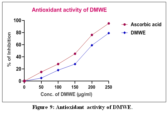 |
Figure 9: Antioxidant activity of DMWE
|
Antioxidant activity of DMWE at different concentrations (20–120µg/mL), expressed as percentage inhibition of free radicals. A concentration-dependent increase in antioxidant activity was observed, with higher inhibition values at elevated concentrations, indicating strong free radical scavenging potential of DMWE. The activity of DMWE was compared with a standard antioxidant, showing a similar trend with slightly lower inhibition values. The information is shown as mean ± SD (n = 3). The statistical value was assessed using one-way ANOVA (*p < 0.05 compared to control).
Hemorrhagic Activity of DMWE
The hemorrhagic potential of Donkey Milk Whey Extract (DMWE) was tested in in vivo dermal hemorrhagic assay and compared with the positive control (2 MHD venom) and negative control (saline). The hemorrhagic lesion was clearly observed in the positive control group, which had a mean hemorrhagic diameter of 10.4 ± 0.6 mm as shown in Fig. 1, indicating a high level of hemorrhagic activity of the venom. The negative control (saline) however did not exhibit any hemorrhagic lesion as the hemorrhagic diameter was negligible at 0.8 ± 0.2 mm. The hemorrhagic response was significantly decreased in a DMWE dose-dependent fashion. The hemorrhagic diameter was reduced to 4.6 ± 0.4 mm at 125 µg, 2.8 ± 0.3 mm at 250 µg, and 1.2 ± 0.2 mm at 375 µg of DMWE. Results of statistical analysis showed that there was significant decrease in hemorrhagic activity in all DMWE treated groups as compared to venom treated positive control (p<0.001). The results show that DMWE has strong protective properties against venom-induced haemorrhagic damage, and is very biocompatible to the skin, making it a promising candidate for haemorrhagic tissue injury control.
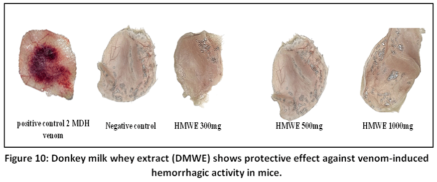 |
Figure 10: Donkey milk whey extract (DMWE) shows protective effect against venom-induced hemorrhagic activity in mice.
|
The representative Dorsal skin images of venom in 2 minimum hemorrhagic doses (2 MHD), saline (Negative control) and Donkey Milk Whey Extract (DMWE) treated with different concentrations (125, 250, and 375 µg) are illustrated. The positive control had a severe haemorrhagic lesion of 10.4 ± 0.6 mm while the negative control had only minimal haemorrhagic response of 0.8 ± 0.2 mm. The hemorrhagic lesion was dramatically decreased in a dose-dependent manner, measuring 4.6 ± 0.4 mm, 2.8 ± 0.3 mm, and 1.2 ± 0.2 mm. The results are shown as mean ± SEM (n = 3). Statistical significance was determined using one-way ANOVA and Duncan’s Multiple Range Test (p < 0.001).
Discussion
The presence of such encrypted bioactive peptides synthesized by whey proteins could be the cause of the dual hemostatic and antithrombotic effect of DMWE. The previous studies indicate that milk-derived peptides have cardioprotective properties, including ACE inhibition, platelet aggregation, and control of the coagulation process.39,40Additional evidence supporting these results includes larger evidence that supports that whey peptides provide cardiovascular health benefits and these benefits are antihypertensive and lipid-lowering actions.5,7,16 The liberation of these multi-purpose peptides during digestion facilitates the provision of procoagulant and antithrombotic activities which are easy to achieve through the liberation of these multi-purpose peptides during the digestion process. Several studies have shown that peptides in milk have the potential to improve endogenous antioxidant responses and prevent ROS, which can support the antioxidant activity of DMWE potential.12,14,18,41 The glutathione synthesis in cells is known to be promoted by whey proteins which in turn protect cells against oxidative damage and contributes to the prevention of chronic diseases.16,42 These processes are a strong indication of the radical scavenging activity exhibited in the present study. Moreover, lactoferrin and lysozyme are whey proteins, which are involved in immunological protection and inhibition of pathogens. These proteins do not only have direct antimicrobial effects, but also regulate the inflammatory response by modulating cytokines.23,24 The immunomodulatory nature of whey proteins as observed in this study is in line with previous reports that have highlighted the role of whey proteins in the promotion of both the innate and adaptive immunity.5,16These results are complimentary of the earlier studies that found donkey milk prevent damage of cells by oxidative stress and increase the activity of antioxidant enzymes. Earlier studies confirm that whey proteins such as lactoferrin and lysozyme play a crucial role in immunological protection and elimination of pathogens. These results coincide with the immunomodulatory and antibacterial properties depicted in this study. Besides direct antibacterial activity, such proteins regulate the inflammatory processes. The anti-inflammatory effect that has been achieved by inhibiting the activity of enzymes that break carbohydrates is correlated with the prior studies of milk-derived peptides that have strong inhibitory action against NF-KB signaling and cytokine production. Recent researches have revealed the antiprolliferative and apoptotic properties of milk-derived peptides, indicating that they can have anticancer applications.17 Its dual property of control and protection of donkey milk whey highlights its potential use as a base to prepare special medical nutrition and nutraceuticals.
Conclusion
The experimental evidence of Donkey Milk Whey Extract (DMWE) indicates a complicated sequence of bioactivity with a functional duality. The extract has a balancing effect of hemostatic control by acting as a localized procoagulant, thereby greatly decreasing the bleeding times, and as a powerful antiplatelet and thrombolytic agent. This counterintuitive ability, revealed by the ability to modulate prothrombin time and block agonist-induced platelet aggregation, indicates that DMWE has concealed bioactive peptides that can alleviate pathogenic thrombosis without interfering with important healing mechanisms. Besides cardiovascular health, DMWE has a tremendous potential of metabolic control. Its dose-dependent inactivation of α-amylase and α-glucosidase indicates a capability to resemble clinical inhibitors, which provides a powerful approach of controlling postprandial hyperglycemia. The extract is a complete nutraceutical, combined with large antioxidant potential, due to sulfur-containing amino acids, and intrinsic immunomodulatory properties. Simply put, the range of proteomic compositions of donkey milk whey presents a multifaceted array of physiological reactions that traverse the protection of cells to the regulation of metabolism on a global scale. These results support the importance of studying DMWE as a medical food base with certain properties, which will provide a complex solution to the multifactorial cardiovascular and metabolic issues.
Acknowledgement
The authors acknowledge Tumkur University and the Karnataka State Minority Welfare Department, Government of Karnataka, India.
Funding Sources
This work is supported by MPhil – PhD Fellowship Scheme of Directorate of Minorities, Government of Karnataka. Ms. Asma Tabassum got the fellowship under the 2021 MPhil–PhD Fellowship Renewal Students Scheme (Application Reference No. MDB015220000078.
Conflict of Interest
The authors do not have any conflict of interest.
Data Availability Statement
This statement does not apply to this article.
Ethics Statement
The Institutional Human Ethical Committee of Tumkur University, Tumakuru (IHEC-TUT No: 03/Res/2024-2025) accepted the designated human blood utilized for all the tests, which were carried out in accordance with ethical guidelines.
Informed Consent Statement
All healthy adult blood donors were informed about the study and signed informed consent forms before blood samples were taken. All participants were given information regarding the purpose of the study, the procedures of the study, the potential risks and benefits of the study, confidentiality of the participants personal information, and the right to end the study at any time. All the procedures were conducted on human subjects following the ethical principles of the Declaration of Helsinki.
Clinical Trial Registration
This research does not involve any clinical trials.
Permission to reproduce material from other sources
Not Applicable.
Author Contributions
- Asma Tabassum: Designed the study, Performed the experiment, Collected data, Carried out analysis and interpretation, Prepared original manuscript
- Rajesh Medar: Investigation, Methodology, Data curation, Format analysis
- Bhagyalakshmi Manjappa: Research supervision, Project administration, Review, Editing.
References
- Martini M, Altomonte I, Tricò D, Lapenta R, Salari F. Current Knowledge on Functionality and Potential Therapeutic Uses of Donkey Milk. Animals. 2021;11(5):1382. doi:10.3390/ani11051382
CrossRef - Fantuz F, Ferraro S, Todini L, Piloni R, Mariani P, Salimei E. Donkey milk concentration of calcium, phosphorus, potassium, sodium and magnesium. Int Dairy J. 2012;24(2):143-145. doi:10.1016/j.idairyj.2011.10.013
CrossRef - Garhwal R, Sangwan K, Mehra R, et al. A Systematic Review of the Bioactive Components, Nutritional Qualities and Potential Therapeutic Applications of Donkey Milk. J Equine Vet Sci. 2022;115:104006. doi:10.1016/j.jevs.2022.104006
CrossRef - Xu Q, Wei L, Chen X, et al. Nutritional Composition and Biological Activities of Donkey Milk: A Narrative Review. Foods. 2025;14(13):2337. doi:10.3390/foods14132337
CrossRef - Korhonen H. Milk-derived bioactive peptides: From science to applications. J Funct Foods. 2009;1(2):177-187. doi:10.1016/j.jff.2009.01.007
CrossRef - Yalcin A. Emerging Therapeutic Potential of Whey Proteins and Peptides. Curr Pharm Des. 2006;12(13):1637-1643. doi:10.2174/138161206776843296
CrossRef - Mansinhbhai CH, Sakure A, Maurya R, et al. Significance of whey protein hydrolysate on anti-oxidative, ACE-inhibitory and anti-inflammatory activities and release of peptides with biofunctionality: an in vitro and in silico approach. J Food Sci Technol. 2022;59(7):2629-2642. doi:10.1007/s13197-021-05282-3
CrossRef - Zhang X, Jiang G, Ji C, et al. Comparative Whey Proteome Profiling of Donkey Milk With Human and Cow Milk. Front Nutr. 2022;9. doi:10.3389/fnut.2022.911454
CrossRef - Li M, Yu H, Chen J, et al. Novel insights into whey protein differences between donkey and bovine milk. Food Chem. 2021;365:130397. doi:10.1016/j.foodchem.2021.130397
CrossRef - Li Y, Ma Q, Liu G, Wang C. Effects of donkey milk on oxidative stress and inflammatory response. J Food Biochem. 2022;46(4). doi:10.1111/jfbc.13935
CrossRef - Du X, Jing H, Wang L, Huang X, Wang X, Wang H. Characterization of structure, physicochemical properties, and hypoglycemic activity of goat milk whey protein hydrolysate processed with different proteases. LWT. 2022;159:113257. doi:10.1016/j.lwt.2022.113257
CrossRef - Das B, Basaiawmoit B, Sakure AA, et al. Production and Characterization of Antidiabetic and Antioxidative Peptides from Cheddar Cheese Whey with Anti-Inflammatory Activity: In Vitro, in Silico and Molecular Dynamic Study. Food Biotechnol. 2025;39(4):351-387. doi:10.1080/08905436.2025.2581887
CrossRef - Nongonierma AB, FitzGerald RJ. The scientific evidence for the role of milk protein-derived bioactive peptides in humans: A Review. J Funct Foods. 2015;17:640-656. doi:10.1016/j.jff.2015.06.021
CrossRef - Sarmadi B, Ismail A, Yusof L, Faridz Mod Yunoh M. Mechanism of action of cocoa on bone metabolism in calcium- and estrogen-deficient rat model of osteoporosis: Evidence for site and dose-related responses and involvement of IGF-I. J Funct Foods. 2020;66:103793. doi:10.1016/j.jff.2020.103793
CrossRef - FitzGerald RJ, Murray BA, Walsh DJ. Hypotensive Peptides from Milk Proteins. J Nutr. 2004;134(4):980S-988S. doi:10.1093/jn/134.4.980S
CrossRef - Pihlanto A. Antioxidative peptides derived from milk proteins. Int Dairy J. 2006;16(11):1306-1314. doi:10.1016/j.idairyj.2006.06.005
CrossRef - Meisel H. Biochemical Properties of Peptides Encrypted in Bovine Milk Proteins. Curr Med Chem. 2005;12(16):1905-1919. doi:10.2174/0929867054546618
CrossRef - Mao X, Gu J, Sun Y, et al. Anti-proliferative and anti-tumour effect of active components in donkey milk on A549 human lung cancer cells. Int Dairy J. 2009;19(11):703-708. doi:10.1016/j.idairyj.2009.05.007
CrossRef - Polidori P, Vincenzetti S. Use of Donkey Milk in Children with Cow’s Milk Protein Allergy. Foods. 2013;2(2):151-159. doi:10.3390/foods2020151
CrossRef - Tidona F, Sekse C, Criscione A, et al. Antimicrobial effect of donkeys’ milk digested in vitro with human gastrointestinal enzymes. Int Dairy J. 2011;21(3):158-165. doi:10.1016/j.idairyj.2010.10.008
CrossRef - Lacroix IME, Li-Chan ECY. Inhibition of Dipeptidyl Peptidase (DPP)-IV and α-Glucosidase Activities by Pepsin-Treated Whey Proteins. J Agric Food Chem. 2013;61(31):7500-7506. doi:10.1021/jf401000s
CrossRef - Lacroix IME, Li‐Chan ECY. Overview of food products and dietary constituents with antidiabetic properties and their putative mechanisms of action: A natural approach to complement pharmacotherapy in the management of diabetes. Mol Nutr Food Res. 2014;58(1):61-78. doi:10.1002/mnfr.201300223
CrossRef - Brumini D, Criscione A, Bordonaro S, Vegarud GE, Marletta D. Whey proteins and their antimicrobial properties in donkey milk: a brief review. Dairy Sci Technol. 2016;96(1):1-14. doi:10.1007/s13594-015-0246-1
CrossRef - Giansanti F, Panella G, Leboffe L, Antonini G. Lactoferrin from Milk: Nutraceutical and Pharmacological Properties. Pharmaceuticals. 2016;9(4):61. doi:10.3390/ph9040061
CrossRef - Marco ML, Heeney D, Binda S, et al. Health benefits of fermented foods: microbiota and beyond. Curr Opin Biotechnol. 2017;44:94-102. doi:10.1016/j.copbio.2016.11.010
CrossRef - Zhang L, Chu J, Hao W, et al. Gut Microbiota and Type 2 Diabetes Mellitus: Association, Mechanism, and Translational Applications. Mediators Inflamm. 2021;2021(1). doi:10.1155/2021/5110276
CrossRef - Embleton ND, Berrington JE. Clinical Trials of Lactoferrin in the Newborn: Effects on Infection and the Gut Microbiome. In: 2020:141-151. doi:10.1159/000505334
CrossRef - Ardlie NG, Han P. Enzymatic Basis for Platelet Aggregation and Release: The Significance of the ‘Platelet Atmosphere’ and the Relationship between Platelet Function and Blood Coagulation. Br J Haematol. 1974;26(3):331-356. doi:10.1111/j.1365-2141.1974.tb00477.x
CrossRef - Manjappa B, Gangaraju S, Girish KS, et al. Momordica charantia seed extract exhibits strong anticoagulant effect by specifically interfering in intrinsic pathway of blood coagulation and dissolves fibrin clot. Blood Coagulation & Fibrinolysis. 2015;26(2):191-199. doi:10.1097/MBC.0000000000000191
CrossRef - Prasad S, Kashyap RS, Deopujari JY, Purohit HJ, Taori GM, Daginawala HF. Development of an in vitro model to study clot lysis activity of thrombolytic drugs. Thromb J. 2006;4(1):14. doi:10.1186/1477-9560-4-14
CrossRef - Denis C, Methia N, Frenette PS, et al. A mouse model of severe von Willebrand disease: Defects in hemostasis and thrombosis. Proceedings of the National Academy of Sciences. 1998;95(16):9524-9529. doi:10.1073/pnas.95.16.9524
CrossRef - Chelladurai GRM, Chinnachamy C. Alpha amylase and Alpha glucosidase inhibitory effects of aqueous stem extract of Salacia oblonga and its GC-MS analysis. Brazilian Journal of Pharmaceutical Sciences. 2018;54(1). doi:10.1590/s2175-97902018000117151
CrossRef - Nater UM, La Marca R, Florin L, et al. Stress-induced changes in human salivary alpha-amylase activity—associations with adrenergic activity. Psychoneuroendocrinology. 2006;31(1):49-58. doi:10.1016/j.psyneuen.2005.05.010
CrossRef - Tolmie M, Bester MJ, Apostolides Z. Inhibition of α‐glucosidase and α‐amylase by herbal compounds for the treatment of type 2 diabetes: A validation of in silico reverse docking with in vitro enzyme assays. J Diabetes. 2021;13(10):779-791. doi:10.1111/1753-0407.13163
CrossRef - Abulkhair HS. α-Glucosidase-targeting 1,2,4-triazole antidiabetic candidates: comparative analysis and future perspectives. Future Med Chem. 2025;17(24):3025-3040. doi:10.1080/17568919.2025.2587561
CrossRef - Lebovitz HE. ALPHA-GLUCOSIDASE INHIBITORS. Endocrinol Metab Clin North Am. 1997;26(3):539-551. doi:10.1016/S0889-8529(05)70266-8
CrossRef - Xue Y, Zeng L, Yan Q, Jiang Z. Efficient Production of a Novel Subtilisin-like Serine Protease in Aspergillus niger for the Preparation of DPP-IV Inhibitory Activity Peptides from Protein Byproducts. J Agric Food Chem. 2025;73(35):22014-22026. doi:10.1021/acs.jafc.5c06047
CrossRef - Okoh S, Asekun O, Familoni O, Afolayan A. Antioxidant and Free Radical Scavenging Capacity of Seed and Shell Essential Oils Extracted from Abrus precatorius (L). Antioxidants. 2014;3(2):278-287. doi:10.3390/antiox3020278
CrossRef - Lyons KE, Ryan CA, Dempsey EM, Ross RP, Stanton C. Breast Milk, a Source of Beneficial Microbes and Associated Benefits for Infant Health. Nutrients. 2020;12(4):1039. doi:10.3390/nu12041039
CrossRef - Udenigwe CC, Aluko RE. Food Protein‐Derived Bioactive Peptides: Production, Processing, and Potential Health Benefits. J Food Sci. 2012;77(1). doi:10.1111/j.1750-3841.2011.02455.x
CrossRef - Corrochano AR, Sariçay Y, Arranz E, Kelly PM, Buckin V, Giblin L. Comparison of antioxidant activities of bovine whey proteins before and after simulated gastrointestinal digestion. J Dairy Sci. 2019;102(1):54-67. doi:10.3168/jds.2018-14581
CrossRef - Zemel MB, Miller SL. Dietary Calcium and Dairy Modulation of Adiposity and Obesity Risk. Nutr Rev. 2004;62(4):125-131. doi:10.1111/j.1753-4887.2004.tb00034.x
CrossRef
Abbreviations:
ACE: Angiotensin-Converting Enzyme
AMC: 7-Amino-4-methylcoumarin
ANOVA: Analysis of Variance
APTT: Activated Partial Thromboplastin Time
CaCl₂: Calcium Chloride
CMWE: Crude Milk Whey Extract
DMRT: Duncan’s Multiple Range Test
DMWE: Donkey Milk Whey Extract
DMSO: Dimethyl Sulfoxide
DNS: 3,5-Dinitrosalicylic Acid
DPPH: 2,2-Diphenyl-1-picrylhydrazyl
DPP-IV: Dipeptidyl Peptidase-IV
GC-MS: Gas Chromatography–Mass Spectrometry
HCl: Hydrochloric Acid
IC₅₀: Half Maximal Inhibitory Concentration
IGF-I: Insulin-like Growth Factor-I
INR: International Normalized Ratio
MHD: Minimum Hemorrhagic Dose
mM: Millimolar
min: Minute
mU: Milliunit
MWE: Milk Whey Extract
µg: Microgram
µL: Microliter
µM: Micromolar
NF-κB: Nuclear Factor Kappa B
nm: Nanometer
OD: Optical Density
PBS: Phosphate Buffered Saline
pH: Potential of Hydrogen
pNA: p-Nitroaniline
PPP: Platelet-Poor Plasma
PRP: Platelet-Rich Plasma
PT: Prothrombin Time
RBC: Red Blood Cells
ROS: Reactive Oxygen Species
rpm: Revolutions Per Minute
s: Seconds
SD: Standard Deviation
SEM: Standard Error of Mean
UV-Vis: Ultraviolet–Visible Spectrophotometry
°C: Degree Celsius
 PlumX:
 Views: 391 Views
  PDF Downloads:
8
PDF Downloads:
8 Accepted on: 21-07-2026
Second Review by: Dr. Sarraa Dhiaa Kasim
Final Approval by: Dr. Wagih Ghannam