
Aseptic In vitro Synthesis of Pinus gerardiana Ectomycorrhizae with Amanita ceciliae and Lactarius sanguifluus
 and Anand Sagar
 Department of Biosciences, Himachal Pradesh University, Summer Hill, Shimla (H.P.), india.
Corresponding Author E-mail:aksbios@hpuniv.ac.in
DOI : http://dx.doi.org/10.13005/bbra/3181
Download this article as:
![]()
The present investigations are aimed to synthesize in vitro ectomycorrhizae between Pinus gerardiana and two gilled ectomycorrhizal (ECM) mushrooms (Amanita ceciliae and Lactarius sanguifluus). To carry out in vitro synthesis, pure cultures of ECM mushrooms (A. ceciliae and L. sanguifluus) were isolated on Potato Dextrose Agar (PDA) and Modified Melin-Norkans (MMN) Medium respectively. The synthesis was achieved successfully in the surfaced sterilized seedlings of P. gerardiana geminated under aseptic conditions by using vermiculite, peat, medium for ECM fungi and inoculum of each fungus in the test tubes. Mycorrhization was checked periodically in the test tubes. P. gerardiana seedlings were lifted from test tubes after five months to observe ectomycorrhizae formation on the root system with A. ceciliae and L. sanguifluus. The synthesized ectomycorrhizae were dark brown in case of A. ceciliae whereas in case of L. sanguifluus the colour of ECM roots was yellowish brown. Anatomy of synthesized ectomycorrhizae with both ECM fungi showed fully developed fungal mantle and Hartig net. The seedlings with ECM synthesis showed a significant effect on the growth and development.
KEYWORDS:Amanita ceciliae; Ectomycorrhiza; In vitro synthesis; Lactarius sanguifluus; Pinus gerardiana
Introduction
The ECM fungi form symbiotic interactions with roots of forest trees and play a vital role to bring about various benefits to the plant species, including enhancement in the roots absorption surface area 1, increased uptake of various nutrients 2, resistance to pathogenic microorganisms 3 and drought stress 4. ECM symbiosis also help to increase the growth and nutrient contents of plants growing in nutrient deficient soils 5 and in return, the photosynthetic host tree species supply carbohydrates to their non-photosynthetic ECM fungal partners.
The roots of a single tree in natural forest ecosystems are almost invariably associated with many different ECM fungal species 6. Therefore host specificity of ECM fungi is an important part of forest ecosystems, which can be utilized as a tool for managing different forest resources 7. Natural hosts of ECM mushroom Tricholoma matsutake include Pinus sylvestris and P. abies 8,9, P. densiflora and Quercus mongolica in northeastern China and Korean peninsula 10,11, P. armandii, P. densata, P. wallichiana, P. yunnanensis, Casta-nopsis orthacantha, Q. aquifolioides, Q. pannosa, Q. guyavifolia, Q. semecarpifolia, Lithocarpus spp., and Pasania spp. in southwestern China and Bhutan 8,11 whereas Amanita muscaria, Laccaria laccata, Pisolithus sp. and Suillus brevipes have been reported to produce ectomycorrhizae with P. patula 12.
Many systems of in vitro ECM synthesis have been developed by various researchers to examine the potential of different ECM fungi to form symbiosis with different forest tree species 13-18. ECM synthesis investigations are helpful to decide fungus and host plant compatibility for morphological, anatomical and physiological investigations 19.
Pinus gerardiana is an important edible nut yielding conifer and is well known in dry fruit trade. The trade of its seeds significantly contributes to the annual income of most of the tribal families living in the area of its distribution. In district Kinnaur of Himachal Pradesh alone approximate export value of its annual produce around 18 crores rupees. It clearly reflects that the tree is not only ecologically important to the area but also has direct relevance to the economic status of the people residing over there 20. Hence keeping into consideration the immense economic as well as ecological importance of P. gerardiana, the present investigation is aims to know about the ECM symbiosis between two gilled mushrooms and P. gerardiana also provide morphological and anatomical account of the synthesized ECM roots. These ECM fungi could be a very useful tool for the artificial inoculation in the nurseries to develop inoculated ECM seedling which further could also show better establishment in its natural habitats and helps in the reforestation and regeneration programs of P. gerardiana in Himachal Pradesh, India.
MATERIALS AND METHODS
Collection and isolation of pure culture of ECM fungi
The sporophore of A. ceciliae and L. sanguifluus were collected from district Kinnaur of Himachal Pradesh. The cultures were isolated from pileus region of the fresh sporophore of A. ceciliae and L. sanguifluus. Pileus was pulled gently apart and exposed interior tissue was removed with the help of sterilized scalpel and transferred into Petri plates containing PDA and MMN Medium. Then they were incubated at 250C and observed regularly for the appearance of culture. The well-grown colonies were subcultured on a PDA medium to obtain pure cultures.
Surface sterilization and seeds germination
Seeds of P. gerardiana were washed for 4 hours under the running tap H2O and then treated with 10% H2O2 for 10 minutes. The H2O2 was drained and seeds were rinsed in sterilized distilled H2O for 2 minutes. Then they were treated with 10% NaClO and Tween-20 and kept for 30 minutes. After that washed 5-6 times using sterilized distilled H2O. Then seeds were placed in sterilized H2O for 24 hours. After that seeds were put on sterilized wet filter paper in petri-plates and placed in low temperature for 24 hours before put in the seed germinator.
In vitro ECM synthesis
ECM synthesis was carried out by using in vitro synthesis experiments 21. For this 70ml of fungal nutrient medium, 90ml of vermiculite and 10ml peat was used in each 200ml test tube. The mixture of test tubes was autoclaved and then for treatments a disc of 5mm fungal culture was placed in test tube under aseptic conditions. For both treatments and uninoculated controls 10 replicates were used and treatments were kept for mycelium colonization. After that aseptically germinated P. gerardiana seedling was put into each treatments and control placed in the growth chamber and observed regularly for ECM synthesis. After 5 months seedlings pulled out from test tubes and checked for mycorrhization by following the methods of Fortin et al. 22.
Confirmation of ECM synthesis
The confirmation of ECM synthesis was done following Moore et al. 23. For this a minute inoculum of vermiculite, peat mixture was removed from test tubes and inoculated on Petri plates containing nutrient medium. The synthesized ECM roots were detached from root system of the seedlings, surface sterilized and small pieces of ECM roots were inoculated on nutrient medium. Then Petri plates were observed for the mycelial growth and fungal colonies formed from both were compared with original ECM fungal culture colonies to confirm the ECM synthesis.
Morpho-anatomical characterization of ectomycorrhiza
Morphological as well as anatomical detail of ECM roots was made by following Zak 24. The various growth characteristics e.g. form of ectomycorrhiza, colour of the fungal mantle, thickness of mantle etc. were recorded. The fixation ECM roots was done in F.A.A. solution and then preserved by using 70% alcohol. Anatomical details were worked out in both fresh as well as from preserved material. The sectioning of ECM roots was done following Johansen 25.
Effect of in vitro ECM synthesis on seedlings growth
The five P. gerardiana seedlings were lifted randomly from each treatment and control. The observations on various growth parameters e.g. shoot length, root length, total number of short roots, shoot fresh weight, root fresh weight, shoot dry weight and root dry weight were recorded.
Statistical analysis of the data
The data was statistically analyzed, with the help of ANOVA test and Tukey’s multiple compression test was used to determine HSD (honestly significant difference) values for significance among various mean values.
Results
Isolation of pure culture and sub culturing of ECM fungi
The pure cultures of A. ceciliae was isolated on PDA medium. The colonies form dense mycelial mat and are white in colour. The pure culture of L. sanguifluus has been isolated on MMN medium. The colony of mycelium form concentric zones during its growth. The margin of the colony was irregular. Sub culturing of ECM mushrooms was carried out on PDA medium.
In vitro ECM synthesis
The pure cultures of ECM mushrooms (A. ceciliae and L. sanguifluus) were anlysed for their potential of ECM synthesis with the roots of P. gerardiana seedlings (Figure 1A and Figure 2A). The experiments revealed the successful in vitro mycorrhization between the host and two ECM mushrooms tested. The treated seedling showed the formation of many branched short lateral roots which ultimately leads to the development of ectomycorrhizae (Figure 1B and Figure 2B). The A. ceciliae and L. sanguifluus formed dark brown (Figures 1C,D) and yellowish brown types (Figures 2C,D) of ECM roots respectively. The description of synthesized ECM roots is given in Table 1.
Table 1: Morpho-anatomical description of Pinus gerardiana ECM roots synthesized with Amanita ceciliae and Lactarius sanguifluus
|
Sr. No. |
|
Ectomycorrhizal Mushrooms |
|
|
Amanita ceciliae |
Lactarius sanguifluus |
||
|
|
Macroscopic Characteristics |
||
|
1 |
Colour |
Dark Brown |
Yellowish Brown |
|
2 |
Shape |
Branched, Monopodial |
dichotomously branched |
|
3 |
Texture |
Smooth to loosely wooly |
Smooth to shiny |
|
4 |
Odour and taste |
Not distinct |
Not distinct |
|
5 |
Emanating Hyphae |
Many long irregularly radiating outwards |
Infrequent |
|
6 |
Root Hairs |
Absent |
Absent |
|
|
Microscopic Characteristics |
||
|
7 |
Thickness of Mantle |
25-30 µm |
15-20 µm |
|
8 |
Degree of development of “Hartig net” |
Well developed |
Well developed |
Effect of in vitro ECM Synthesis on the seedlings growth
The seedlings of P. gerardiana were evaluated for various growth characteristics after the completion of in vitro experiments with both ECM fungi tested. The results on different growth characteristics are presented in Table 2. The results revealed that in both treatments (A. ceciliae and L. sanguifluus) there was a significant difference(P<0.01 and P<0.05) on the overall growth characteristics of the seedlings as compared to the control (Table 2). Therefore in vitro ECM synthesis have significant effect on the seedlings growth and development.
The anatomical details of synthesized ECM roots with both ECM mushrooms tested showed the formation fungal mantle and Hartig net (Figures 1F and Figures 2F). In case of A. ceciliae, the thickness of fungal mantle was 25-30 µm whereas in case of L. sanguifluus it was 15-20 µm (Table 1). The seedlings kept as control were non-mycorrhizal with prominent root hairs and without ECM anatomical features in the transverse section of their roots. The culture colonies of A. ceciliae was reisolated on PDA and Hagem’s nutrient medium from both synthesized ECM roots and peat moss, vermiculite mixture (Figure 1E). Similarly the culture colonies of L. sanguifluus was reisolated on PDA and MMN nutrient medium from both synthesized ECM roots and peat moss, vermiculite mixture (Figure 2E)
The culture colonies of A. ceciliae and L. sanguifluus reisolated were found to have similar characteristics while compared with the culture colonies isolated from the fresh sporophores of both ECM mushrooms. Thus both (A. ceciliae and L. sanguifluus) confirming the synthesis of ECM roots with P. gerardiana seedlings.
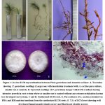 |
Figure 1: In vitro ECM mycorrhization between Pinus gerardiana and Amanita ceciliaes: A. Test tubes showing P. gerardiana seedlings (Larger one with inoculation treatment with A. ceciliae pure culture, smaller one is control).
|
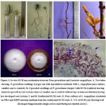 |
Figure 2: In vitro ECM mycorrhization between Pinus gerardiana and Lactarius sanguifluus: A. Test tubes showing P. gerardiana seedlings (Larger one with inoculation treatment with L. sanguifluus pure culture, smaller one is control).
|
Table 2: Effect of artificial inoculation on the growth of Pinus gerardiana seedlings.
|
Sr. No. |
Growth Characteristics |
Inoculated Treatments (cm ± S.D.) |
Un-inoculated (cm ± S.D.) |
|
|
|
|
A. ceciliae |
L. sanguifluus |
Control |
|
1 |
Root Length (cm) |
41.30±1.53** |
51.24±1.22** |
34.58±3.38 |
|
2 |
Shoot Length (cm) |
15.06±1.24** |
14.38±1.27** |
8.88±1.21 |
|
3 |
Total No. of Short Roots |
73.60±7.81** |
62.40±5.00** |
12.60±2.30 |
|
4 |
Root Fresh Weight (mg) |
1.64±0.36** |
2.59±0.34** |
0.23±0.06 |
|
5 |
Shoot Fresh Weight (mg) |
2.64±0.60** |
2.36±0.55** |
0.98±0.13 |
|
6 |
Root Dry Weight (mg) |
0.27±0.10** |
0.48±0.09** |
0.09±0.02 |
|
7 |
Shoot Dry Weight (mg) |
0.83±0.23** |
0.78±0.25* |
0.35±0.04 |
** p<0.01; * p≤0.05; NS; Non-Significant differences as revealed through one way ANOVA and Tukey’s HSD multiple comparison test.
Discussion
During the present study two ECM mushrooms A. ceciliae and L. sanguifluus were isolated into pure culture on PDA and MMN medium respectively. The findings of the present study are supported by the results of several previous researches that reported mycelial growth of ECM fungi on various nutrient media. Phlebopus portentosus isolated on (Murashige and Skoog Agar) MS medium 26, Scleroderma sinnamariense prefer (Fungus-Host) FH medium for mycelial growth 27, Rhizopogon roseolus and Tricholoma matsutake cultures show best mycelia growth on PDA medium 28, Pisolithus microcarpus was isolated on (Pridham-Gottlieb Modifled by Kuek) PGK medium 29, Similarly Endo et al. 30 isolated specimens of B. Edulis on MA medium and subculturing was done successfully on (Malt Extract Agar) MA and (Malt Yeast Agar) MYA nutrient media. However, Agueda et al. 31 isolated Boletus aereus, B. edulis, B. pinophilus and B. reticulatus on BAF nutrient medium. The pure cultures are greatly affected by the changes made in cultivation medium and most of the ECM Boletale mushrooms favour media containing malt such as MA and MMN 29.
The genus Pinus is a host for numerous ECM fungi and these fungi made symbiotic association with the roots of this conifer 32. During the present investigations in vitro ectomycorrhizae of P. gerardiana were synthesized with two ECM mushrooms (A. ceciliae and L. sanguifluus). Similarly mycorrhization between Picea abies and Cenococcum geophilum was achieved in asceptic conditions by inoculating mycelum on MMN medium 33. Vaario et al. 34 achived ECM in vitro synthesis between Abies firma and Pisolithus tinctorius. Pinus densiflora is found to be formed ECM association in laboratory conditions with broad range of ECM fungi 35,36.
In Tibet, Tricholoma matsutake synthesized mycorrhizae in coniferous and fagaceous hosts tree in natural habitat and synthesized Hartig net in the ECM roots of P. densiflora 35. In Canada T. matsutake synthesized mycorrhizae in pine and oak hosts trees 37 and also formed typical Hartig net in P. densiflora ECM roots.
In similar study Laiye et al. 38 utilized vermiculite, modified MMN medium and peat moss for the in vitro synthesis of ECM roots in the two Larch spp. seedlings with various ECM fungi. More recently, ECM in vitro synthesis of Cedrus deodara with Rhizopogon himalayensis 17, Pinus gerardiana with Boletus edulis and Suillus scibricus 18 was achived successfully.
The successes was achieved to germinate spores 39, obtaining pure culture mycelia, in vitro synthesis of ectomycorrhizae 40 and artificial shiro formation in potted culture system containing granite based soil substrate 41.
ECM fungi had a wide range of host specificities as reported by Kumla et al. 42 and synthesized in vitro ectomycorrhizae in two tree species viz. Eucalyptus camaldulensis and Pinus kesiya with single ECM fungus Pisolithus orientalis. Pure culture of P. tinctorius had potential for ECM association with Pinus taiwanensis in aseptic systems, but the association has not been reported in natural conditions 43. Gomes et al. 44 reported that cultures of P. arhizus isolated from fruiting bodies form symbiotic association with Quercus suber and also had the potential to synthesize mycorrhiza with Arbutus unedo under laboratory and nursery conditions. The ectomycorrhizae were appears mainly on short lateral apices 45.
The results of the morpho-anatomical study were similar to the previous reports of Samson and Fortin 46 to synthesized grayish brown to pinkish grey, whitish and yellow coloured ECM roots in Larix laricina with two ECM fungal genus Fuscoboletinus and Suillus. In vitro synthesis of ECM roots between S. sibiricus and P. wallichiana was achieved successfully in vessels and formed ECM roots were bifurcate to coralloid and creamish yellow in colour 47. Garcia-Rodriguez et al. 48 carried out synthesis to ECM roots in Eucalyptus urophylla and Pinus greggii with Pisolithus tinctorius, the synthesized ectomycorrhizae were simple, 1.0-2.6 mm long, yellow brown to bright yellow in colour and fungal mantle was 15.0 µm in thickness whereas ECM roots formed in Pinus greggii were bright yellow to yellowish brown in colour, dichotomously divided and rarely monopodial,.
The ECM roots produced during in vitro experiments in the present study showed similar anatonical features as that observed in many previous studies carried out by Kumla et al. 42 and observed that Pisolithus orientalis has ability to synthesized fungal mantles under in vitro conditions with Pinus kesiya and Eucalyptus camaldulensis.The both fungal mantles were plectenchymatous and similar to the mantle synthesized by Pisolithus tinctorius with Abies firma and Quercus ilex, Q. coccifera 49, Pisolithus microcarpus with Eucalyptus grandis 50 and Pisolithus aurantioscabrosus with Shorea sp. 51, Cedrus deodara with Rhizopogon himalayensis 17, Suillus scibiricus, Boletus edulis with Pinus gerardinan 18. Similar fungal mantle is synthesized by Boletus edulis with Cistus spp. 31 and Picea abies 52.
The culture of A. ceciliae and L. sanguifluus were reisolated from both synthesis substrate mixture as well as from synthesised ECM roots. They have similar characteristics as that of original cultures isolated from the fruiting bodies, thus validate the successful in vitro synthesis of ectomycorrhizae with these two ECM mushrooms. Similarly, Scleroderma aurantium was reisolated from both peat vermiculite mixture and synthesized ECM roots on MMN nutrient medium 53. The fungal cultures were also reisolated from both synthesis experiment substrate mixture and from synthesized ECM roots to verify symbiotic interactions 47,16,17.
Conclusion
The present study has synthesized ECM roots of P. gerardiana under laboratory conditions by using in vitro experiments with the pure cultures of two ECM mushrooms (A. ceciliae and L. sanguifluus). The synthesized ectomycorrhizae with A. ceciliae and L. sanguifluus were dark brown and yellowish brown in colour respectively. The T.S. of ECM roots showed a typical ECM anatomy.
Acknowledgments
The authors are thankful to Department of Bio-Sciences, Himachal Pradesh University, Summer Hill, Shimla H.P. for providing the related support to compile this research work.
Conflıct of Interest
The authors declare that there is no conflict of interest
Fundıng Sources
There are no funding Sources
References
- Harley J.L., Smith S.E. Mycorrhizal Symbiosis. Academic, New York. 1983.
- Landeweert R., Hoffland E., Finlay R.D., Kuyper T.W., van Breemen N. Linking plants to rocks: ectomycorrhizal fungi mobilize nutrients from minerals. Ecol. Evol. 2001; 16: 248-254.
CrossRef - Branzanti M.B., Rocca E., Pisi A. Effect of ectomycorrhizal fungi on chestnut ink disease. Mycorrhiza. 1999; 9: 103-109.
CrossRef - Morte A., Lovisolo C., Schubert A. Effect of drought stress on growth and water relations of the mycorrhizal association Helianthemum almeriense – Terfezia claveryi. Mycorrhiza. 2000; 10: 115-119.
CrossRef - Jones M.D., Durall D.M., Tinker P.B. Fluxes of carbon and phosphorus between symbionts in willow ectomycorrhizas and their changes with time. New Phytol. 1991; 119: 99-106.
CrossRef - Dahlberg A. Community ecology of ectomycorrhizal fungi: an advancing interdisciplinary field. New Phytol. 2001; 150: 555-562.
CrossRef - Smith S.E., Read D. Mycorrhizal Simbiosis 3rd Academic Press. Elsevier, Great Britain. 2008.
- Matsushita N., Kikuchi K., Sasaki Y., Guerin-Laguette A., Vaario L.M., Suzuki K., Lapeyrie F., Intini M. Genetic relationship of Tricholoma matsutake and nauseosum from the Northern Hemisphere based on analyses of ribosomal DNA spacer regions. Mycoscience. 2005; 46(2): 90-96.
CrossRef - Vaario L.M., Pennanen T., Sarjala T., Savonen E.M., Heinonsalo J. Ectomycorrhization of Tricholoma matsutake and two major conifers in Finland: an assessment of in vitro mycorrhiza formation. Mycorrhiza. 2010; 20: 511-518.
CrossRef - Murata H., Babasaki K., Saegusa T., Takemoto K., Yamada A., Ohta A. Traceability of Asian matsutake, specialty mushrooms produced by the ectomycorrhizal basidiomycete Tricholoma matsutake, on the basis of retroelement-based DNA markers. Environ. Microbiol. 2008; 74: 2023-2031.
CrossRef - Yamanaka K., Aimi T., Wan J., Cao H., Chen M. Species of host trees associated with Tricholoma matsutake and closely allied in Asia (in Japanese). Sci. Technol. 2011; 19: 79-87.
- Mohan V., Natarajan K., Ingleby K. Anatomical studies on ectomycorrhizas. II. The ectomycorrhizas produced by Amanita muscaria, Laccaria laccata and Suillus brevipes on Pinus patula. Mycorrhiza. 1993; 3: 43-49.
CrossRef - Rincon A., Alvarez I.F., Pera J. Inoculation of containerized Pinus pinea seedlings with seven ectomycorrhizal fungi. Mycorrhiza. 2001; 11: 265-271.
CrossRef - Eros-Honti Z., Jakucs E. Characterization of beech ectomycorrhizae formed by species of the Pachyphloeus–Amylascus Mycorrhiza. 2009; 19: 337-345.
CrossRef - Geng L.Y., Wang X.H., Yu F.Q., Deng X.J., Tian X.F., Shi X.F., Xie X.D., Liu P.G., Shen Y.Y. Mycorrhizal synthesis of Tuber indicum with two indigenous hosts, Castanea mollissima and Pinus armandii. Mycorrhiza. 2009; 19: 461-467.
CrossRef - Kumar S., Sagar A., Sehgal A.K. Ectomycorrhizal synthesis of Lactarius sanguifluus (Paulet) Fr. with Abies pindrow Royle Ex D. Don. J. Biotech. Biosci. 2019; 7(6): 89-92.
- Singh L., Tapwal A., Thakur J.S., Lakhanpal T.N. Studies on the nutritional requirement and synthesis in vitro of mycorrhiza of Cedrus deodara with Rhizopogon himalayensis. Kavaka. 2020; 54: 24-29.
CrossRef - Sehgal A.K., Sagar A. In vitro mycorrhization of two wild edible bolete species with Pinus gerardiana – an economically high altitude conifer. Biotech. Res. Asia. 2022; 19(4): 1009-1018.
CrossRef - Giomaro G.M., Sisti D., Zambonelli A. Cultivation of edible ectomycorrhizal fungi by in vitro mycorrhizal synthesis. In: In vitro culture of mycorrhizas. Soil Biology (Declerck S., Strullu D.G., Fortin J.A. eds), Springer. 2005; pp. 253-267.
CrossRef - Kapoor K.S., Kumar S., Singh O. Chilgoza (Pinus gerardiana Wall): Champion of the rocky mountains. HFRI, Conifer Campus , Panthaghati, Shimla, H.P. 2003; Bro. No. 8.
- Molina R. Pure culture synthesis and host specificity of red alder mycorrhizae. J. Bot. 1979; 59: 1223-1228.
CrossRef - Fortin J.A., Piche Y., Lalonde M. Technique for the observation of early morphological changes during ectomycorrhiza formation. J. Bot. 1980; 58: 361-365.
CrossRef - Moore L.M., Jansen A.E., van Griensven L.J.L.D. Pure culture synthesis of ectomycorrhizas with Cantharellus cibarius. Acta Bot. Neerlan. 1989; 38(3): 273-278.
CrossRef - Zak B. Characterization and identification of Douglas fir mycorrhizae. In: Mycorrhizae (Hacskaylo E. Ed.), S. Department of Agriculture. Forest Service. Misc. Publication 1189. U.S. Printing Office. Washington, D.C. 1971; pp. 38-53.
- Johansen D.A. Plant microtechnique. Tata McGraw-Hill Publishing Company, Bombay, New Delhi. 1940.
- Kumla J., Danell E., Bussaban B., Lumyong S. Suitable growth conditions and nutrition factors on in vitro culture of Phlebopus portentosus (Boletales). Mai. J. Sci. 2011; 38: 156-159.
- Siri-in J., Kumla J., Suwannarach N., Lumyong S. Culture condition and some properties of pure culture of ectomycorrhizal fungus, Scleroderma sinnamariense. Mai. J. Sci. 2014; 41: 275-285.
- Islam F., Ohga S. Effects of media formulation on the growth and morphology of ectomycorrhizae and their association with host plant. International Scholarly Research Notices Agronomy. 2013; 1-12.
CrossRef - Rossi M.J., Oliveira V.L. Growth of the ectomycorrhizal fungus Pisolithus microcarpus in different nutritional conditions. J. Microbiol. 2011; 42: 624-632.
CrossRef - Endo N., Kawamura F., Kitahara R., Sakuma D., Fukuda M., Yamada, A. Synthesis of Japanese Boletus edulis ectomycorrhizae with Japanese red pine. Mycoscience. 2014; 55: 405-416.
CrossRef - Agueda B., Parlade J., Fernandez-Toiran L.M., Cisneros O., de Miguel A.M., Modrego M.P., Martinez-Pena F., Pera J. Mycorrhizal synthesis between Boletus edulis species complex and rockroses (Cistus ). Mycorrhiza. 2008; 18: 443-449.
CrossRef - Hawley G.L., Taylor A.F.S., Dames J.F. Ectomycorrhizas in association with Pinus patula in Sabie. South Afri. J. Sci. 2008; 104: 273-283.
- Stulten C.H., Kong F.X., Hampp R. Isolation and regeneration of protoplasts from the ectomycorrhizal ascomycete Cenococcum geophilum Mycorrhiza. 1996; 5: 259-266.
CrossRef - Vaario L.M., Tanaka M., Ide Y., Gill W.M., Suzuki K. In vitro ectomycorrhiza formation between Abies firma and Pisolithus tinctorius. 1999; 9: 177-183.
CrossRef - Yamada, A., Kobayashi H., Murata H., Kalmis E., Kalyoncu F et al. In vitro ectomycorrhizal specificity between the Asian red pine Pinus densiflora and Tricholoma matsutake and allied species from worldwide Pinaceae and Fagaceae forests. Mycorrhiza. 2010; 20: 333-339.
CrossRef - Endo N., Gisusi S., Fukuda M., Yamada A. In vitro mycorrhization and acclimatization of Amanita caesareoides and its relatives on Pinus densiflora. 2013; 23: 303-315.
CrossRef - Chapela I.H., Garbelotto M. Phylogeography and evolution in matsutake and close allies inferred by analyses of ITS sequences and AFLPs. Mycologia. 2004; 96: 730-741.
CrossRef - Laiye Q., Quoreshi A.M., Koj I., Yutaka T., Ryo F., Takayoshi K. In vitro ectomycorrhiza formation on two Larch species of seedlings with six different fungal species. J. For. Res. 2003; 6: 65-73.
- Murata H., Ohta A., Yamada A., Horimai Y., Katahata S., Yamaguchi M., Neda H. Monokaryotic hyphae germinated from a single spore of the ectomycorrhizal basidiomycete Tricholoma matsutake. Mycoscience. 2015; 56: 287-292.
CrossRef - Yamada A., Maeda K., Ohmasa M. Ectomycorrhiza formation of Tricholoma matsutake isolates on seedlings of Pinus densiflora in vitro. Mycoscience. 1999; 40: 455-463.
CrossRef - Yamada A., Maeda K., Kobayashi H., Murata H. Ectomycorrhizal symbiosis in vitro between Tricholoma matsutake and Pinus densiflora seedlings that resembles naturally occurring ‘shiro’. Mycorrhiza. 2006; 16: 111-116.
CrossRef - Kumla J., Suwannarach N., Lumyong S. Characterization of Pisolithus orientalis in culture and in vitro mycorrhization with Eucalyptus camaldulensis and Pinus kesiya. Mycosphere. 2016; 7(9): 1414-1424.
CrossRef - Hung L.L., Chien C.Y. Two new mycorrhizal syntheses: Pisolithus tinctorius and Suillus bovinus with Taiwan red pine. Mycologia. 1979; 71: 202-206.
CrossRef - Gomes F., Machado H., Martin E.S., Portugal A., Canhoto J.M. Mycorrhizal synthesis between Pisolithus arhizus and adult clones of Arbutus unedo in vitro and in nursery. For. Res. 2013; 24: 659-670.
CrossRef - Chilvers G.A., Gust L.W. Comparison between the growth rates of mycorrhizas, uninfected roots and a mycorrhizal fungus of Eucalyptus st-johnii T. Bak. New Phytol. 1982; 91: 453-466.
CrossRef - Samson J., Fortin J.A. Structural characterization of Fuscoboletinus and Suillus ectomycorrhizae synthesized on Larix laricina. Mycologia. 1988; 80: 382-392.
CrossRef - Sagar A., Lakhanpal T.N. Pure culture synthesis of Pinus wallichiana ectomycorrhiza with Suillus sibiricus. Phytopathol. 2005; 58: 323-325.
- Garcia-Rodriguez J.L., Perez-Moreno J., Aldrete A., Cetina-Alcala, V.M., Vaquera-Huerta H. Characterization of the wild ectomycorrhizal fungus Pisolithus tinctorius (Pers.) Coker et Couch in culture and in symbiosis with Eucalypt and Pine. 2006; 40: 665-676.
- Diez J., Horumbia M. New report of the mycorrhizal association between Pisolithus tinctorius (Sclerodermataceae, Basidiomycota) and Quercus coccifera (Fagaceae, Angiospermae). Mycol. 2011; 32: 95-102.
CrossRef - Costa M.D., Campos A.N.R., Santos M.L., Borges AC. 2010. In vitro ectomycorrhiza formation by monokaryoic and dikaryotic isolates of Pisolithus microcarpus in Eucalyptus grandis. Revista Arvore. 34: 377-387.
CrossRef - Watling R., Taylor A., See L.S., Sims K., Alexander I. A rain-forest Pisolithus: its taxomomy and ecology. Nova Hed. 1995; 61: 417-429.
- Franz F., Acker G. Rhizomorphs of Picea abies ectomycorrhizae: ultrastructural aspects and element analysis (EELS and ESI) on hyphal inclusions. Nov Hed. 1995; 60: 253-267.
- Richter D.L., Bruhn J.N. Pure culture synthesis of Pinus resinosa ectomycorrhizae with Scleroderma aurantium. Mycologia. 1986; 78: 139-142.
CrossRef
Accepted on: 19-11-2023
Second Review by: Dr. Chimdesa Adugna
Final Approval by: Dr. Ali Mohamed Elshafei


![]()
![]()
