BisphenolSInducedApoptosis inMaleGermCells: A MajorConsequence of Exhausted Antioxidants
 Kumari Pragya1, Manish Kumar Sharma2, Priya Khangrawat1, Sharey Sharey1 and Anil Kumar Chandolia3
 1Department of Zoology, Reproductive Physiology Lab, University of Rajasthan, Jaipur, India
2Department of Zoology, Raj Rishi Government College, Alwar, India
3Department of Zoology, B.B.D. Government College, Jaipur, India
Corresponding Author’s E-mail: drseemaa07@gmail.com
Download this article as:
ABSTRACT:Bisphenol S (BPS) is used to make hard plastics and synthetic fibers for clothing. It is now increasingly used as an alternative for BPA in food packaging materials, baby bottles, food containers and thermal paper. Although BPS has higher molecular stability than BPA, it has been reported in human serum globally. There are evidences that suggesting its potential role in testicular function and spermatogenesis. In the present study, BPS induced oxidative stress in testicular tissues were investigated for its impending role in activation of pro-apoptotic factors. Wistar albino rats were randomly distributed into five groups consisting of Group A: control; Group B-D: 100, 500, 1000 µg/kg body weight/day for 45 days; Group E: 1000 µg/kg body weight/day + 8 mg/kg body weight of Vit-E for 45 days. Oxidative stress parameters i.e. LPO, GSH, GPx, SOD and CAT were examined in testicular tissues. Apoptotic markers i.e. p53, BCL-2 and Caspase-3 were evaluated to draw a parallel between performance of antioxidants and upregulation of expression of apoptotic markers. Result showed dose dependent increase in LPO of testicular tissues exceeding up to 37% in Group D. GPx activity decline by 13-14% while GSH showed attempt to maintain testicular level at lower dose and Vit-E supplemented group. Likewise, nearly 40% decline was noted in activity of catalase in groups C-D. There was a clear equivalence between BPS induced oxidative stress and expression of Caspase-3 and BCL-2. In conclusion, BPS appeared to generate high oxidative stress in testicular tissue leading to equivalently higher activation of pro-apoptotic factors in germ cells. Loss of germ cells in high dose groups indicate excessive elimination of germ cells either due to non- transformability or loss of cellular integrity.
KEYWORDS:Bisphenol S; Caspase-3; Oxidative stress; Pro-apoptotic factors; Testicular function
Introduction
Following major health concerns around use of bisphenol A (BPA), bisphenol S (BPS) was adopted as a replacement. The reason for replacement was its greater stability and less likely to leach out monomers into packaged foods and drinks.1 It is also reported to be thousands of times lower potency rate comparing to BPA.2 Based on its properties and relative safety, it has been adopted in manufacturing of thermal paper, plastic coating, food containers, synthetic fibers for cloths etc. Astonishingly, despite being a safer option BPS was claimed by some previous studies to be more toxic (specifically in reproductive system) than BPA.1-3 One of the studies has mentioned that BPS exerts disruptive effects in various biological systems that are either similar or even more prominent than those posed by BPA.4
Antioxidants play vital role in minimizing damages to cellular protein, lipids, and genetic materials induced by reactive oxygen species (ROS) and reactive nitrogen oxide species (RNOS).5 Nonetheless, excessive formation of ROS can disrupt redox homeostasis and create imbalance between ROS and antioxidants.6 Most bisphenols are known to disrupt redox balances in various tissues or organs; such as BPA,7 BPS,8 BPF,9 etc. Generation of excessive oxidative radicals in vital organs can trigger severe injuries leading to serious health issues. Reproductive system has been reported to be one of the prime targets of bisphenols, due to its estrogenic mimicking ability.10 Male reproductive system is rather more sensitive to bisphenols comparing to female equivalents.11 High oxidative stress has adverse repercussions on spermatogenesis. Previous studies have also noted potentially harmful impact of excessive ROS on male fertility and sperm characteristics.12 A study by Sharma et al.,13 reported that oxidative stress induced apoptosis in germ cells during spermatogenesis led to idiopathic infertility.
There is a growing sense of apprehension that persistence of endocrine disrupting agents in environment is the cause of increase in number of infertilities around the globe. Bisphenol A has already been researched extensively for its adverse role in male reproductive system.14 Not far behind, BPS has also been reported with hypo-spermatogenesis and mitochondrial dysfunction in rats.15 The method of dysregulation and limiting effect of BPS during spermatogenesis is however opaque. Nevertheless, BPS is known to generate extraordinary ROS in the testicular tissues.16 Therefore, the present study investigates role of BPS induced oxidative stress on important apoptotic biomarkers in testicular tissues of Wistar albino rat.
Materials and Methods
Test material
The test chemical bisphenol S (BPS/4,4’-Sulfonyldiphenol, 4-Hydroxyphenyl sulfone) of molecular weight 250.27 g/mol was purchased from Sigma Aldrich (CAS Number; 80-09- 1) (Merck, MO, USA).
Test animal and ethical approval
Male Wistar albino rats (Rattus norvegicus) of age 3 months weighing in range of 150- 200 g were used in the present study. Rats of proven fertility through mating prior to experiment commencement, were housed in polypropylene cages (43×27×15 cm) and provided with drinking water ad libitum. Veterinary supervision was facilitated around the clock for seven days a week, in the university departmental animal facility. Day and night schedule were strictly followed for 12 h:12 h light and dark ratio. Experiments were performed under guidance of Committee for the Purpose of Control and Supervision of Experiments on Animals.17
Experimental design
Experimental groups were constituted based on daily oral doses of BPS. Equal number of rats (n=5) were randomly distributed in to five groups. Group A: sham treated with equal amount of ddH2O used to dissolve BPS; Group B: treated with 100 µg/kg body weight/day of BPS; Group C: treated with 500 µg/kg body weight/day of BPS; Group D: treated with 1000 µg/kg body weight/day of BPS; and Group E: treated with 1000 µg/kg body weight/day of BPS + 80 mg/kg body weight/day of Vitamin E. Respective treatments in all groups were continued for 45 consecutive days, on the 46th day animals were euthanized for further investigations. Doses adopted in the present study were in accordance to the OECD guidelines for testing of chemicals.18 The dose selection for current study was also referred from Azevedo et al.,19 and Mao et al.20
Estimation of oxidative stress in testicular tissues
To estimate oxidative stress in testicular tissues, portions of tissues were dissected out, and weighed. Portion of tissue was homogenized in chilled 0.05 M potassium phosphate buffer (PPB) for measurement of activities of antioxidants. To process investigation, 1ml of homogenate mixed with trichloroacetic acid (10%) was centrifuged at 40000g for 1h. Following centrifugation supernatant was carefully collected and used in estimation of LPO, GPx, and CAT.
Lipid peroxidation
The LPO was measured in testicular tissues of test animals and control through method explained by Ohkawa.21 A 50μl supernatant was mixed with 2μl of butylated hydroxytoluene dissolved in methanol, followed by addition of 50μl of acid reagent (phosphoric acid 1M) and TBA solution. The solution was mixed properly and later incubated for 1 h at 60 ºC. Sample supernatant was added to microplate (75μl each) and observed on microplate reader (Bio Rad Laboratories Inc., CA, USA) at 532 nm.
Glutathione peroxidase
Activity of GPx was measured in testicular tissues by method of Wood.22 In short, a cuvette containing 700μl of phosphate buffer (pH 7) was added with 25μl of glutathione reductase (GR) solution (100 U/ml), 25μl of sodium azide (0.12 M), 50μl of Na2 EDTA (0.15 mM), 50μl of β NADPH (3 mM) and finally 50 µl of sample supernatant was added. Reaction mixture was mixed properly and equilibrated at 25 ºC. As soon as hydrogen peroxide (7.5 mM) was added reaction initiated, this phenomenon of NADPH to NADP conversion was recorded at 340 nm at every 1 min interval for next 5 min. The enzyme activity was expressed as nmol NADPH oxidized/min/mg protein using 6.22 mM- 1 cm- 1 as molar extinction coefficient.
Catalase
Activity of CAT was investigated in testis according to the method explained in Aebi.23 Briefly, a 475μl of phosphate buffer (pH 7), was added with 250μl H2O2 and 25μl of test sample. Control solution mixture was made by replacing 25μl of sample to ddH2O of equal volume in the previous mixture. Later, decomposition of H2O2 was measured at 240 nm and expressed in U/mg protein where U represents μmol H2O2 decomposed/min.
Glutathione
Testicular GSH was estimated according to the method explained by Hissin and Hilf.24 Briefly, a 250 mg of tissue pellet was resuspended in metaphosphoric acid (25%) and potassium phosphate buffer (pH 8). The sample was later sonicated and centrifuged at 30000 g for 30 min. Supernatant was collected (200μl) and incubated with potassium phosphate EDTA buffer (pH 8) (1.7 ml) and o-phthaldialdehyde (1 mg/ml) (100μl). Later reaction mixture was read spectrophotometrically at 350 nm excitation wavelength.
Superoxide dismutase
Estimation of superoxide dismutase (SOD) in testicular tissues was carried out according to Marklund and Marklund.25 In short, a 2.85 g of tris was dissolved in 1 l of ddH2O and later added with 1.11g of EDTA-Na2 (Tris-EDTA buffer). Likewise, a 0.6 ml concentrated HCl was diluted in 1 l ddH2O and later 0.25 g of pyrogallol was added (Pyrogallol solution). Tris-EDTA buffer was used to set zero in the spectrophotometer at the wavelength of 420 nm. Later, test sample was added to pyrogallol solution and read spectrophotometrically at 420 nm at 0- and 1-min post addition.
Estimation of p53, BCL-2 and caspase 3
Tissue slides were prepared for antibody staining through graded rehydration and washing in wash buffer for 10 min. slides were blocked by peroxidase blocking reagent for 5 min. This was followed by incubation with primary antibody of investigated biomarkers (p53, BCL2 and Caspase 3) (Invitrogen, MD, USA). Slides were washed properly post-incubation with PBS buffer and later treated with Histostain-Plus, HRP and secondary antibody (dilution 60:40) (ThermoFisher Scientific, MA, USA). After adding secondary antibody slides were allowed to rest at room temperature for 30 min. Subsequently, DAB reagent was added for 30 s and observed under microscope. These slides were further counterstained with haematoxylin and later washed in graded alcohol and cleared in xylene. A drop of DPX was poured over the tissue and a coverslip was placed and later evaluated for intensity of brown-dark bluish colour in the tissue for positive cells.
Statistical analysis
The mean values were compared using respective standard error mean (SEM) followed by statistical comparison between control and test groups for evaluation of significant changes in values by one-way analysis of variance (ANOVA) test along with Tukey’s and Dunnett’s multiple comparisons (MINITAB, Pennsylvania, US). Significant variation were assessed based on confidence intervals (CIs) of 95%, 99%, and 99.99%, regarded as significant, highly significant and extremely significant, respectively.
Results
Oxidative stress in testicular tissue
Lipid peroxidation of testicular tissue
Lipid peroxidation in testicular tissues of BPS treated rats showed significant elevation when compared with Group A (control). Maximum level of LPO was noted in Group D which was measured as 135.38±4.08 nm/mg against 109.05±3.66 nM/mg tissue of control. Although Group B showed no statistically significant increase in LPO when compared with control, nevertheless, at least 7-8% increase was noted in the group. This indicates a positive direct impact of BPS on the LPO of testicular tissue (Figure 1). While administration of Vit-E reflected slightly better toleration against concurrent doses of BPS.
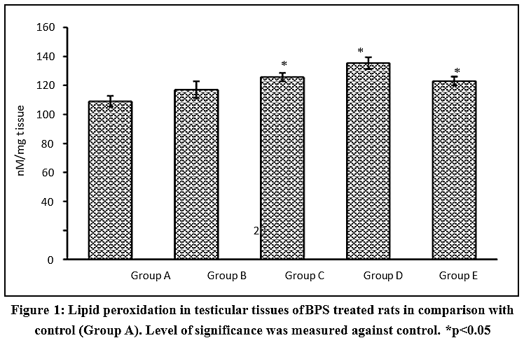 |
Figure 1: Lipid peroxidation in testicular tissues of BPS treated rats in comparison with control (Group A). Level of significance was measured against control. *p<0.05 Click here to View Figure |
Glutathione and glutathione peroxidase
Glutathione (GSH) level in testicular tissues indicated remarkable alterations in response to BPS treatment. Although there was significant decline in groups C-D, the depreciation however, was not dose dependent. Group B showed slightly higher GSH level (14.93±1.99 µM/g) than control (14.52±0.99 µM/g) with abnormally high SEM (Figure 2A). This reflects attempts of involuntary homeostasis against insistent generation of oxidative radicals. Highly unlikely, Group C indicated higher loss of GSH in testicular tissues comparing to both maximum dose Group D and Group A (control). Similar, results were also observed for Group E which indicted lower GSH level but slightly better than Group C (~10%) (Figure 2A).
Glutathione peroxidase (GPx) which is the main actor in reducing harmful hydrogen peroxides is assisted by GSH (hydrogen donor). Therefore, level of GSH in testicular tissues determined activity of GPx. Results showed minimum activity of GPx in Group D (3.16±0.13 nmol/min/mg protein) comparing to control (4.03±0.13 nmol/min/mg protein) and other test groups (Figure 2B). Whereas, groups C and E indicated slightly higher (13-14%) activity comparing to Group D but still remained significantly lower than the control. Interestingly, activity of GPx in Group B was found unaltered and SEM remained low, indicating minimum variation in activities of GPx within group.
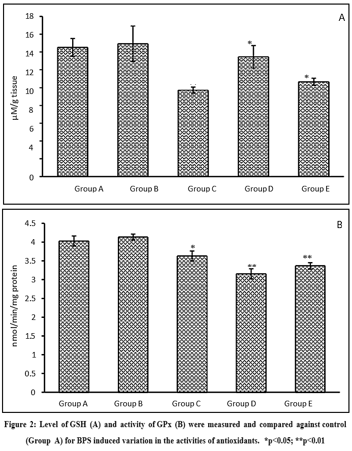 |
Figure 2: Level of GSH (A) and activity of GPx (B) were measured and compared against control (Group A) for BPS induced variation in the activities of antioxidants. *p<0.05; **p<0.01 |
Response of superoxide dismutase and catalase
Harmful oxygen molecules get neutralized by balanced activities of SOD. The currents study noted significant decline in activities of SOD in all test groups. Most importantly, the decline was dose dependent, where, high dose induced greater decline. More accurately, Group B showed 18% decline, Group C showed 24%, and Group D revealed 37% decline comparing to control (Figure 3A). Administration of Vit-E in Group E (5.32±0.37 U/mg protein) indicated better toleration in activities of SOD despite high dose of BPS but remined significantly low comparing to control (6.78±0.12 U/mg protein). The level of activities in Group E was in parallel with Group B.
Activity of catalase which is an important antioxidant in neutralizing hydrogen peroxide at high turnover rate, reduced significantly in groups C and D, when compared with control (Figure 3B). Interestingly, Group B showed no alteration in activity regardless of daily doses of 100 µg/kg body weight. While groups C and D noted with >40% decline in activity of catalase. Where Group D noted with maximum decline in activity which was measured as 2.56±0.35 U/mg protein against control’s 5.72±0.16 U/mg protein catalase activity (Figure 3B). However, catalase activity in Group E showed better toleration when compared with Group D (>30%).
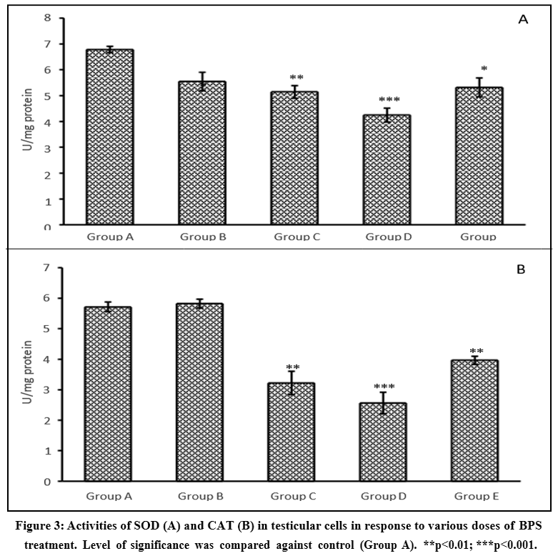 |
Figure 3: Activities of SOD (A) and CAT (B) in testicular cells in response to various doses of BPS treatment. Level of significance was compared against control (Group A). **p<0.01; ***p<0.001. |
Linear regression analysis of antioxidants against lipid peroxidation in testis
Interesting result was observed when linear and logarithmic regression were analyzed between variables of antioxidants and LPO in response to BPS. Result showed a positive relatedness between both variable (R2=0.618) which indicate cumulative exertions of investigated antioxidants in reducing BPS induced oxidative insults. However, logarithmic regression showed slowing pace of effectiveness of antioxidants, indicating higher lipid peroxidation in testicular tissues if the daily doses were to exceed from the 45 days experimental period (Figure 4).
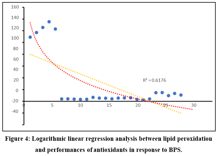 |
Figure 4: Logarithmic linear regression analysis between lipid peroxidation and performances of antioxidants in response to BPS. |
Alterations in apoptotic markers
p53 expression
Group A in the present study revealed few positive p53 cells near the basal lamina. These cells were identified as spermatogonial cells and were limited in numbers, remaining germ cells were found mostly negative (Figure 5A). In contrast, Group B showed spermatogonial cells, Leydig cells and other interstitial cells positive for p53 expression (Figure 5B). Group C indicated persistence of positive interstitial cells, along with the spermatogonial cells. This group was exclusively spotted with p53 positive spermatids (Figure 5C). Group D showed relatively lower positive spermatogonial cells, due to loss of spermatogenic cells and disruption of spermatogenesis. Leydig cell was invariably low, thus, similar results were also observed for interstitial cells as for the germ cells (Figure 5D). Presence of Leydig cells in Group E indicated better toleration against BPS induced effects recorded in Group D. However, Leydig cells showed positive expression for p53 along with precursor spermatogonial cells (Figure 5E).
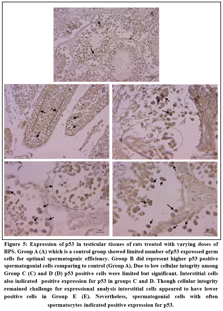 |
Figure 5: Expression of p53 in testicular tissues of rats treated with varying doses of BPS. Group A (A) which is a control group showed limited number of p53 expressed germ cells for optimal spermatogenic efficiency. Group B did represent higher p53 positive spermatogonial cells comparing to control (Group A). |
BCL-2 expression
Expression of BCL-2 in normal spermatogenesis is common to avoid excessive elimination of germ cells. The results showed number of spermatogonial cells and spermatocytes positive for BCL-2 in Group A (Figure 6A). While in Group B number of BCL-
2 positive cells increased substantially. It also indicated positive expression among spermatids indicating demand of suppression of pro-apoptotic protein (Figure 6B). The same were relatively low in Group C and Group D1 due excessive loss of germ cells (Figures 6C-D). Interestingly, Group E showed substantial retention of expression of BCL-2 despite daily exposure of 1000 µg/kg b.w. of BPS, showing better toleration against induced damages when compared with Group D (Figure 6E).
Caspase 3 expression
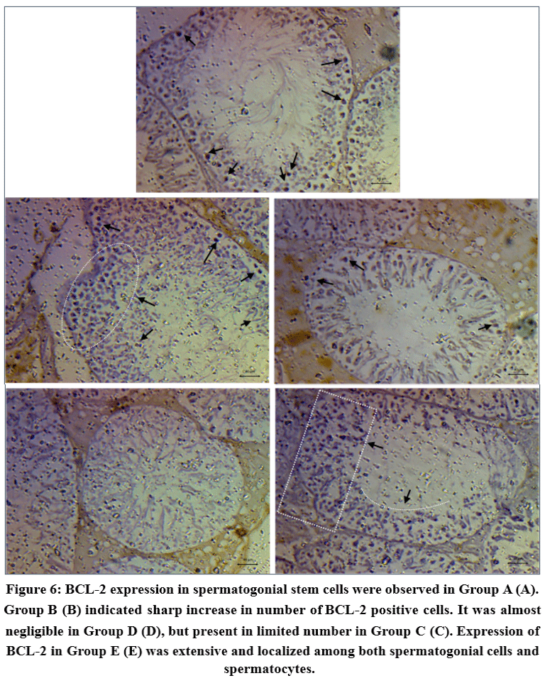 |
Figure 6: BCL-2 expression in spermatogonial stem cells were observed in Group A (A). Group B (B) indicated sharp increase in number of BCL-2 positive cells. |
Results of the present study showed a constant present of caspase 3 positive spermatogonia in Group A, nonetheless, its presence in Group B increased substantially (Figure 7A-B). It was remarkable to note that caspase-3 expression was mostly localized among spermatogonial cells and rarely positive in successor germ cells. Interestingly, number of caspase-3 positive spermatogonial cells almost disappeared from seminiferous tubules among groups C and D (Figure 7C-D). This could be due to loss of germ cells and spermatogenesis. Group E, showed high number of caspase-3 positive germ cells with clear distinction of spermatogonial localization and limited expression among spermatocytes (Figure 7E)
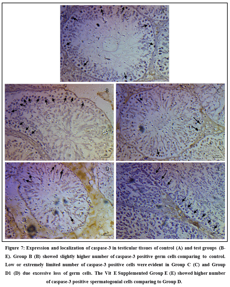 |
Figure 7: Expression and localization of caspase-3 in testicular tissues of control (A) and test groups (B-E). Group B (B) showed slightly higher number of caspase-3 positive germ cells comparing to control. |
Discussion
Decades long global use of BPA worried scientific communities of potential repercussions on human health. Its adversities have already started making headlines and begun to create anxiety in general population. Industries and manufacturers are in desperate need to find a suitable alternative of BPA with minimum or no health and safety concerns. Emergence of BPS is one of those attempts to dispose of immediate human BPA exposures. In this attempt BPS has seen remarkable market growth in last one decade. According to Chem Analyst BPS current market (2023 evaluation) is about 185000 tonnes and expected to grow at the rate of 4.2% in next 10 years.26 BPS in now common in baby milk-bottles, water bottles, lunch boxes, food containers, food packaging materials, thermal papers, medical devices, etc. where BPA was traditionally used. Condition suddenly changed when reports begin to surface that BPS causes hormonal and obesogenic effects on human and potentially these effects were worse than BPA.1
To this day, there are almost 2200 papers published on BPS (NLM database) among which 1427 papers are associating it with some health issue. Strikingly, NLM database also reveals that among 1427 papers where BPS is associated with diseases, almost 900 papers found strong evidence of its role in disrupting reproductive system. These data indicate that BPS does not only have similar impact to that of the BPA but also prone to cause unknown damages in future if persisted in the environment for as long as BPA. Previous study claimed that BPS works via different pathways than BPA,1 thus expected to causes various health related issues not conventional to BPA. In the present study, attempt was made to evaluate specifics of damages BPS causes to testicular tissues in a doses-dependent manner. Since BPA is also strongly associated with testicular damages and fertility limiting effect, evaluation of oxidative stress caused by BPS and its consequential impact apoptotic markers would be of extreme significance.
The BPS induced oxidative stress at the investigated doses showed considerable increase in lipid peroxidation in testicular tissues specifically at doses 500 µg/kg b.w./day and above. It showed that free radicals generated by BPS exposure caused excessive lipid oxidation and formation of hydrogen peroxide. The result was in accordance to previous studies, indicating disruption of lipid metabolism,27 and increase in lipid peroxidation,28 by BPS at various doses in several species. Interestingly, lipid peroxidation in group of animals administered with Vit-E concurrently with 1000 µg/kg b.w./day of BPS showed limited toleration. Generation of excessive hydrogen peroxide by BPS exposure was also evident in activities of GPx in the testicular tissues. As results from current study suggested that dose over 500 µg/kg b.w./day could significantly reduce activity of GPx and level of GSH in testis. GSH is required by GPx as a cofactor to reduce hydrogen peroxide, thus, decline in activity of GPx is well predicted looking at the highly significant decline in levels of GSH in testis. There are many studies that indicated BPS induced oxidative stress causes damages in various organs.29,30 Interestingly, there are evidences that suggested decline of testicular GSH upon BPA exposure,31,32 however, BPS’s role in alteration of GSH level in testis is still opaque. Based on the results, the current study assumes that BPS affects testicular GSH homeostasis by potentially high production of hydrogen peroxide and subsequently equivalent utilization in GPx activities. There was some toleration observed in group of animals treated with simultaneous doses of Vit-E, although, the activity of GPX or level of testicular GSH was hardly within control range.
Two major antioxidants superoxide dismutase and catalase also found to be extremely affected by the daily doses of BPS, specifically by doses 500 and 1000 µg/kg b.w./day. Both antioxidants showed direct dose dependent effect of BPS on their activities. Which confirmed their protective role in the BPS induced oxidative testicular damages. The present study confirmed that generation of superoxide and hydrogen peroxide is an affirmative phenomenon in response to BPS exposure. However, use of supplementary antioxidants may significantly play additive role in enhancing rate of activity of both SOD and CAT. The current study showed over 20-30% improvement in activity of both antioxidants when provided daily supplementation of Vit-E. A recent study by Pu et al.,33 reported that administration of BPS (10 µg/l and 100 µg/l) in crayfish significantly decline activities of CAT and SOD in intestinal tissues. Now this is remarkable despite being different species and tissue type, BPS retains its ability to generate oxidative radicals and appeared to be dynamic in targeting organs, unlike BPA. Which if proven true, may deteriorate overall health further in long-term exposure. The present study predicted further depreciation in activity of antioxidants if the exposure periods were to exceed from 45 days.
Excessive generation of hydrogen peroxide induce apoptosis in affected cells depending on the concentration. Where low concentration led to caspase-dependent and high concentration causes caspase independent activation of apoptotic cells death.34 The present study showed higher expression of caspase 3 in testicular tissues of group of animals exposed to 100 µg/kg b.w./day of BPS. Surprisingly, the expression drastically declined in animals exposed to 500 and 1000 µg/kg b.w./day of BPS. It showed that low dose of BPS activates caspase dependent pathway of apoptosis and higher doses activates independent pathway. It was also interesting to witness re-emergence of caspase-3 expression in group of animals supplemented with Vit-E. Therefore, it appeared that relatively higher activities of antioxidants in group of animals treated with 100 µg/kg b.w./day of BPS and supplementation of Vit-E concurrently with 1000 µg/kg b.w./day of BPS most likely avoided caspase independent apoptotic cell deaths in seminiferous tubules. However, this claim is arguable as number of germ cells in high dose groups were limited, therefore, number of positive cells were also significantly low. It was remarkable to note that spermatogonial cells were the prime location of caspase-3 activity in both groups. Previous studies have reported that caspase dependent apoptosis is a well-orchestrated cellular destruction with specific proteolytic cascades,35 while, independent pathway just evades the caspase signaling system, through other pathways such as BCL-2.36
BCL-2 expression system is extremely important in immunoregulation through T cell differentiation. It is also considered as anti-apoptotic,37 due to its ability to regulate cell death by resisting stimuli that usually promote apoptosis.38 Interestingly, high number of germ cells were found positive for BCL-2 in testicular tissues of animals administered with 100 µg/kg b.w./day of BPS. This expression was also evident in group of animals treated with 500 µg/kg b.w./day of BPS. Localization of these positive cells were wide which included spermatogonia, spermatocytes and spermatids. Similar results were also witnessed in group supplemented with Vit-E. Although BPS is likely to avoid caspase independent pathways, it showed that simultaneous activation of both caspase- 3 and BCL-2 may also be feasible if cellular integrity is beyond repair. It is reported that there is signaling cross talked between death receptor induced apoptotic signal and intrinsic apoptotic program.39 In some cases, caspase is responsible to cleave BCL-2 (BID-BCL-2 family member) to truncated form causing amplification of apoptotic program through intrinsic pathway.40 It is likely that BPS promotes range of apoptotic pathways based on dose strength.
The present study observed large number of germ cells undergone p53 mediated apoptotic consequence regardless of doses of BPS. Spermatogonial cells, spermatocytes and Leydig cells were found positive of p53 expression in all three test groups. Previous studies reported that p53 activation is mediated by oxidative stress and oxidative stress is regulated by p53 with the support of GPx.41,42 This supports earlier assumption in the present study that BPS generate excessive hydrogen peroxide, which affirmatively affects GPx and GSH cycle leading to regulation of p53. Upregulation of caspase- 3 and BCL-2 in response to BPS exposure indicated a pro-apoptotic event in testicular cells. With higher expression of p53, it was revealed that BPS led to direct transcriptional activation of pro-apoptotic factors, which usually expressed in non-transforming cells.43 This indicated a potential cell cycle arrest during spermatogenesis. Although there is evidence that BPS causes cell cycle arrest (CCA), nonetheless, BPA has been reported to cause CCA in various cells.44,45 A study by Yin et al.,46 reported that BPA and its analogues cause differential cell cycle arrest in testicular cells.
Conclusion
Conclusively, BPS affirmatively generates high oxidative stress in testicular cells leading to significant depreciation in activities of important antioxidants. Low activity of GPx and CAT indicated excessive generation of hydrogen peroxide in all BPS treated groups. However, 500 and 1000 µg/kg b.w./day doses of BPS caused immense damage to germ cells through apoptotic elimination of germ cells. It appeared that BPS was able to upregulate caspase-3, and BCL-2 simultaneously, specifically in groups administered with 100 µg/kg b.w./day doses of BPS and group of animals supplemented with Vit-E while concurrently administered with 1000 µg/kg b.w./day doses of BPS. This showed excessive resistance and concurrent removal of damaged germ cells. Spermatogonial cells were the prime target of BPS, however, spermatocytes and spermatids were also found positive for pro-apoptotic markers. Direct transcriptional activation of apoptosis was also found active in BPS treated groups, indicating germ cells undergone cell cycle arrest.
Acknowledgement
We would like to thank Centre for Advances Studies, Department of Zoology, University of Rajasthan, Jaipur, India for granting access to the central instrumental facilities.
Funding Sources
We would like to thank Council of Scientific and Industrial Research (CSIR), New Delhi, India for financial support obtained as Junior and Senior Research Fellowship (Grant Scheme No.: 09/149(0737)/2019-EMR-I).
Conflict of Interest
The authors do not have any conflict of interest.
Data Availability Statement
This statement does not apply to this article.
Ethics Statement
Clearance was acquired in advance from Institutional Animal Ethics Committee (IAEC) for the experiments carried out in the present study. All parameters were conducted in strict guidance of Committee for the Purpose of Control and Supervision of Experiments and Animals.
Informed Consent Statement
This study did not involve human participants, and therefore, informed consent was not required.
Clinical Trial Registration
This research does not involve any clinical trials.
Permission to reproduce material from other sources
Not Applicable.
Author Contributions
- Seema Srivastava: Conceptualization, Methodology, Writing – Original Draft interpretation of the data overall Supervision
- Kumari Pragya: Experimentation, data collection, analysis compilation of the entire work.
- Manish Kumar Sharma: writing and editing.
- Priya Khangrawat: Helped in bringing and rearing animals.
- Sharey Sharey: Assisted in experimentation
- Anil Kumar Chandolia: Helped in bringing and rearing animals.
References
- Thoene M, Dzika E, Gonkowski S, Wojtkiewicz J. Bisphenol S in food causes hormonal and obesogenic effects comparable to or worse than Bisphenol A: a literature review. Nutrients. 2020;12(2):532.
CrossRef - Qiu W, Zhan H, Hu J, et al. The occurrence, potential toxicity, and toxicity mechanism of bisphenol S, a substitute of bisphenol A: a critical review of recent progress. Ecotoxicol Environ Saf. 2019;173:192-202.
CrossRef - Kinch CD, Ibhazehiebo K, Jeong JH, et al. Low-dose exposure to bisphenol A and replacement bisphenol S induces precocious hypothalamic neurogenesis in embryonic zebrafish. Proc Natl Acad Sci U S A. 2015;112(5):1475-1480.
CrossRef - Rochester JR, Bolden AL. Bisphenol S and F: a systematic review and comparison of the hormonal activity of bisphenol A substitutes. Environ Health Perspect. 2015;123(7):643-650.
CrossRef - Stone WL, Pham T, Mohiuddin SS. Biochemistry, antioxidants. In: StatPearls [Internet]. StatPearls Publishing; 2024. Accessed August 22, 2024. https://www.ncbi.nlm.nih.gov/books/NBK541064/
- Auten RL, Davis JM. Oxygen toxicity and reactive oxygen species: the devil is in the details. Pediatr Res. 2009;66(2):121-127.
CrossRef - Mukherjee U, Das S, Ghosh S, Maitra S. Reproductive toxicity of bisphenol A, at environmentally relevant concentrations, on ovarian redox balance, maturational response, and intra-oocyte signalling events in Labeo bata. Sci Total Environ. 2024;906:167415.
CrossRef - Sahu A, Verma R. Bisphenol S dysregulates thyroid hormone homeostasis; testicular survival, redox and metabolic status: ameliorative actions of melatonin. Environ Toxicol Pharmacol. 2023;104:104300.
CrossRef - Liu J, Lin Y, Peng C, et al. Bisphenol F induced hyperglycemia via activation of oxidative stress-responsive miR-200 family in the pancreas. Ecotoxicol Environ Saf. 2023;255:114769.
CrossRef - Matuszczak E, Komarowska MD, Debek W, Hermanowicz A. The impact of bisphenol A on fertility, reproductive system, and development: a review of the literature. Int J Endocrinol. 2019;2019:4068717.
CrossRef - Peña-Corona SI, Vásquez Aguire WS, Vargas D, Juárez I, Mendoza-Rodríguez CA. Effects of bisphenols on Blood-Testis Barrier protein expression in vitro: a systematic review and meta-analysis. Reprod Toxicol. 2021;103:139-148.
CrossRef - Hussain T, Kandeel M, Metwally E, et al. Unraveling the harmful effect of oxidative stress on male fertility: a mechanistic insight. Front Endocrinol (Lausanne). 2023;14:1070692.
CrossRef - Sharma P, Kaushal N, Saleth LR, et al. Oxidative stress-induced apoptosis and autophagy: balancing the contrary forces in spermatogenesis. Biochim Biophys Acta Mol Basis Dis. 2023;1869(5):166742.
CrossRef - Presunto M, Mariana M, Lorigo M, Cairrao E. The effects of bisphenol A on human male infertility: a review of current epidemiological studies. Int J Mol Sci. 2023;24(15):12417.
CrossRef - Darghouthi M, Rezg R, Boughmadi O, Mornagui B. Low-dose bisphenol S exposure induces hypospermatogenesis and mitochondrial dysfunction in rats: a possible implication of StAR protein. Reprod Toxicol. 2022;107:104-111.
CrossRef - Fenclová T, Chemek M, Havránková J, et al. Effect of Bisphenol S on testicular tissue after low-dose lactation exposure. Environ Pollut. 2022;315:120114.
CrossRef - Committee for the Purpose of Control and Supervision of Experiments on Animals (CPCSEA). Guidelines on the Regulation of Scientific Experiments on Animals. Ministry of Environment and Forests; 2010.
- Organisation for Economic Co-operation and Development (OECD). Test No. 407: Repeated Dose 28-Day Oral Toxicity Study in Rodents. OECD Publishing; 2008. OECD Guidelines for the Testing of Chemicals, Section 4.
- Azevedo LF, Hornos Carneiro MF, Dechandt CRP, et al. Global liver proteomic analysis of Wistar rats chronically exposed to low-levels of bisphenol A and S. Environ Res. 2020;182:109080.
CrossRef - Mao W, Mao L, Zhao N, et al. Disposition of Bisphenol S metabolites in Sprague-Dawley rats. Sci Total Environ. 2022;811:152288.
CrossRef - Ohkawa H, Ohishi N, Yagi K. Assay for lipid peroxides in animal tissues by thiobarbituric acid reaction. Anal Biochem. 1979;95(2):351-358.
CrossRef - Wood JL. Metabolic conjugation and metabolic hydrolysis. In: Fishman WH, ed. Metabolic Conjugation and Metabolic Hydrolysis. Vol 2. Academic Press; 1970:61-299.
- Aebi H. Catalase. In: Bergmeyer HU, ed. Methods of Enzymatic Analysis. 5th ed. Academic Press; 1974:273-278.
CrossRef - Hissin PJ, Hilf R. A fluorometric method for determination of oxidized and reduced glutathione in tissues. Anal Biochem. 1976;74(1):214-226.
CrossRef - Marklund S, Marklund G. Involvement of the superoxide anion radical in the autooxidation of pyrogallol and a convenient assay for superoxide dismutase. Eur J Biochem. 1974;47(3):469-474.
CrossRef - Bisphenol S market analysis: industry market size, plant capacity, production, operating efficiency, demand & supply, end user industries, sales channel, regional demand, company share, manufacturing process, 2015-2033. Accessed August 22, 2024. https://www.chemanalyst.com/industry-report/bisphenol-s-290
- Li J, Li Z, Zhu Y, et al. Bisphenol S remodels red blood cell membrane lipids by altering plasma lipid levels, causing the risk of venous thrombosis in SD rats and zebrafish embryos. Environ Int. 2023;182:108331.
CrossRef - Maćczak A, Cyrkler M, Bukowska B, Michałowicz J. Bisphenol A, bisphenol S, bisphenol F and bisphenol AF induce different oxidative stress and damage in human red blood cells (in vitro study). Toxicol In Vitro. 2017;41:143-149.
CrossRef - Mohan S, Jacob J, Malini NA, et al. Biochemical responses and antioxidant defense mechanisms in Channa striatus exposed to Bisphenol S. J Biochem Mol Toxicol. 2024;38(2):e23651.
CrossRef - Kaptaner B, Yılmaz C, Aykut H, et al. Bisphenol S leads to cytotoxicity-induced antioxidant responses and oxidative stress in isolated rainbow trout (Oncorhynchus mykiss) hepatocytes. Mol Biol Rep. 2021;48(12):7657-7666.
CrossRef - Srivastava S, Sharma MK, Sharey. Variation in the oxidative status of testicular tissues during critical pubertal age under influence of Bisphenol A. Biomed Pharmacol J. 2022;15(4):2191-2198.
CrossRef - Santiago J, Silva JV, Santos MAS, Fardilha M. Fighting bisphenol A-induced male infertility: the power of antioxidants. Antioxidants (Basel). 2021;10(2):289.
CrossRef - Pu C, Liu Y, Ma J, et al. Bisphenol S exposed changes in intestinal microflora and metabolomics of freshwater crayfish, Procambarus clarkii. Aquat Toxicol. 2024;272:106957.
CrossRef - Xiang J, Wan C, Guo R, Guo D. Is hydrogen peroxide a suitable apoptosis inducer for all cell types? Biomed Res Int. 2016;2016:7343965.
CrossRef - Nakajima YI, Kuranaga E. Caspase-dependent non-apoptotic processes in development. Cell Death Differ. 2017;24(8):1422-1430.
CrossRef - Kögel D, Prehn JHM. Caspase-independent cell death mechanisms. In: Madame Curie Bioscience Database [Internet]. Landes Bioscience; 2000-2013. Accessed August 22, 2024. https://www.ncbi.nlm.nih.gov/books/NBK6197/
- Opferman JT, Kothari A. Anti-apoptotic BCL-2 family members in development. Cell Death Differ. 2018;25(1):37-45.
CrossRef - Pollard TD, Earnshaw WC, Lippincott-Schwartz J, Johnson DA, eds. Programmed cell death. In: Cell Biology. 3rd ed. Elsevier; 2017:797-815.
CrossRef - Li H, Zhu H, Xu CJ, Yuan J. Cleavage of BID by caspase 8 mediates the mitochondrial damage in the Fas pathway of apoptosis. Cell. 1998;94(4):491-501.
CrossRef - Gross A, Yin XM, Wang K, et al. Caspase cleaved BID targets mitochondria and is required for cytochrome c release, while BCL-XL prevents this release but not tumor necrosis factor-R1/Fas death. J Biol Chem. 1999;274(2):1156-1163.
CrossRef - Cano CE, Gommeaux J, Pietri S, et al. Tumor protein 53-induced nuclear protein 1 is a major mediator of p53 antioxidant function. Cancer Res. 2009;69(1):219-226.
CrossRef - Tan M, Li S, Swaroop M, et al. Transcriptional activation of the human glutathione peroxidase promoter by p53. J Biol Chem. 1999;274(18):12061-12066.
CrossRef - Aubrey BJ, Kelly GL, Janic A, et al. How does p53 induce apoptosis and how does this relate to p53-mediated tumour suppression? Cell Death Differ. 2018;25(1):104-113.
CrossRef - Bilancio A, Bontempo P, Di Donato M, et al. Bisphenol A induces cell cycle arrest in primary and prostate cancer cells through EGFR/ERK/p53 signaling pathway activation. Oncotarget. 2017;8(69):115620-115631.
CrossRef - Kozieł-Leszczyńska MJ, Piastowska-Ciesielska AW. Bisphenols and their role in female infertility and hormone-related cancer. Endocrine. 2026;91(1):32.
CrossRef - Yin L, Hu C, Yu XJ. High-content analysis of testicular toxicity of BPA and its selected analogs in mouse spermatogonial, Sertoli cells, and Leydig cells revealed BPAF induced unique multinucleation phenotype associated with the increased DNA synthesis. Toxicol In Vitro. 2023;89:105589.
CrossRef
Accepted on: 11-09-2026
Second Review by: Dr. Daya Shankar Gautam
Final Approval by: Dr. Hifzur R. Siddique