Integrative Liquid Biopsy and Pharmacogenomics for Monitoring Treatment Response and Guiding Precision Therapy in Triple-Negative Breast Cancer: A Mechanistic and Clinical Review
 , Jagannadham Nuthana Yashwanth, Erothi Balakoti*and Donka Devi
 Pharmacology Division, AU College of Pharmaceutical Sciences, Andhra University, Visakhapatnam, India
Corresponding Author’s E-mail: 625209525006@andhrauniversity.edu.in
Download this article as:
ABSTRACT:Triple-negative breast cancer (TNBC) is a biologically heterogeneous and clinically aggressive breast cancer subtype in which therapeutic resistance often emerges despite initial chemosensitivity. Conventional tissue biomarkers remain essential for diagnosis and initial treatment selection, but they provide limited information about temporal tumour evolution, molecular residual disease, clonal resistance and patient-specific toxicity risk. Liquid biopsy and pharmacogenomics offer complementary precision-oncology tools for addressing this gap. Circulating tumour DNA (ctDNA) can track tumour burden, molecular response and acquired resistance; circulating tumour cells (CTCs) can reveal viable metastatic phenotypes and epithelial–mesenchymal plasticity; extracellular vesicles and circulating RNAs may reflect tumour–stromal communication and adaptive resistance signalling. Pharmacogenomics adds a second layer by identifying tumour vulnerabilities such as BRCA1/2-associated homologous recombination deficiency and host determinants of drug toxicity such as DPYD and UGT1A1 variation. This review synthesizes mechanistic and translational evidence supporting integrated liquid biopsy–pharmacogenomics approaches in TNBC. Current clinical utility is strongest for BRCA1/2-directed PARP inhibition, PD-L1-guided immunotherapy in eligible settings, DPYD-guided fluoropyrimidine safety, UGT1A1-informed sacituzumab govitecan toxicity surveillance and ctDNA-based molecular residual disease risk assessment. However, most liquid-biopsy-guided treatment adaptation remains investigational. Future progress requires prospective trials demonstrating that biomarker-guided escalation, de-escalation or therapy switching improves survival, reduces toxicity or prevents molecular relapse.
KEYWORDS:BRCA1/2; DPYD; Immunotherapy; Molecular residual disease; Pharmacogenomics;Poly (ADP-ribose) polymerase (PARP) inhibitors; Precision oncology; Sacituzumab govitecan; UGT1A1
Introduction
Triple-negative breast cancer is defined clinically by the absence of estrogen receptor, progesterone receptor and HER2 overexpression or amplification. Although this negative definition is useful for therapeutic classification, it does not capture the biological complexity of the disease. Transcriptomic studies have shown that TNBC includes distinct molecular states, including basal-like immune-active, mesenchymal, luminal androgen receptor and DNA-repair-deficient phenotypes, each associated with different patterns of proliferation, immune infiltration, metastatic behaviour and treatment sensitivity.1 Therefore, TNBC should not be viewed as a single uniform entity, but as a biologically unstable disease shaped by genomic instability, tumour microenvironmental pressure and therapy-driven clonal selection.
The therapeutic landscape of TNBC has expanded beyond conventional chemotherapy. Pembrolizumab combined with chemotherapy has improved outcomes in high-risk early TNBC and in PD-L1-positive metastatic TNBC.2 PARP inhibitors exploit homologous recombination repair deficiency in germline BRCA1/2-mutated HER2-negative breast cancer through synthetic lethality.3 Antibody–drug conjugates such as sacituzumab govitecan have improved outcomes in previously treated metastatic TNBC by delivering the topoisomerase I inhibitor SN-38 through Trop-2-directed targeting, and first-line indications have now expanded in selected metastatic settings.4 Trastuzumab deruxtecan has further changed the therapeutic relevance of low HER2 expression in metastatic breast cancer by demonstrating that HER2-low disease can be pharmacologically exploitable when a potent payload and bystander effect are present.5
Despite these advances, relapse remains frequent because residual tumour clones may survive treatment through multiple mechanisms, including DNA-repair restoration, immune escape, lineage plasticity, altered drug transport, payload resistance, stromal shielding and pharmacokinetic underexposure. This makes TNBC a dynamic treatment-monitoring problem rather than a one-time biomarker-matching problem. A baseline tissue biopsy can define initial eligibility for therapy, but it is spatially limited, poorly suited to repeated sampling and unable to capture evolving micrometastatic disease, treatment-induced clonal selection or acquired resistance.6 Serial liquid biopsy addresses this clinical gap directly. Because plasma can be sampled before each cycle, after surgery and during surveillance, ctDNA and related analytes provide a real-time readout of molecular tumour burden that tissue cannot supply. In TNBC, where clonal evolution can be rapid, this temporal resolution is the principal practical advantage over static tissue genotyping.
Liquid biopsy enables serial, minimally invasive assessment of tumour-derived material in blood. ctDNA reflects molecular tumour burden and clonal evolution; CTCs represent viable metastatic units; extracellular vesicles and circulating RNAs may capture tumour–stroma communication and resistance signalling. Pharmacogenomics adds a complementary layer by explaining whether a drug is biologically appropriate for the tumour and pharmacologically safe for the patient.7 Therefore, the strongest precision model in TNBC is not “liquid biopsy versus tissue biopsy,” but a combined approach in which tissue profiling defines baseline therapeutic vulnerability, liquid biopsy monitors whether that vulnerability is being suppressed or bypassed, and pharmacogenomics refines treatment choice, dosing and toxicity prevention.8
Biological rationale for integrating liquid biopsy and pharmacogenomics
TNBC progression is governed by three interacting biological processes: tumour-cell intrinsic evolution, host–drug interaction and microenvironmental selection. Liquid biopsy mainly captures tumour-derived signals released into circulation, including DNA fragments, intact tumour cells, extracellular vesicles and RNA cargo.9 Pharmacogenomics captures both tumour-intrinsic vulnerabilities and inherited host determinants of drug metabolism, exposure and toxicity. Their integration is valuable because treatment failure in TNBC often reflects a mismatch between tumour biology, drug mechanism and host pharmacology.10
In clinic, these tools are not used sequentially in isolation. A practical workflow starts with tissue and germline testing to select a regimen, then uses serial ctDNA to ask whether that regimen is suppressing circulating tumour clones, and uses host pharmacogenes to decide whether the planned dose is safe. For example, a patient with residual TNBC after neoadjuvant therapy may be a candidate for adjuvant capecitabine on clinical grounds, yet DPYD genotyping may mandate dose reduction or drug avoidance; the same patient may later receive sacituzumab govitecan, in which UGT1A1 status informs neutropenia risk while rising ctDNA can flag molecular progression before imaging. This simultaneous use of tumour-dynamic and host-static biomarkers is the operational meaning of integration.
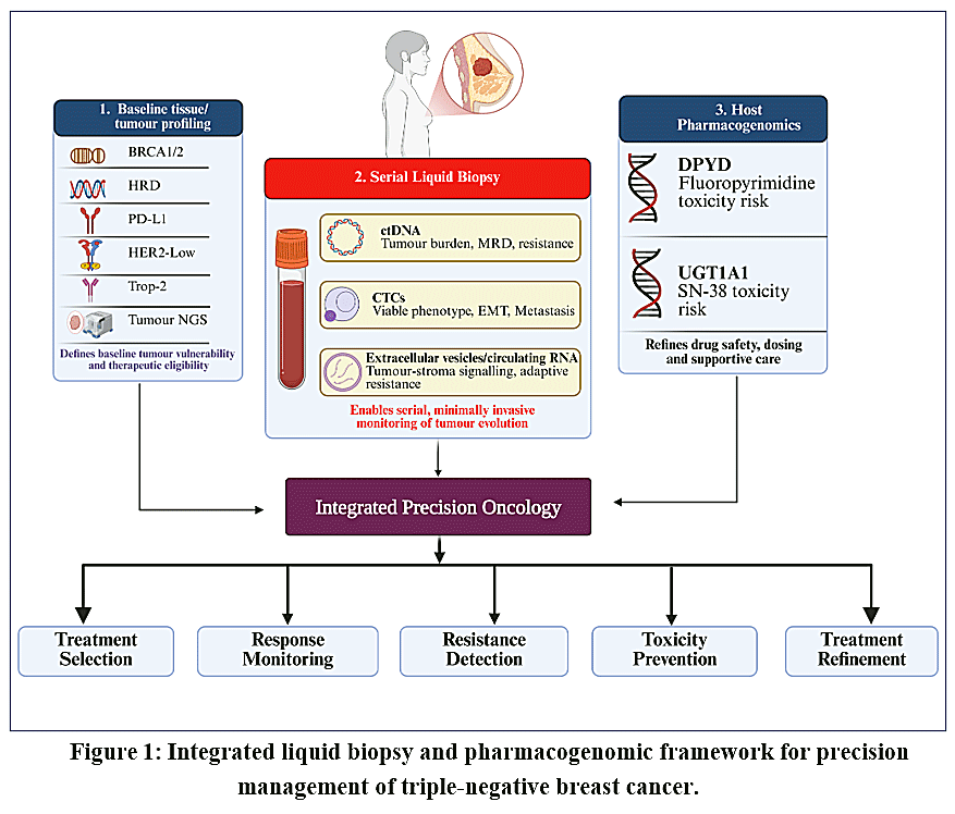 |
Figure 1: Integrated liquid biopsy and pharmacogenomic framework for precision management of triple-negative breast cancer. Click here to View Figure |
Baseline tumour profiling identifies therapeutic vulnerabilities, serial liquid biopsy monitors tumour burden, molecular residual disease and resistance, while host pharmacogenomics supports toxicity risk assessment and safer treatment.
ctDNA is generated when tumour cells release fragmented DNA into circulation through apoptosis, necrosis, immune-mediated cytotoxicity or active secretion. During effective therapy, ctDNA usually declines because drug-sensitive clones are eliminated and viable tumour mass contracts. Persistent or rising ctDNA suggests residual disease, resistant subclones, occult metastasis or tumour compartments that remain pharmacologically protected. Mechanistically, ctDNA is not simply a blood-based tumour marker; it is a dynamic molecular record of clonal suppression or clonal escape under treatment pressure.11
CTCs provide complementary information because they are intact tumour cells rather than fragmented DNA. They can reveal cell-state plasticity, epithelial–mesenchymal transition, stemness features, immune-evasion phenotypes and metastatic competence.12 This is particularly important in TNBC because mesenchymal and hybrid epithelial–mesenchymal states may promote invasion, chemotherapy tolerance and immune escape.13 CTC clusters are especially relevant because multicellular tumour aggregates can survive circulation more efficiently than single cells through preserved cell–cell adhesion, collective migration, anoikis resistance and junctional signalling involving molecules such as plakoglobin.14
Extracellular vesicles and exosomal RNAs add another mechanistic layer by reflecting active tumour–stroma communication. TNBC-derived vesicles may transfer microRNAs, proteins, lipids and nucleic acids that remodel endothelial cells, fibroblasts, immune cells and pre-metastatic niches.15 These vesicles may support angiogenesis, epithelial–mesenchymal transition, macrophage polarization, immune suppression and drug resistance. However, extracellular-vesicle, platelet-educated RNA, methylation and fragmentomic assays remain less standardized than ctDNA assays, and their clinical thresholds are not yet validated for routine decision-making.16 In this review they are discussed only as emerging mechanistic tools so that they do not overshadow clinically mature markers such as ctDNA, BRCA1/2, PD-L1, DPYD and UGT1A1.
Table 1: Mechanistic roles of major liquid biopsy analytes in TNBC
|
Analyte |
Mechanistic signal | Clinical implication | Developmental status |
| ctDNA | Tumour fraction, clonal mutations, molecular residual disease, resistance variants | Response monitoring, relapse-risk assessment, plasma genotyping |
Most clinically advanced17 |
|
CTCs |
Viable tumour-cell phenotype, EMT, stemness, immune escape | Metastatic-risk biology, single-cell profiling, drug-resistance studies | Clinically promising but not standardized18 |
| CTC clusters | Collective migration, plakoglobin-associated adhesion, anoikis resistance | High metastatic competence and poor-prognosis biology |
Mechanistically strong, clinically immature17 |
|
Extracellular vesicles |
Tumour–stroma signalling, immune suppression, angiogenesis, resistance cargo | Exploratory response and recurrence biomarkers | Investigational19 |
| Exosomal microRNA | Post-transcriptional pathway regulation | Candidate predictors of response or relapse |
Requires validation18 |
|
cfRNA / methylation / fragmentomics |
Gene-expression and epigenetic tissue-of-origin signals | Multi-analyte detection and monitoring |
Emerging19 |
Beyond ctDNA, CTCs and extracellular vesicles, emerging liquid-biopsy analytes may provide additional information about systemic tumour biology. Tumour-educated platelets can sequester tumour-derived RNA and nucleic-acid fragments, reflecting a more systemic tumour-associated signal.20 Circulating mRNA and non-coding RNA species may capture transcriptional activity associated with EMT, immune suppression and therapy resistance.21 Methylation and fragmentomic patterns could provide epigenetic and structural data for tissue-of-origin inference and molecular residual disease detection beyond mutations.22 For now, however, these assays should be considered research tools rather than routine clinical decision markers.
ctDNA as a dynamic pharmacodynamic biomarker
The value of ctDNA in TNBC lies in its temporal resolution. Tissue sequencing identifies the molecular state of a tumour at one time point, whereas serial ctDNA shows how tumour clones respond and adapt during treatment. This distinction is crucial because TNBC can undergo rapid clonal selection under the pressure of chemotherapy, immunotherapy, PARP inhibition or antibody–drug conjugates.3
During neoadjuvant therapy, ctDNA clearance can be interpreted as a molecular pharmacodynamic response. If cytotoxic therapy induces tumour-cell apoptosis and reduces viable tumour burden, the mutant allele fraction should decline over serial samples. Conversely, persistent ctDNA after early cycles may indicate drug-tolerant persister cells, poor intratumoural drug penetration, defective immune-mediated clearance, stromal protection or expansion of pre-existing resistant subclones. In high-risk HER2-negative breast cancer cohorts, serial ctDNA analysis has shown clinically meaningful association with treatment response and residual disease, supporting its role as a real-time marker of molecular response.23
Post-treatment ctDNA positivity has even greater biological significance. After surgery and systemic therapy, detectable ctDNA suggests that tumour DNA is still being released from residual viable disease, usually below radiological detection. In BRE12-158, detection of ctDNA and CTCs after neoadjuvant chemotherapy was associated with inferior outcomes in patients with early-stage TNBC and residual disease.24 Mechanistically, this means that pathology identifies residual disease in breast or lymph-node tissue, whereas ctDNA identifies systemic molecular persistence.
However, ctDNA should not be interpreted as a flawless binary marker. False negatives may occur in low-shedding tumours, isolated central nervous system disease, very low-volume residual disease or after transient cytoreduction. False positives may occur because of clonal hematopoiesis if paired leukocyte sequencing is not performed.25 Therefore, ctDNA should be integrated with residual cancer burden, nodal status, tumour subtype, imaging and treatment context rather than used as a standalone substitute for standard clinical assessment.
The c-TRAK-TN trial illustrates the difference between prognostic validity and clinical actionability. The study showed that ctDNA surveillance is feasible in high-risk early TNBC, but many patients had radiologically detectable metastatic disease when ctDNA became positive, and pembrolizumab triggered by ctDNA positivity did not produce sustained ctDNA clearance in the treated subset.26 This does not weaken the biological value of ctDNA; rather, it shows that ctDNA-guided intervention must be earlier, more sensitive and therapeutically matched to the mechanism of relapse. A positive ctDNA result is a molecular warning signal, but clinical benefit requires an effective intervention before overt metastatic progression.
Mechanisms of liquid biopsy-detected resistance
Liquid biopsy is particularly valuable for studying resistance evolution because TNBC resistance rarely arises from a single mechanism. Under therapy pressure, tumour clones may acquire or select adaptive states that restore DNA repair, evade immune attack, reduce target expression, increase drug efflux or alter payload sensitivity.27
In PARP inhibitor-treated BRCA-mutated TNBC, ctDNA can reveal BRCA1/2 reversion mutations that restore the open reading frame and partially recover homologous recombination repair. This directly reverses the synthetic-lethal vulnerability created by BRCA loss. Additional resistance mechanisms include replication-fork stabilization, reduced PARP trapping, loss of 53BP1-Shieldin pathway function, increased drug-efflux transporter activity and restoration of DNA-end resection. Because these mechanisms can emerge subclonally, plasma sequencing may detect them before a single-site tissue biopsy captures the resistant population.28
In immunotherapy-treated TNBC, persistent or rising ctDNA may reflect immune escape rather than simple chemotherapy resistance. Tumour cells may evade immune pressure through impaired antigen presentation, HLA loss, beta-2 microglobulin disruption, JAK/STAT pathway alteration, interferon-signalling defects, T-cell exclusion, regulatory myeloid expansion or T-cell exhaustion.29 Therefore, ctDNA kinetics under immunotherapy should be interpreted differently from kinetics under cytotoxic therapy. Early transient ctDNA fluctuations may occur during tumour inflammation or immune-mediated killing, whereas sustained ctDNA rise is more concerning for molecular progression.30
In antibody–drug conjugate-treated TNBC, resistance can occur at multiple pharmacological levels. Reduced antigen expression can limit antibody binding. Altered internalization or lysosomal trafficking can reduce payload release. Efflux transporter activity can decrease intracellular payload retention.31 Topoisomerase I pathway alteration or enhanced DNA-damage tolerance can reduce sensitivity to SN-38 or deruxtecan payloads. For sacituzumab govitecan, resistance may involve reduced Trop-2-mediated delivery or altered SN-38 sensitivity. For trastuzumab deruxtecan in HER2-low disease, resistance may emerge through HER2 expression heterogeneity, impaired payload release or selection of HER2-nonexpressing clones. These mechanisms make ADC resistance biologically suitable for longitudinal plasma monitoring, but routine ctDNA-guided ADC switching remains investigational.32
Pharmacogenomics: separating tumour vulnerability from host toxicity
A common limitation in oncology biomarker discussions is the failure to distinguish tumour pharmacogenomics from host pharmacogenomics.33 Tumour pharmacogenomics predicts whether the cancer has a druggable vulnerability. Host pharmacogenomics predicts whether the patient can metabolize or tolerate the drug safely.34 Both are essential, but they answer different clinical questions.
BRCA1/2 mutations are tumour-vulnerability biomarkers. Germline BRCA1/2 loss impairs homologous recombination repair, forcing tumour cells to rely on alternative and often error-prone DNA-repair pathways.35 PARP inhibition exploits this dependency by trapping PARP on DNA, increasing replication-fork collapse and generating double-strand breaks that BRCA-deficient cells cannot efficiently repair.36 In the adjuvant setting, olaparib improved outcomes in high-risk germline BRCA-mutated HER2-negative early breast cancer, supporting BRCA1/2 as a clinically actionable pharmacogenomic marker.37
DPYD and UGT1A1 are host-safety biomarkers. DPYD encodes dihydropyrimidine dehydrogenase, the rate-limiting enzyme in fluoropyrimidine catabolism. Reduced DPD activity can cause excessive fluoropyrimidine exposure, resulting in severe diarrhea, mucositis, neutropenia, neurotoxicity and potentially fatal toxicity. Therefore, DPYD testing informs capecitabine safety rather than capecitabine efficacy.38 UGT1A1 glucuronidates SN-38, the active payload released from sacituzumab govitecan. Reduced UGT1A1 activity, especially UGT1A1*28 homozygosity, can increase SN-38 exposure and heighten neutropenia or diarrhea risk. Thus, UGT1A1 status supports toxicity anticipation, dose-management readiness and supportive-care planning rather than routine exclusion from therapy.39 Other host pharmacogenomic candidates, such as CYP450 enzymes, glutathione-S-transferases, ABC transporters and DNA-repair polymorphisms, may also affect chemotherapy exposure or toxicity, but are not as clinically developed as DPYD-guided fluoropyrimidine safety and UGT1A1-informed SN-38 toxicity surveillance.40
Table 2: Tumour-directed versus host-directed pharmacogenomics in TNBC
|
Biomarker type |
Example | Primary question answered | Clinical meaning |
| Tumour vulnerability | BRCA1/2 mutation | Is the tumour sensitive to PARP inhibition or DNA-damaging therapy? |
Guides drug selection41 |
|
Tumour immune phenotype |
PD-L1 CPS | Is immune checkpoint blockade more likely to help? | Guides pembrolizumab eligibility in metastatic TNBC42 |
| Tumour target expression | HER2-low, Trop-2 | Can an ADC deliver payload to tumour cells? |
Guides or supports ADC strategy43 |
|
Host metabolism |
DPYD | Will fluoropyrimidine exposure become dangerous? | Guides dose reduction, avoidance or monitoring43,44 |
| Host glucuronidation | UGT1A1 | Will SN-38 toxicity risk be increased? |
Guides monitoring and supportive care43,44 |
|
Dynamic resistance |
ctDNA-detected reversion or pathway bypass | Has the tumour escaped the original vulnerability? |
Guides re-biopsy, therapy sequencing or trial selection41,43 |
Table 3: Pharmacogenomic variants, associated drugs, guidelines and recommended action
|
Gene / variant |
Associated drug(s) | Guideline or label source | Recommended action |
| BRCA1/2 germline pathogenic variant | Olaparib, talazoparib; informs platinum rationale | OlympiA; FDA/EMA PARP labels; ASCO/NCCN guidance |
PARP inhibition in eligible HER2-negative disease; genetic counselling |
|
HRD genomic scar (not current HR function) |
Platinum agents; investigational PARP combinations | Context-dependent; not a stand-alone PARP-selection assay |
Interpret with functional repair markers and early ctDNA clearance; not a current-vulnerability marker |
|
DPYD no-function alleles (e.g. *2A, *13) — poor metabolizer |
Capecitabine, 5-fluorouracil | CPIC (2018; allele tables revised 2024) | Avoid if activity score is 0; if unavoidable, markedly reduced dose with intensive monitoring |
| DPYD decreased-function alleles (HapB3/c.1129-5923C>G, c.2846A>T) — intermediate metabolizer | Capecitabine, 5-fluorouracil | CPIC; FDA safety communications; national guidelines |
Reduce starting dose (~50% for activity score 1.0–1.5); titrate by toxicity; consider TDM |
|
UGT1A1*28/*28 or *6/*6 (poor metabolizer) |
Sacituzumab govitecan (SN-38); irinotecan | FDA Trodelvy label; CPIC (Level A / in development); DPWG UGT1A1–irinotecan45 | Do not automatically exclude therapy; intensify blood-count and diarrhea surveillance; prepare for early dose modification |
| PD-L1 CPS (companion diagnostic) | Pembrolizumab (+ chemotherapy; + sacituzumab govitecan in approved first-line subsets) | KEYNOTE-355 label; current FDA first-line indications |
Eligibility in metastatic TNBC where a CPS threshold is specified; not a monitoring biomarker |
DNA-repair pharmacogenomics: BRCA, HRD and platinum/PARP sensitivity
DNA-repair deficiency is one of the most important therapeutic vulnerabilities in TNBC. BRCA1/2-deficient tumours are unable to repair double-strand breaks through high-fidelity homologous recombination. This creates dependence on PARP-mediated repair and alternative DNA-repair pathways. PARP inhibitors exploit this dependency by trapping PARP on damaged DNA, intensifying replication-associated DNA damage and promoting synthetic lethality.46
However, homologous recombination deficiency is not identical to BRCA mutation. Genomic HRD scars reflect historical DNA-repair deficiency, not necessarily current functional HR impairment. A tumour may retain an HRD scar but restore homologous recombination through BRCA reversion mutations, 53BP1 pathway loss, restoration of end resection or replication-fork protection. This explains why HRD assays may be prognostic of chemotherapy responsiveness without consistently predicting preferential benefit from platinum or PARP inhibition in every clinical context.47
Platinum therapy creates interstrand and intrastrand DNA crosslinks.48 BRCA-deficient tumour cells are less able to repair these lesions, making platinum biologically rational in DNA-repair-deficient TNBC. Yet platinum sensitivity is influenced by more than BRCA or HRD scores. Drug influx, detoxification, nucleotide excision repair, cell-cycle state, apoptotic priming and stromal drug penetration also influence response. Therefore, a more mechanistically complete approach would combine baseline BRCA/HRD status with early ctDNA clearance and functional repair markers such as RAD51 foci. Static HRD status identifies historical vulnerability; dynamic ctDNA kinetics may indicate whether the vulnerability is actually being exploited during treatment.49
Immunotherapy biomarkers and liquid biopsy response logic
Pembrolizumab has reshaped TNBC treatment, but immune response remains difficult to predict. PD-L1 combined positive score is clinically useful in metastatic TNBC, yet it is an imperfect proxy for immune responsiveness.3 PD-L1 expression reflects an adaptive immune-resistance state driven partly by interferon-γ signalling, but response also depends on tumour antigenicity, antigen presentation, cytotoxic T-cell infiltration, myeloid suppression, stromal exclusion, vascular normalization, microbiome effects and prior treatment exposure.50
ctDNA can help interpret immunotherapy response because molecular tumour burden may change before radiographic burden. A sustained decline in ctDNA during chemoimmunotherapy suggests effective tumour clearance. Persistent ctDNA despite radiographic stability may identify molecular non-response. Conversely, apparent radiographic enlargement with falling ctDNA may support immune-related inflammatory change rather than true progression.8 Nevertheless, ctDNA alone cannot define immunotherapy benefit because immune-cell infiltration, necrosis and tumour shedding can transiently alter cell-free DNA levels.
A more informative immunotherapy-monitoring model would combine ctDNA kinetics with PD-L1 CPS, tumour-infiltrating lymphocytes, interferon-response signatures, antigen-presentation markers and immune-cell methylation signals. However, such models remain investigational. At present, the translational value of ctDNA in immunotherapy-treated TNBC is strongest as a response-monitoring and trial-enrichment tool rather than as a standalone treatment-switching marker.51
Antibody–drug conjugates: mechanistic pharmacology and pharmacogenomic safety
Sacituzumab govitecan links an anti-Trop-2 antibody to SN-38 through a hydrolysable linker. Its activity depends on antigen binding, internalization, linker cleavage, intracellular and extracellular payload release, topoisomerase I inhibition and bystander killing. Because SN-38 can diffuse into neighbouring cells, sacituzumab govitecan may retain activity even when target expression is spatially heterogeneous. This is particularly important in TNBC, where antigen density can vary between lesions and evolve under treatment pressure.3
UGT1A1 is mechanistically relevant because SN-38 is detoxified through glucuronidation. Reduced UGT1A1 function can increase active SN-38 exposure, predisposing patients to neutropenia and diarrhea. The clinical implication is not automatic drug avoidance, but proactive toxicity management: baseline risk assessment, close blood-count monitoring, early diarrhea treatment, dose modification when necessary and consideration of supportive measures in high-risk patients.52 The original accelerated and subsequent regular FDA approvals of sacituzumab govitecan in previously treated metastatic TNBC, and later first-line indications in selected unresectable locally advanced or metastatic TNBC, establish the regulatory context in which UGT1A1-informed safety planning is now applied.53,54
Trastuzumab deruxtecan illustrates a related ADC principle. HER2-low tumours were historically treated as HER2-negative because HER2 expression was insufficient for conventional HER2-targeted strategies. However, trastuzumab deruxtecan has a highly potent topoisomerase I payload and bystander effect, allowing activity in HER2-low metastatic breast cancer.55 For TNBC, this shifts the conceptual meaning of receptor expression: antigen expression should be interpreted as a quantitative drug-delivery variable rather than a simple positive/negative status.
Capecitabine and DPYD: precision safety after residual disease
Capecitabine is clinically relevant in TNBC patients with residual invasive disease after neoadjuvant chemotherapy. The CREATE-X trial supports post-neoadjuvant capecitabine benefit in this setting, especially in the TNBC subset.56 Mechanistically, capecitabine is converted to 5-fluorouracil, which inhibits thymidylate synthase and incorporates toxic metabolites into RNA and DNA, disrupting nucleotide synthesis and tumour-cell proliferation.57
The pharmacogenomic risk lies in catabolism. If DPD activity is reduced because of pathogenic DPYD variants, fluoropyrimidine metabolites accumulate and can cause severe systemic toxicity. This is a clear example of why efficacy biomarkers and safety biomarkers must be integrated. A patient may be clinically appropriate for capecitabine because of residual TNBC, yet pharmacogenomically vulnerable to serious toxicity.58 CPIC assigns an activity score to DPYD diplotypes and recommends avoidance or substantial dose reduction in poor metabolizers and a reduced starting dose with subsequent titration in intermediate metabolizers.38,59 Precision therapy therefore requires both disease-risk assessment and drug-safety assessment before the first capecitabine dose.
Integrated decision framework for TNBC
A clinically useful integrated model should assign each biomarker to a specific decision point rather than treating all biomarkers as equally actionable.
At baseline, tissue biopsy confirms TNBC phenotype, PD-L1 status, HER2-low status and tumour genomic alterations. Germline testing identifies BRCA1/2 and other inherited variants relevant to DNA-repair biology and familial risk. Baseline ctDNA establishes tumour shedding and provides a molecular reference for serial monitoring.60
During neoadjuvant therapy, serial ctDNA can measure pharmacodynamic response. Persistent ctDNA should not automatically change treatment outside a trial, but it should identify patients for adaptive escalation studies. ctDNA clearance may indicate strong molecular response, but it should not yet justify treatment de-escalation outside prospective clinical trials.61,62
After surgery, ctDNA positivity indicates molecular residual disease and should be interpreted alongside residual cancer burden, nodal status and systemic therapy exposure. In metastatic disease, plasma ctDNA can complement tissue sequencing by identifying actionable alterations, resistance mechanisms or clonal evolution when tissue biopsy is unavailable or outdated. Pharmacogenomic markers then refine drug selection and safety: BRCA1/2 for PARP inhibitors, PD-L1 for pembrolizumab-based therapy where indicated, DPYD for capecitabine safety, UGT1A1 for sacituzumab govitecan toxicity risk and HER2-low tissue status for trastuzumab deruxtecan eligibility.63,64
Table 4: Integrated biomarker-guided precision pathway in TNBC
|
Clinical point |
Liquid biopsy input | Pharmacogenomic input | Therapeutic implication |
| Diagnosis | Baseline ctDNA detectability and tumour fraction | Germline BRCA1/2, tissue PD-L1, HER2-low, tumour NGS |
Establish eligibility for chemotherapy, immunotherapy, PARP inhibition or trial enrolment10,42,65 |
|
Early neoadjuvant therapy |
ctDNA clearance or persistence | BRCA/HRD and immune context | Identify molecular response or non-response10,66 |
| Post-surgery | ctDNA MRD status | DPYD for capecitabine; BRCA1/2 for olaparib |
Guide adjuvant risk stratification and safety planning10,65 |
|
Metastatic therapy |
Plasma genotyping and response tracking | PD-L1, BRCA1/2, UGT1A1, HER2-low | Support therapy sequencing and toxicity monitoring42,66 |
| Progression | Emerging ctDNA resistance variants | Reversion mutations, pathway bypass, host toxicity profile |
Inform re-biopsy, clinical trial selection or therapeutic switch10,64 |
Translational limitations and future priorities
The major barrier is not biomarker discovery but clinical utility. A biomarker may predict relapse without improving survival if no effective early intervention exists. c-TRAK-TN demonstrated this gap: ctDNA detection was feasible, but intervention triggered by ctDNA positivity did not clearly reverse molecular relapse.26 Future trials must therefore test whether biomarker-guided treatment changes outcomes, not merely whether biomarkers predict poor prognosis.
Assay standardization is equally important and remains incomplete. ctDNA studies should report tumour-informed versus tumour-naïve design, limit of detection, variant filtering, clonal hematopoiesis control, sampling interval and preanalytical handling.67 False-negative MRD results are biologically expected in low-shedding primary tumours, isolated CNS disease and after transient cytoreduction; they create a risk of false reassurance if ctDNA is used as a substitute for imaging or clinical follow-up. False-positive calls arise chiefly from clonal hematopoiesis of indeterminate potential, laboratory contamination and variant misclassification when a matched leukocyte control is omitted. These error modes argue for tumour-informed assays with paired buffy-coat sequencing rather than unfiltered plasma-only panels when MRD decisions are contemplated. CTC and extracellular-vesicle studies should likewise standardize enrichment methods, marker panels, storage conditions and analytical thresholds. Multi-omic models should avoid overfitting through external validation, locked algorithms, calibration analysis and clinically interpretable cut-offs.68
Logistical and economic hurdles are now as important as analytical ones. Serial tumour-informed ctDNA assays are costly, turnaround times of one to three weeks can outpace cycle-by-cycle decision needs, and reimbursement is inconsistent outside selected trial or high-risk surveillance settings. Multi-omic screening that layers plasma sequencing, methylation, fragmentomics, CTC isolation and pharmacogene panels multiplies both cost and interpretative burden without proven incremental survival gain. Implementation also requires phlebotomy infrastructure, rapid sample processing to limit leukocyte lysis, laboratory accreditation and clinician training in how to act—or not act—on a molecular result. Until adaptive trials show that these costs buy fewer toxic deaths, fewer futile cycles or longer invasive disease-free survival, routine multi-omic surveillance cannot be presented as standard of care.
The most promising future approach is an adaptive precision model: tissue and germline testing at baseline, serial ctDNA for molecular response, CTC and extracellular-vesicle profiling for resistance biology in research settings, pharmacogenomic safety markers before toxicity-prone drugs, and randomized treatment adaptation based on molecular response or MRD status. Such models should evaluate invasive disease-free survival, overall survival, toxicity reduction, quality of life, cost-effectiveness and safe de-escalation.69
Conclusion
TNBC requires dynamic precision oncology because its biology changes under treatment pressure. Liquid biopsy captures tumour evolution, residual disease and resistance, while pharmacogenomics explains drug sensitivity and patient-specific toxicity risk. ctDNA currently has the strongest evidence for monitoring molecular response and molecular residual disease, whereas CTCs, extracellular vesicles and fragmentomic approaches provide mechanistic insight but remain less clinically mature. The markers with the firmest present-day clinical foothold are germline BRCA1/2 testing for PARP-inhibitor selection, PD-L1 CPS for pembrolizumab eligibility where labelled, pre-treatment DPYD genotyping before fluoropyrimidines, and UGT1A1-informed surveillance during sacituzumab govitecan. Serial ctDNA is best used for MRD risk stratification and resistance tracking in trials, not yet as an automatic switch for standard therapy. The next step is prospective demonstration that these combined decisions improve survival, reduce harm or safely personalize treatment intensity, together with assay standardization and defined intervention thresholds.
Acknowledgement
Authors would like to acknowledge AU College of Pharmaceutical Sciences.
Funding Sources
The author(s) received no financial support for the research, authorship, and/or publication of this article.
Conflict of Interest
The authors do not have any conflict of interest.
Data Availability Statement
This statement does not apply to this article.
Ethics Statement
This research did not involve human participants, animal subjects, or any material that requires ethical approval.
Informed Consent Statement
This study did not involve human participants, and therefore, informed consent was not required.
Clinical Trial Registration
This research does not involve any clinical trials.
Permission to reproduce material from other sources
Not Applicable.
Author Contributions:
- Chinni Krishna Khandavalli: Supervision, review and editing
- Jagannadham Nuthana Yashwanth: Supervision, review and editing
- Erothi Balakoti: Conceptualisation, writing—original draft
- Donka Devi: Supervision, review and editing
References
- Clark CA, Yang ES. Harnessing DNA repair defects to augment immune-based therapies in triple-negative breast cancer. Front Oncol. 2021;11:703802.
CrossRef - Vagia E, Mahalingam D, Cristofanilli M. The landscape of targeted therapies in TNBC. Cancers (Basel). 2020;12(4):916.
CrossRef - Li Y, Zhang H, Merkher Y, et al. Recent advances in therapeutic strategies for triple-negative breast cancer. J Hematol Oncol. 2022;15(1):121.
CrossRef - Li Y, Zhan Z, Yin X, Fu S, Deng X. Targeted therapeutic strategies for triple-negative breast cancer. Front Oncol. 2021;11:731535.
CrossRef - Kang S, Kim S. HER2-low breast cancer: now and in the future. Cancer Res Treat. 2024;56(3):700-20.
CrossRef - Fountzilas E, Tsimberidou AM, Vo HH, Kurzrock R. Clinical trial design in the era of precision medicine. Genome Med. 2022;14(1):101.
CrossRef - McLeod HL. Cancer pharmacogenomics: early promise, but concerted effort needed. Science. 2013;339(6127):1563-6.
CrossRef - Nikanjam M, Kato S, Kurzrock R. Liquid biopsy: current technology and clinical applications. J Hematol Oncol. 2022;15(1):131.
CrossRef - Ma L, Guo H, Zhao Y, et al. Liquid biopsy in cancer: current status, challenges and future prospects. Signal Transduct Target Ther. 2024;9(1):336.
CrossRef - Lehmann BD, Colaprico A, Silva TC, et al. Multi-omics analysis identifies therapeutic vulnerabilities in triple-negative breast cancer subtypes. Nat Commun. 2021;12(1):6276.
CrossRef - Nakamura Y, Watanabe J, Akazawa N, et al. CtDNA-based molecular residual disease and survival in resectable colorectal cancer. Nat Med. 2024;30(11):3272-83.
CrossRef - Linkner TR, Nagy ZB, Kalmár A, et al. Circulating tumor cells: indicators of cancer progression, plasticity and utility for therapies. Pathol Oncol Res. 2025;31:1612181.
CrossRef - Zhao N, Powell RT, Yuan X, et al. Morphological screening of mesenchymal mammary tumor organoids to identify drugs that reverse epithelial-mesenchymal transition. Nat Commun. 2021;12(1):4262.
CrossRef - Nasr M, Lynch CC. How circulating tumor cluster biology contributes to the metastatic cascade. Cancer Metastasis Rev. 2023;42(4):1133-46.
CrossRef - Clancy J, D’Souza-Schorey C. Tumor-derived extracellular vesicles: multifunctional entities in the tumor microenvironment. Annu Rev Pathol. 2023;18:205-29.
CrossRef - Zhou B, Xu K, Zheng X, et al. Application of exosomes as liquid biopsy in clinical diagnosis. Signal Transduct Target Ther. 2020;5(1):144.
CrossRef - Lone SN, Nisar S, Masoodi T, et al. Liquid biopsy: a step closer to transform diagnosis, prognosis and future of cancer treatments. Mol Cancer. 2022;21(1):79.
CrossRef - Brogna MR, Ferrara G, Varone V, et al. Liquid biopsy in triple-negative breast cancer: a promising tool for diagnosis, prognosis, and treatment monitoring. Front Oncol. 2025;15:1642521.
CrossRef - Caputo V, Ciardiello F, Corte CMD, et al. Diagnostic value of liquid biopsy in the era of precision medicine. Explor Target Antitumor Ther. 2023;4(1):102-38.
CrossRef - Kwon W, Lee M, Ahn E, et al. Tumor-educated platelets in cancer diagnostics and prognostics. Int J Cancer. 2026.
- Kim KD, Yoo BC. Circulating RNA as a functional component of liquid biopsy in cancer. Int J Mol Sci. 2026;27(5):2403.
CrossRef - Keller L, Belloum Y, Wikman H, Pantel K. Clinical relevance of blood-based ctDNA analysis: mutation detection and beyond. Br J Cancer. 2021;124(2):345-58.
CrossRef - García-Murillas I, Schiavon G, Weigelt B, et al. Mutation tracking in circulating tumor DNA predicts relapse in early breast cancer. Sci Transl Med. 2015;7(302):302ra133.
CrossRef - Stecklein SR, Kimler BF, Yoder R, et al. CtDNA and residual cancer burden are prognostic in triple-negative breast cancer patients with residual disease. NPJ Breast Cancer. 2023;9(1):10.
CrossRef - Peng Y, Mei W, Ma K, Zeng C. Circulating tumor DNA and minimal residual disease in solid tumors. Front Oncol. 2021;11:763790.
CrossRef - Turner NC, Swift C, Jenkins B, et al. Results of the c-TRAK TN trial. Ann Oncol. 2023;34(2):200-11.
CrossRef - Vasan N, Baselga J, Hyman DM. A view on drug resistance in cancer. Nature. 2019;575(7782):299-309.
CrossRef - Li X, Zou L. BRCAness, DNA gaps, and gain and loss of PARP inhibitor-induced synthetic lethality. J Clin Invest. 2024;134(14):e181062.
CrossRef - Liu D, Jenkins RW, Sullivan RJ. Mechanisms of resistance to immune checkpoint blockade. Am J Clin Dermatol. 2019;20(1):41-54.
CrossRef - Schroeder C, Gatidis S, Kelemen O, et al. Tumour-informed liquid biopsies to monitor advanced melanoma patients under immune checkpoint inhibition. Nat Commun. 2024;15(1):8750.
CrossRef - Wu M, Huang W, Yang N, Liu Y. Learn from antibody-drug conjugates: consideration in the future construction of peptide-drug conjugates. Exp Hematol Oncol. 2022;11(1):93.
CrossRef - Ocaña A, Amir E, Pandiella A. HER2 heterogeneity and resistance to anti-HER2 antibody-drug conjugates. Breast Cancer Res. 2020;22(1):15.
CrossRef - Smith AD, Roda D, Yap TA. Strategies for modern biomarker and drug development in oncology. J Hematol Oncol. 2014;7:70.
CrossRef - Rodríguez-Antona C, Tarón M. Pharmacogenomic biomarkers for personalized cancer treatment. J Intern Med. 2015;277(2):201-17.
CrossRef - Zhou Y, Tao L, Qiu J, et al. Tumor biomarkers for diagnosis, prognosis and targeted therapy. Signal Transduct Target Ther. 2024;9(1):132.
CrossRef - Dibitetto D, Widmer CA, Rottenberg S. PARPi, BRCA, and gaps: controversies and future research. Trends Cancer. 2024;10(9):857-69.
CrossRef - Tutt A, Garber JE, Kaufman B, et al. Adjuvant olaparib for patients with BRCA1- or BRCA2-mutated breast cancer. N Engl J Med. 2021;384(25):2394-405.
CrossRef - Amstutz U, Henricks LM, Offer SM, et al. Clinical Pharmacogenetics Implementation Consortium (CPIC) guideline for dihydropyrimidine dehydrogenase genotype and fluoropyrimidine dosing: 2017 update. Clin Pharmacol Ther. 2018;103(2):210-16. Allele definition and functionality tables updated January 2024.
CrossRef - Goedhart T, Guchelaar H. Pre-therapeutic UGT1A1 genotyping in breast cancer patients receiving sacituzumab govitecan to improve safety: a meta-analysis and recommendation. Clin Transl Sci. 2025;19(1):e70462.
CrossRef - Gummadi A, Guddati AK. Genetic polymorphisms in pharmaceuticals and chemotherapy. World J Oncol. 2021;12(5):149-54.
CrossRef - Reizine N, O’Donnell PH. Modern developments in germline pharmacogenomics for oncology prescribing. CA Cancer J Clin. 2022;72(4):315-32.
CrossRef - Zhu S, Wu Y, Song B, et al. Recent advances in targeted strategies for triple-negative breast cancer. J Hematol Oncol. 2023;16(1):100.
CrossRef - Maslarinou A, Manolopoulos VG, Ragia G. Pharmacogenomic-guided dosing of fluoropyrimidines beyond DPYD. Front Pharmacol. 2023;14:1184523.
CrossRef - Wheeler HE, Maitland ML, Dolan ME, Cox NJ, Ratain MJ. Cancer pharmacogenomics: strategies and challenges. Nat Rev Genet. 2013;14(1):23-34.
CrossRef - Hulshof EC, Deenen MJ, Guchelaar HJ, Gelderblom H, Dutch Pharmacogenetics Working Group. DPWG guideline for the gene-drug interaction between UGT1A1 and irinotecan. Eur J Hum Genet. 2023;31(9):982-7.
CrossRef - Kolesnichenko M, Scheidereit C. Synthetic lethality by PARP inhibitors. Signal Transduct Target Ther. 2024;9:179.
CrossRef - Chiang Y, Lin P, Cheng W. Homologous recombination deficiency assays in epithelial ovarian cancer. Front Oncol. 2021;11:675972.
CrossRef - Li S, Wang L, Wang Y, et al. The synthetic lethality of targeting cell cycle checkpoints and PARPs in cancer treatment. J Hematol Oncol. 2022;15(1):147.
CrossRef - Villacampa G, Llop-Guevara A, Filmann N, et al. RAD51 testing in patients with early HER2-negative breast cancer and homologous recombination deficiency. Clin Cancer Res. 2025;31(5):808-14.
CrossRef - Tufail M, Jiang C, Li N. Immune evasion in cancer: mechanisms and cutting-edge therapeutic approaches. Signal Transduct Target Ther. 2025;10(1):227.
CrossRef - O’Meara TA, Tolaney SM. Tumor mutational burden as a predictor of immunotherapy response in breast cancer. Oncotarget. 2021;12(5):394-400.
CrossRef - Rugo HS, Tolaney SM, Loirat D, et al. Safety analyses from the phase 3 ASCENT trial of sacituzumab govitecan in metastatic triple-negative breast cancer. NPJ Breast Cancer. 2022;8(1):98.
CrossRef - U.S. Food and Drug Administration. FDA grants regular approval to sacituzumab govitecan for locally advanced or metastatic triple-negative breast cancer. 2021. Updated indications including first-line metastatic TNBC (ASCENT-03 / ASCENT-04), June 2026.
- Bardia A, Hurvitz SA, Tolaney SM, et al. Sacituzumab govitecan in metastatic triple-negative breast cancer (ASCENT). N Engl J Med. 2021;384(16):1529-41.
CrossRef - Modi S, Jacot W, Yamashita T, et al. Trastuzumab deruxtecan in previously treated HER2-low advanced breast cancer. N Engl J Med. 2022;387(1):9-20.
CrossRef - Masuda N, Lee SJ, Ohtani S, et al. Adjuvant capecitabine for breast cancer after preoperative chemotherapy. N Engl J Med. 2017;376(22):2147-59.
CrossRef - Longley DB, Harkin DP, Johnston PG. 5-Fluorouracil: mechanisms of action and clinical strategies. Nat Rev Cancer. 2003;3(5):330-8.
CrossRef - Al-Mahayri ZN, Patrinos GP, Ali BR. Toxicity and pharmacogenomic biomarkers in breast cancer chemotherapy. Front Pharmacol. 2020;11:445.
CrossRef - CPIC. Guideline for fluoropyrimidines and DPYD. https://cpicpgx.org/guidelines/guideline-for-fluoropyrimidines-and-dpyd/. Allele tables updated January 2024.
- Turner NC, Swift C, Jenkins B, et al. Primary results of the cTRAK TN trial. Cancer Res. 2022;82(4 Suppl):GS3-06.
CrossRef - Thompson JC, Scholes D, Carpenter EL, Aggarwal C. Molecular response assessment using circulating tumor DNA in advanced solid tumors. Br J Cancer. 2023;129(12):1893-902.
CrossRef - Bartolomucci A, Nóbrega M, Ferrier T, et al. Circulating tumor DNA to monitor treatment response in solid tumors and advance precision oncology. NPJ Precis Oncol. 2025;9(1):84.
CrossRef - Katre RL, Giri A. Pharmacogenomics in oncology: a step toward personalized cancer therapy. 2026. doi:10.5281/zenodo.18900901.
- Guo L, Kong D, Liu J, et al. Breast cancer heterogeneity and its implication in personalized precision therapy. Exp Hematol Oncol. 2023;12(1):3.
CrossRef - Jacene HA, Dietsche E, Specht JM. The current and future roles of precision oncology in advanced breast cancer. J Nucl Med. 2024;65(3):349-56.
CrossRef - Zagami P, Carey LA. Triple negative breast cancer: pitfalls and progress. NPJ Breast Cancer. 2022;8(1):95.
CrossRef - Zhang Q, Luo J, Wu S, et al. Prognostic and predictive impact of circulating tumor DNA in patients with advanced cancers treated with immune checkpoint blockade. Cancer Discov. 2020;10(12):1842-53.
CrossRef - Meo AD, Bartlett J, Cheng Y, Pasic M, Yousef GM. Liquid biopsy: a step forward towards precision medicine in urologic malignancies. Mol Cancer. 2017;16(1):80.
CrossRef - Malone ER, Oliva M, Sabatini P, Stockley T, Siu LL. Molecular profiling for precision cancer therapies. Genome Med. 2020;12(1):8.
CrossRef
Accepted on: 18-09-2026
Second Review by: Dr. Ramya Sri
Final Approval by: Dr. Wagih Ghannam