Comparative Phytochemical Profiling and Antioxidant Activity of Selected Plants of the genus Rotheca Raf. (Lamiaceae)
 , Jinu John3 and Rogimon Plammoottil Thomas1*
 1Department of Botany, CMS College Kottayam (Autonomous), Kerala, India
2Department of Botany, B. C. M. College Kottayam (Autonomous), Kerala, India
3Department of Biotechnology, CMS College Kottayam (Autonomous), Kerala, India
Corresponding author’s E-mail: rogimon@cmscollege.ac.in
Download this article as:
ABSTRACT:Comparative phytochemical screening and antioxidant evaluation were performed on leaf extracts of Rotheca serrata (L.) Steane & Mabb. and Rotheca microphylla (Blume) Callm. & Phillipson (Lamiaceae). Standard qualitative assays indicated the presence of alkaloids, tannins, flavonoids, glycosides, sugars, anthraquinones, steroids, terpenoids and amino acids in both species. Antioxidant activity was assessed using DPPH radical scavenging, nitric oxide scavenging, phosphomolybdenum reducing power and ferric-reducing antioxidant power (FRAP) assays. In the DPPH assay, IC50 values were 66.1 µg·mL-1 for R. serrata and 74.6 µg·mL-1 for R. microphylla; in the nitric oxide assay, IC50 values were 41.7 µg·mL−1 and 57.6 µg·mL−1, respectively, demonstrating greater radical-scavenging potency for R. serrata. In reducing-power assays, R. microphylla exhibited higher phosphomolybdenum reducing capacity, whereas R. serrata showed stronger FRAP activity. Total phenolic content measured by the Folin–Ciocalteu method was higher in R. serrata (746.26 ± 8.42 µg GAE·g−1 extract) than in R. microphylla (661.58 ± 13.08 µg GAE·g−1 extract), which correlates with the marginally greater overall antioxidant activity observed for R. serrata. These findings identify both Rotheca species as promising sources of natural antioxidants. The study's novelty lies in providing the first comparative antioxidant and phytochemical profiling of R. serrata and R. microphylla, establishing a quantitative link between phenolic content and antioxidant efficacy and generating baseline data that support isolation and structural elucidation of bioactive constituents and subsequent in vivo and mechanistic investigations. The results have potential implications for the development of plant-derived antioxidant agents and for ethnopharmacological validation of Rotheca species.
KEYWORDS:Antioxidants; Clerodendrum serratum; Phytochemistry; Rotheca microphylla; Rotheca serrata; Total phenolic content
Introduction
Members of the plant kingdom produce a wide array of secondary metabolites: alkaloids, flavonoids, phenolics, steroids, tannins, terpenoids and others that are responsible for their medicinal, culinary and traditional uses. These compounds contribute not only to organoleptic properties but also to plant defence, pollination, and allelopathy, and they serve as a rich source of lead molecules for drug discovery. The Lamiaceae is a largely aromatic family with many species of medicinal and ethnobotanical importance. Within this family, the genus Rotheca Raf. (APG classification) comprises roughly 36 species distributed across tropical and subtropical Asia and Africa. In Kerala, India, two Rotheca species occur: Rotheca serrata (L.) Steane & Mabb., the type species widely cited for therapeutic applications,1 and Rotheca microphylla (Blume) Callm. & Phillipson, commonly cultivated as an ornamental, though reported as medicinal in Myanmar and parts of Africa.2 Ethnobotanical records and experimental studies document diverse traditional uses of R. serrata for treatment of fever, respiratory disorders, inflammation, infections and malaria, among them, supporting its potential as a source of bioactive phytochemicals.3-5
Reactive oxygen species and other free radicals are normal by-products of endogenous metabolism and are also generated by exogenous stressors such as radiation, pollution, and tobacco smoke. While they play physiological roles in signalling, immunity, and programmed cell death, excessive free radicals induce oxidative stress that damages DNA, proteins, and lipids and is implicated in cancer, cardiovascular disease, neurodegeneration and inflammatory disorders.6 Antioxidants, many of which are plant-derived phytochemicals, which neutralize free radicals and thereby mitigate oxidative damage. Phenolic compounds in particular are ubiquitous antioxidants that make an important contribution to dietary antioxidant intake and to the therapeutic properties of many plants.
Given the reported ethnopharmacological uses of Rotheca species and the known variability of phytochemical profiles, a comparative phytochemical and antioxidant analysis of R. serrata and R. microphylla can identify bioactive constituents and inform their therapeutic and commercial potential. This study therefore examines and compares the qualitative and quantitative phytochemical composition and in vitro antioxidant activities of R. serrata and R. microphylla collected from Kerala, India.
Materials and Methods
Collection of plant materials
Fresh plant materials of Rotheca serrata (L.) Steane & Mabb. and Rotheca microphylla (Blume) Callm. & Phillipson were collected from Chithirapuram, Idukki District, Kerala, India (10.037117° N, 77.047123° E) and Ananganadi, Palakkad District, Kerala, India (10.825292° N, 76.332659° E), respectively, during January 2024, taxonomically identified and authenticated by Dr Rogimon P Thomas, Department of Botany, CMS College, Kottayam. Shade-dried leaves were ground into fine powder and stored in airtight containers with proper labelling for downstream uses.
Preparation of plant extracts
The methanolic and petroleum ether leaf extracts of both plants were prepared using the ultrasonication method. 10 g of leaf powder was added to 100 mL of solvent and sonicated for 20 minutes. After ultrasonication, the extracts were filtered and used for the study. The chemicals and reagents used in the study are of analytical grade and were procured from Merck and SRL.
Qualitative Phytochemical Analysis
Both methanolic and petroleum ether extracts of the plants were tested for the presence of various phytochemicals using the following standard tests.
Test for Alkaloids: Dragendorff’s test
Add 2 mL of Conc. HCl to 5 mL of extract. Add 1 mL of Dragendorff‟s reagent to it. Appearance of an orange or red precipitate indicates the presence of alkaloids.
Test for Tannins: FeCl3 Test
Add 3 drops of 5% FeCl3 to 1 mL of extract. Formation of a greenish-black precipitate indicates the presence of tannins in the extract
Test for Flavonoids: Shibata’s reaction
Warm 3 mL of extract with three pieces of Magnesium turnings and mix it with 3 drops of HCl; the appearance of pink/ orange colouration indicates the presence of flavonoids.
Test for Saponins: Frothing test
Mix 1ml of extract and 3 mL of water in a test tube and shake the mix vigorously. Persistent frothing for a minimum of 5 minutes can be considered as an indication of the presence of saponins in the extract.
Test for Glycosides: Fehling’s Test
Add 10 mL of 50% HCl to 2 mL of the extract in a test tube. Heat the mixture in a boiling water bath for 30 minutes. Add 5 mL of Fehling’s solution (mixture of solution A and solution B) and boil the mixture for 5 minutes. A brick-red precipitate indicates the presence of glycosides in the extract.
Test for Cardiac glycosides: Keller-Kiliani Test
Mix 2 mL of the extract with 1 mL of glacial acetic acid, add a few drops of FeCl3 and conc. H2SO4 to it. The appearance of green-blue colour indicates the presence of cardiac glycosides in the extract.
Test for Sugars: Molisch’s test
Treat 2 mL of extract solution with a few drops of 15% ethanolic alpha-naphthol solution in a test tube and then add 2 mL of concentrated H2SO4 along the sides of the test tube. The formation of a reddish violet ring at the junction of two layers indicates the presence of carbohydrates.
Test for Anthraquinones: Borntrager’s Test
Shake 5 mL of dried extract with 3 mL petroleum ether. Add 2 mL 25% ammonia solution to the filtrate. Red colouration can be an indication of anthraquinones in the extract.
Test for Steroids: Liebermann’s Burchard Test
Add 2 mL of acetic anhydride to 0.5g of methanolic extract and then 2 mL of H2SO4. The change of colour from violet to blue or green in the sample indicates the presence of steroids.
Test for Terpenoids: Salkowski Test
Shake 5 mL of extract with 2 mL of chloroform and add 2 mL of conc. H2SO4 along the sides of the test tube. A reddish-brown colouration at the interface can be considered as the presence of terpenoids in the extract.
Test for Amino acids: Ninhydrin test
Heat the extract with the Ninhydrin reagent for a few minutes. Development of purple colour indicates the presence of amino acids.
Antioxidant assay
DPPH Free Radical Scavenging Assay
Antioxidant activity of extracts was tested by DPPH free radical scavenging assay method.7 Distinct concentrations of methanolic plant extracts were mixed with 3 mL of 0.004% methanolic solution containing DPPH and incubated in darkness for 30 minutes at 370C. After incubation, the absorbance was measured at 515 nm using UV-Visible spectrophotometer (Systronics- 116). The DPPH radical scavenging activity of extracts was calculated with reference to the absorbance of the control (mixture of methanol (5 mL) and DPPH solution (3 mL)) by using the following equation;
Where, A(c)= Absorbance of control, A(s)= Absorbance of Sample. The DPPH free radical scavenging of standard antioxidant was also analysed by replacing the extract with Vitamin C. IC50 values were calculated as the concentration of extract or standard required to get a % inhibition of 50% by graphical method.
Nitric Oxide Scavenging Assay
Nitric oxide scavenging potential of the plant extract was determined using standard procedure.8 Different concentrations of methanolic plant extracts, mixed with 10 mM Phosphate buffer saline and 2 mL of 10 mM Sodium nitroprusside, were incubated for 2 hours 30 minutes at room temperature. Following incubation, 0.5 mL of the reaction mixture was transferred into fresh tubes and combined with 1 mL of Sulfanilic acid reagent (0.33% in 20% glacial acetic acid) and allowed to stand for 5 minutes for completing diazotization reaction. 1 mL of 0.1% N-1-naphthylethylenediamine dihydrochloride was added to each test tube, mixed well and allowed to stand for 30 minutes and absorbance was measured at 546 nm using UV-Visible spectrophotometer (Systronics- 116). A mixture of 1 mL ethanol and 2 mL of phosphate buffer saline was considered as blank. The nitric oxide scavenging activity of extracts was calculated with reference to the absorbance of the control (without extract) by using the equation.
Where A(con)= absorbance of control, A(test)= absorbance of extract sample. Nitric oxide scavenging activity of the standard antioxidant was also evaluated by replacing the extract with Vitamin C. IC50 values were calculated as the concentration of extract or standard required to get a % inhibition of 50% by graphical method.
Phosphomolybdenum Reducing Power Assay
Antioxidant activity of plant extracts was evaluated using Phosphomolybdenum reducing power assay.9 Varying concentrations of plant extracts were mixed with the reagent solution (0.6 M Sulphuric acid, 28 mM Sodium phosphate and 4 mM ammonium molybdate) in 1:10 ratio. The mixture was incubated in a boiling water bath for 90 minutes. The mixture-containing tubes were allowed to cool, and absorbance was recorded at 765 nm using UV-Visible spectrophotometer (Systronics- 116). A mixture of ethanol and reagent (1:10 ratio) was considered as control. The antioxidant activity using the phosphomolybdenum reducing power assay was also tested using standard antioxidant (Vitamin C).
Ferric reducing antioxidant power assay
The determination of reducing power based on reduction of Fe was done as per the standard procedure.10 Different concentrations of extracts were made by mixing the methanolic extracts with 0.2 M phosphate buffer (pH= 6.6). 2 mL of 1% potassium ferri-cyanide was added to each test tube, mixed well using a vortex mixture and incubated for 20 minutes at 50oC. After incubation, 2 mL of 10% trichloroacetic acid was added, and the reaction mixture was centrifuged at 3000 rpm for 10 minutes. 2 mL of supernatant was collected and mixed with 1 mL distilled water and 1 mL freshly prepared 0.1% Ferric chloride. Absorbance was recorded at 700 nm using UV-Visible spectrophotometer (Systronics- 116).
Determination of Total Phenolic content
The total phenolic content in the sample was quantified by colorimetric method using Folin-Ciocalteu reagent. 1 mL of diluted Folin-Ciocalteu reagent (in 1:1 ratio with deionized water) combined with 2 mL of diluted extract (1:1 ratio of extract and water). After 2 minutes, 1 mL of 20% Sodium carbonate was added and incubated for 30 minutes in the dark. After incubation, the absorbance was measured at 765 nm using a UV-Visible spectrophotometer (Systronics- 116). The reaction mixture without extract was considered as blank. A standard graph was constructed with different concentrations of gallic acid.11
Results
Phytochemical screening
The observations of the qualitative phytochemical analysis revealed some notable differences between the petroleum ether and methanolic leaf extracts of R. serrata and R. microphylla. The methanolic extracts showed the presence of more phytochemicals compared to those of petroleum ether extracts, because alkaloids, flavonoids, tannins, glycosides, sugars, etc, can be effectively extracted in a polar solvent like methanol.
Alkaloids were present in all the extracts, although their strong presence was observed in methanolic extracts. Tannins and flavonoids were also strongly present in the methanolic extracts of both species. The presence of terpenoids and steroids was observed in both extracts of the two plants. Cardiac glycosides and saponins were not observed in any of the extracts.
The observations of preliminary phytochemical analysis of the selected plants provide a way towards detailed and quantitative analysis of the leaf extracts, which is needed to study the biological applications of the selected plants.
Table 1: Preliminary Phytochemical screening of selected plants of genus Rotheca
| Phytochemical constituents | R. serrata | R. microphylla | ||
| Petroleum Ether Extract | Methanolic Extract | Petroleum Ether Extract | Methanolic Extract | |
| Alkaloids | + | ++ | + | ++ |
| Tannins | – | ++ | – | ++ |
| Flavonoids | – | ++ | – | ++ |
| Saponins | – | – | – | – |
| Glycosides | – | + | – | + |
| Cardiac glycosides | – | – | – | – |
| Sugars | – | + | – | + |
| Anthraquinones | + | – | + | – |
| Steroids | – | + | + | + |
| Terpenoids | ++ | + | + | + |
| Amino acids | – | + | – | + |
-: absence, +: presence, ++: strong presence
Anti-oxidant assay
DPPH radical scavenging assay
DPPH is a chemically stable free radical that possesses a deep violet colour. Antioxidant compounds can reduce DPPH to a yellow coloured non radical, which is called as diphenyl picryl hydrazine. When DPPH is mixed with samples having antioxidant potential, the absorbance will decrease with increasing concentration of extracts. This discoloration can be considered as the free radical scavenging ability of the extract. While calculating the percentage of inhibition, higher concentrations of antioxidants show a higher percentage of inhibition. The results of this experiment showed the antioxidant potential of extracts of R. serrata and R. microphylla in a concentration-dependent manner (Table 2). Extracts of R. serrata exhibited a greater scavenging activity compared to that of R. microphylla (Figure 1). Both the plants exhibited a result similar to that of, Vitamin C (Standard), which showed the maximum percentage of inhibition. IC50 values observed for each sample are 12.9 µg/mL for Vitamin C, and 66.1µg/mL and 74.6 µg/mL for R. serrata and R. microphylla, respectively.
Nitric oxide scavenging assay
Nitric oxide scavenging assay is a widely adopted method to determine the antioxidant potential of plant extracts, because elevated levels of nitric oxides can result in oxidative stress and inflammation in the human body. Bioactive antioxidant chemicals in plants are capable of scavenging the nitric oxides in the body and thereby mitigating the harmful effects of it. In this experiment nitric oxide is produced using Sodium nitroprusside and it can be measured using Griess reagent using spectrophotometer in 546 nm. If the sample has the antioxidant ability the absorbance will decrease with increase the concentration of the extracts. Here in the case of R. serrata and R. microphylla there was a concentration- depended decrease in the absorbance and percentage of inhibition were given the Table 3 and results are demonstrated in the Figure 2. In all the concentrations R. serrata showed more inhibitory effect than that of R. microphylla. The IC50 values standard was determined and compared it with that of extracts. IC50 value of standard was 27.8 µg/mL which was lower relative to that of R. serrata and R. microphylla i.e., 41.7 µg/mL and 57.6 µg/mL, respectively.
Phosphomolybdenum Reducing Power Assay
Phosphomolybdenum Reducing Power Assay is another method used to measure the antioxidant capacity of plant extracts. In this experiment, Mo6+ is reduced to Mo5+ due to the activity of the antioxidant property of the extracts, with the subsequent production of a green phosphate/Mo5+. The observations of this assay indicate that the leaf extracts of both plants have the reducing power to convert Mo (VI) to Mo(V). and the reducing capacity shows an increase in a concentration-dependent manner (Table 4). Here, the extracts of R. microphylla showed greater activity in all concentrations than that of R. serrata (Figure 3).
Ferric reducing antioxidant power assay
FRAP assay is also a generally used procedure for determining the antioxidant ability of plant extracts. Here, the reducing power of samples to convert Fe3+ to Fe2+is tested. If the sample has antioxidant properties, the amount of Fe2+ will be high, and it is measured by reading the absorbance at 700 nm after mixing with FeCl3. In this experiment, the absorbance was directly proportional to the concentration of extracts of both plants (Table 5). From the observation, it is clear that both the selected plants have reducing power, and the extract of R. serrata showed more activity at all concentrations than that of R. microphylla (Figure 4).
Determination of Total Phenolic Content (TPC)
The TPC of the samples was quantified by the Folin-Ciocalteu reagent method, with gallic acid as the standard reference. The extract of R. serrata showed a TPC of 746.26 ± 8.42 µg GAE/g of extract, and that of R. microphylla was 661.58 ± 13.08 µg GAE/g of extract. The results indicate that both plants have a considerable concentration of phenolic compounds. This high phenolic content can be the reason for the potential antioxidant capacities of both extracts.
Table 2: DPPH radical scavenging assay
| Concentration
(µg/mL) |
% of Inhibition (Mean ± SD); n=3 | ||
| Standard (Vit C) | R. serrata | R. microphylla | |
| Control | 0.00 | 0.00 | 0.00 |
| T1(10) | 34.31 ± 2.06 | 26.59 ± 1.82 | 28.70 ± 3.02 |
| T2(20) | 66.86 ± 1.89 | 32.43 ± 4.66 | 32.63 ± 2.81 |
| T3(50) | 73.08 ± 2.64 | 42.70 ± 3.82 | 41.19 ± 5.01 |
| T4(100) | 79.18 ± 1.02 | 61.93 ± 2.68 | 56.29 ± 3.08 |
| T5(200) | 83.03 ± 2.01 | 78.40 ± 1.05 | 64.59 ± 0.69 |
| T6(500) | 96.48 ± 0.94 | 82.61 ± 2.12 | 78.02 ± 3.06 |
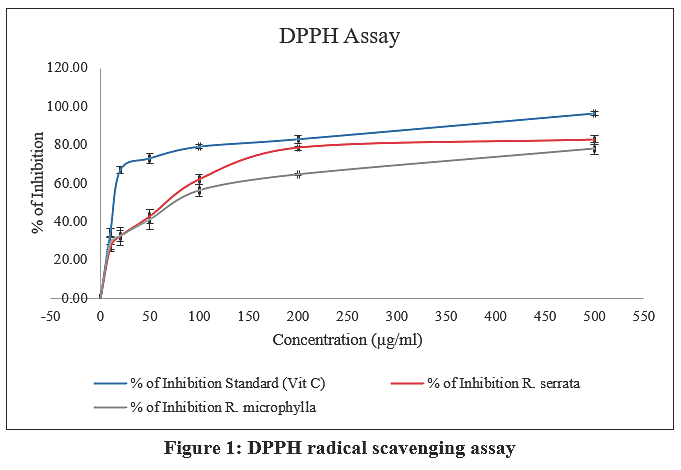 |
Figure 1: DPPH radical scavenging assay
|
Table 3: Nitric Oxide Scavenging Assay
| Concentration
(µg/mL) |
% of Inhibition (Mean ± SD); n=3 | ||
| Standard (Vit C) | R. serrata | R. microphylla | |
| Control | 0.00 | 0.00 | 0.00 |
| T1(10) | 20.63 ± 1.96 | 17.58 ± 0.87 | 15.80 ± 1.12 |
| T2(20) | 36.76 ± 2.05 | 29.52 ± 2.08 | 24.27 ± 2.11 |
| T3(50) | 62.23 ± 0.98 | 55.72 ± 1.62 | 49.05 ± 3.03 |
| T4(100) | 79.67 ± 2.74 | 73.80 ± 2.41 | 66.09 ± 0.81 |
| T5(200) | 84.09 ± 1.02 | 79.42 ± 3.26 | 72.75 ± 2.06 |
| T6(500) | 93.91 ± 1.97 | 81.82 ± 2.91 | 76.93 ± 2.18 |
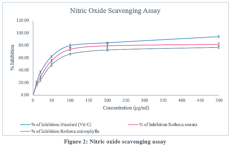 |
Figure 2: Nitric oxide scavenging assay
|
Table 4: Phosphomolybdenum Reducing Power Assay
| Concentration
(µg/ml) |
Absorbance (Mean ± SD); n=3 | ||
| Standard
(Vit C) |
R. serrata | R. microphylla | |
| Control | 0 | 0 | 0 |
| T1(10) | 0.962 ± 0.009 | 0.074 ± 0.011 | 0.091 ± 0.004 |
| T2(20) | 0.114 ± 0.010 | 0.099 ± 0.006 | 0.101 ± 0.012 |
| T3(50) | 0.186 ± 0.012 | 0.147 ± 0.026 | 0.145 ± 0.008 |
| T4(100) | 0.363 ± 0.036 | 0.215 ± 0.012 | 0.245 ± 0.032 |
| T5(200) | 0.522 ± 0.058 | 0.443 ± 0.041 | 0.452 ± 0.016 |
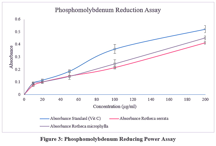 |
Figure 3: Phosphomolybdenum Reducing Power Assay
|
Table 5: Ferric reducing antioxidant power assay
| Concentration
(µg/ml) |
Absorbance (Mean ± SD); n=3 | ||
| Standard
(Vit C) |
R. serrata | R. microphylla | |
| Control | 0 | 0 | 0 |
| T1(10) | 0.962 ± 0.009 | 0.074 ± 0.011 | 0.091 ± 0.004 |
| T2(20) | 0.114 ± 0.010 | 0.099 ± 0.006 | 0.101 ± 0.012 |
| T3(50) | 0.186 ± 0.012 | 0.147 ± 0.026 | 0.145 ± 0.008 |
| T4(100) | 0.363 ± 0.036 | 0.215 ± 0.012 | 0.245 ± 0.032 |
| T5(200) | 0.522 ± 0.058 | 0.443 ± 0.041 | 0.452 ± 0.016 |
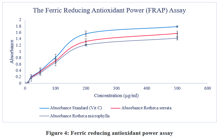 |
Figure 4: Ferric reducing antioxidant power assay
|
Table 6: Determination of Total Phenolic Content
| Concentrations
(µg/ml) |
Absorbance
(Mean ± SD); n=3 |
Phenolic Content (Mean ± SD); n=3 |
| 0 | 0 | |
| 10 | 0.091 ± 0.016 | |
| 20 | 0.202 ± 0.021 | |
| 50 | 0.392 ± 0.008 | |
| 100 | 0.716 ± 0.101 | |
| 400 | 1.262 ± 0.082 | |
| R. serrata | 3.631 ± 0.048
|
746.26 ± 8.42 µg GAE/g of extract |
| R. microphylla | 3.232 ± 1.161 | 661.58 ± 13.08 µg GAE/g of extract |
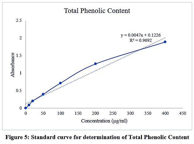 |
Figure 5: Standard curve for determination of Total Phenolic Content
|
Statistical Analysis
All experiments were conducted in triplicate, and values were represented as mean ± standard deviation. Statistical analysis was performed using MS Excel and SPSS, and the significant values were considered as p ≤ 0.001.
Discussion
Natural products remain a prolific source of bioactive molecules for therapeutic development, and plant‑derived formulations are increasingly favoured because of their perceived safety and sustainability relative to many synthetic drugs.12 Phenolic compounds in particular have attracted considerable interest due to their potent antioxidant properties and their documented association with reduced oxidative stress across diverse plant extracts.13 Previous studies have reported strong correlations between phenolic content and antioxidant capacity, underscoring the central role of phenolics in the free‑radical scavenging activity of botanical preparations.14,15
In this study, the methanolic extracts demonstrated superior antioxidant activity compared with extracts obtained using other solvents, indicating greater extraction efficiency for antioxidant constituents in methanol. Chemical fingerprinting by GC‑MS and FTIR of the methanolic fraction revealed triterpenes, phenolic compounds and hydrocarbons as major constituents.4 These compound classes have been implicated in antiplasmodial and antiparasitic activity and are consistent with the traditional antimalarial use of Rotheca species. Complementary phytochemical surveys of Rotheca serrata have reported flavonoids, phenolics, alkaloids, glycosides and terpenoids, and recent work has confirmed significant antioxidant and anti‑inflammatory activities for extracts of this species.3 Antioxidant and anti‑inflammatory actions are mechanistically relevant because they can attenuate oxidative damage and inflammation that exacerbate parasitic and metabolic disorders, thereby enhancing the therapeutic profile of plant extracts.
Our qualitative screening identified multiple bioactive classes in leaves of R. serrata and R. microphylla, including alkaloids, tannins, flavonoids, glycosides, terpenoids, steroids and anthraquinones. The phytochemical profile of R. serrata observed in the present investigation corroborates earlier reports demonstrating the presence of comparable secondary metabolites in this species.16 Such bioactive constituents have been variably linked to antioxidant, antimicrobial, anticancer, anti‑inflammatory and lipid‑lowering effects, highlighting the pharmacological properties of these species.17 Notably, prior studies on C. serratum (syn. R. serrata) also reported a dominance of polyphenolics over other phytochemical classes and attributed several pharmacological effects primarily to polyphenolic constituents or to their combined action with other metabolites.18,19 The absence of saponins in our methanolic leaf extracts agrees with earlier qualitative analyses of C. serratum roots and leaves,20 that similarly failed to detect saponins, though saponins have been reported in some ethanolic extracts of R. microphylla.5 Such interstudy variation likely reflects differences in plant material, extraction solvent and analytical sensitivity.
Quantitatively, total phenolic content was higher in R. serrata (746.26 ± 8.42 µg GAE/mg extract) than in R. microphylla (661.58 ± 13.08 µg GAE/mg extract), and this difference paralleled the modestly greater antioxidant activity observed for R. serrata across DPPH, nitric oxide scavenging, and FRAP assays. These results corroborate the frequently observed positive relationship between phenolic concentration and antioxidant capacity reported for many medicinal plants and support the contention that phenolics are key contributors to the redox properties measured in vitro.21-23
Taken together, the phytochemical composition and antioxidant profile of the studied Rotheca extracts provide a plausible biochemical basis for their traditional and experimentally validated bioactivities. To advance these findings toward therapeutic application, future work should prioritize isolation and structural characterization of the active constituents, determine their individual and synergistic mechanisms and perform in vivo efficacy and toxicity testing alongside pharmacokinetic evaluation. Such studies will be critical for standardizing extracts and assessing their translational potential.
Conclusion
The present investigation demonstrated that both R. serrata and R. microphylla exhibit the presence of various groups of phytochemical compounds and considerable antioxidant potential. Qualitative phytochemical evaluation revealed that both selected plants possess comparable phytochemical composition, indicating the occurrence of similar classes of bioactive constituents. Determination of antioxidant activity with four distinct in vitro assays indicated notable free radical scavenging ability and reducing capacity of both plants. R. serrata showed comparatively higher total phenolic content (TPC) and antioxidant activity in most of the tests performed. R. microphylla also showed antioxidant potential, whereas its activity was mildly lower than that of R. serrata, except for the phosphomolybdenum reducing power assay, indicating variation in the concentration or mechanism of action of specific antioxidant constituents of the plants.
Overall, the observations support that both selected species can be considered as a potential source of antioxidants with significant medicinal and nutraceutical importance. . To advance these findings toward therapeutic application, future work should prioritize isolation and structural characterization of the active constituents, determine their individual and synergistic mechanisms and perform in vivo efficacy and toxicity testing alongside pharmacokinetic evaluation. Such studies will be critical for standardizing extracts and assessing their translational potential.
Acknowledgement
The authors acknowledge the constant support and encouragement by the principal CMS College Kottayam (Autonomous) and B.C.M. College Kottayam (Autonomous). We would like to express our special thanks to Doctor John’s Biotech Centre for Research and Development for providing technical assistance and instrumental facilities for the successful completion of this work.
Funding Sources
The author(s) received no financial support for the research, authorship, and/or publication of this article.
Conflict of Interest
The authors do not have any conflict of interest.
Data Availability Statement
This statement does not apply to this article.
Ethics Statement
This research did not involve human participants, animal subjects, or any material that requires ethical approval.
Informed Consent Statement
This study did not involve human participants, and therefore, informed consent was not required.
Clinical Trial Registration
This research does not involve any clinical trials.
Permission to reproduce material from other sources
Not Applicable
Author Contributions:
- Antu Mariya Jose: Conceptualization, Data Collection, Analysis, Methodology, Writing – Original Draft.
- Jinu John: Editing, Resources.
- Rogimon Plammoottil Thomas: Visualization, Supervision, Project Administration.
References
- Singh MK, Khare G, Iyer SK, Sharwan G, Tripathi DK. Clerodendrum serratum: A clinical approach. J Appl Pharm Sci. 2012;2:11-15.
- DeFilipps RA, Krupnick GA. The medicinal plants of Myanmar. PhytoKeys. 2018; 102:1-341. doi:10.3897/phytokeys.102.24380
CrossRef - Khaidem A, Das MK. Phytochemical screening and antioxidant activity of Rotheca Serrata (L.) Steane & Mabb. Collected From Manipur, Northeast India. Int J Creat Res Thoughts.2025;13(12):877-881.
CrossRef - Chanu WK, Chatterjee A, Singh N, Devi HS, Das MK. Phytochemical screening, antioxidant analyses, and in vitroand in vivo antimalarial activities of herbal medicinal plant – Rotheca serrata (L.) Steane & Mabb. J Ethnopharmacol. 2024;321:117466.
CrossRef - Safeera M, Ansila P.A, Afitha V, et al. In Vitro Antidiabetic Study and Phytochemical Screening of Hydroalcoholic Extract of Rotheca microphylla (Blume) Callm. & Phillipson Leaves. World J Pharm Res. 2026;15(7):771-779.
- Chandimali N, Bak SG, Park EH, et al. Free radicals and their impact on health and antioxidant defenses: a review. Cell Death Discov. 2025;11(1):19. doi:10.1038/s41420-024-02278-8
CrossRef - Blois MS. Antioxidant Determinations by the Use of a Stable Free Radical. Nature. 1958;181(4617):1199-1200. doi:10.1038/1811199a0
CrossRef - Lee H S. Antioxidant activity of browning reaction product isolated from storage-aged orange juice. J Agric Food Chem. 1992;40:550-552. doi:10.1021/jf00016a004
CrossRef - Prieto P, Pineda M, Aguilar M. Spectrophotometric Quantitation of Antioxidant Capacity through the Formation of a Phosphomolybdenum Complex: Specific Application to the Determination of Vitamin E. Anal Biochem. 1999;269(2):337-341. doi:10.1006/abio.1999.4019
CrossRef - Oyaizu M. Studies on products of browning reaction. Antioxidative activities of products of browning reaction prepared from glucosamine. Jpn J Nutr Diet. 1986;44(6):307-315. doi:10.5264/eiyogakuzashi.44.307
CrossRef - Bray H G., Thorpe W V:.Analysis of Phenolic Compounds of Interest in Metabolism. In: Methods of Biochemical Analysis (Glick D. ed). Vol 1. 1st ed. Wiley; 1954:27-52. doi:10.1002/9780470110171.ch2
CrossRef - Bamne F, Sharma P, Shaikh N, Momin M, Khan T, Ali A. Evaluation of antioxidant capacity and safety profile of selected medicinal plant extracts. Biosci Biotechnol Res Asia. 2025;22(3):1087-1098. doi:10.13005/bbra/3425
CrossRef - Robards K, Prenzler PD, Tucker G, Swatsitang P, Glover W. Phenolic compounds and their role in oxidative processes in fruits. Food Chem. 1999;66(4):401-436. doi:10.1016/S0308-8146(99)00093-X
CrossRef - Pan Y, Wang K, Huang S, et al. Antioxidant activity of microwave-assisted extract of longan (Dimocarpus longan) peel. Food Chem. 2008;106(3):1264-1270. doi:10.1016/j.foodchem.2007.07.033
CrossRef - Huang B, Ban X, He J, Tong J, Tian J, Wang Y. Hepatoprotective and antioxidant activity of ethanolic extracts of edible lotus (Nelumbo nucifera) leaves. Food Chem. 2010;120(3):873-878. doi:10.1016/j.foodchem.2009.11.020
CrossRef - Nu KS, Lwin HH. Investigation of morphological, histological characters and qualitative analysis of Rotheca Serrata (L.) Steane & Mabb. J Myanmar Acad Arts Sci. 2021;XIX(4A):359-367.
- Khakhalary S, Narzari S. Phytochemical profiles of leaf extracts of Rotheca serrata (L.) steane & mabb: a medicinal herb of Assam. Indian Bot Soc. 2022;102(4):294-305.
- Mohamed AJ, Mohamed EAH, Aisha AFA, et al. Antioxidant, antiangiogenic and vasorelaxant activities of methanolic extract of Clerodendrum serratum (Spreng.) leaves. J Med Plants Res. 2012;6(3):348-360.doi:10.5897/JMPR11.049
CrossRef - Upadhyay S, Koche V. Antioxidant Activity and Phytochemical Content of Clerodendrum serratum from Different Provinces of Chhattisgarh State, India. Asian J Chem. 2019;31(10):2147-2151. doi:10.14233/ajchem.2019.22056
CrossRef - Yusuf N, Wahab NHA, Maulidiani, et al. Phytochemical analysis and non-enzymatic antioxidant activity of Clerodendrum serratum leaves and stems. Malays J Anal Sci. 2025;29(4):1514
- Gadade JP, Swaroopa A. Patil. Phytochemical paradigm, antioxidant status and their correlation in Rotheca serrata (L.) Steane and Mabb. Ann Phytomedicine. 2019;8(2):156-166.doi: 10.21276/AP.2019.8.2.20
CrossRef - Kaur C, Kapoor HC. Anti-oxidant activity and total phenolic content of some Asian vegetables. Int J Food Sci Technol. 2002;37(2):153-161. doi:10.1046/j.1365-2621.2002.00552.x
CrossRef - Vinson JA, Hontz BA. Phenol Antioxidant Index: Comparative Antioxidant Effectiveness of Red and White Wines. J Agric Food Chem. 1995;43(2):401-403. doi:10.1021/jf00050a027
CrossRef
Abbreviations:
DPPH: 2,2-Diphenyl-1-picrylhydrazyl
FRAP: Ferric Reducing Antioxidant Power
TPC: Total Phenolic content
R. serrata: Rotheca serrata
R. microphylla: Rotheca microphylla
HCl: Hydrochloric acid
FeCl3: Ferric chloride
H2SO4: Sulfuric acid
Vit C: Vitamin C
Accepted on: 30-08-2026
Second Review by: Dr. Nibedita Das
Final Approval by: Dr. Ali Mohamed Elshafei