Advances in in vitro Tumor Models and Genetic Engineering in Cancer Research
 , Gowri Priya Jayasree2, Sneha Ajilal2, Anusha Baby Satravada3, Dhamodharan Prabhu4, , 2*
 1Department of Biotechnology, CEPCI Laboratory and Research Institute, Kollam, Kerala, India
2Department of Biotechnology, Government Arts College, Thiruvananthapuram, Kerala, India
3Computational Oncology, Memorial Sloan Kettering Cancer Centre, New York, USA
4Center for Bioinformatics, Department of Biotechnology, Karpagam Academy of Higher Education, Coimbatore, Tamil Nadu, India
Corresponding author’s E-mail: ramyarprabhu@gmail.com
Download this article as:
ABSTRACT:Cancer stands as one of the leading worldwide health threats because it causes approximately 16% of all global deaths, according to the World Health Organization (WHO, 2025). This review discusses the critical role of various tissue culture techniques in studying fundamental and advanced methods used in cancer research in identifying new therapeutic targets. There are several methods to study cancer cell biology and molecular mechanisms, such as traditional two-dimensional (2D), which has long been the gold standard, and more advanced three-dimensional (3D) culture systems that better reflect molecular characteristics, cell-cell interactions, and signaling pathways. Additionally, genetic engineering tools like Clustered regularly interspaced palindromic repeats-CRISPR-associated protein 9 (CRISPR-Cas9) provide targeted genome editing and RNA interference (RNAi) based approaches such as small interfering RNAs (siRNAs) for the transient gene silencing. These genetic engineering tools offer promising models for investigating the drug response, therapeutic targets, and in cancer research.
KEYWORDS:2D cultures; 3D bioprinting; 3D cultures; CRISPR/Cas9; Small interfering RNAs (siRNAs)
Introduction
Cancer is a deadly disease globally due to the genetic and epigenetic alterations. Cancer is one of the largest global health challenges in the 21st century. The World Health Organization reports that there were 9.7 million cancer deaths in 2022, a number that is predicted to rise to over 35 million new cases by 2050, mainly due to an increasing aging population, unhealthy lifestyles, and environmental exposures.1 Even with considerable improvements in the three pillars of cancer treatment, namely surgery, chemotherapy, and radiotherapy, as well as recent advances in immunotherapy, many cancers remain poorly treatable.2 In preclinical studies, however, existing models show limited relevance to human disease. Conventional two-dimensional (2D) cell cultures cannot mimic the three-dimensional (3D) environment of solid tumors, with their tissue structure and mechanical properties, nor can they account for the complexity and heterogeneity of multicellular tumor ecosystems.3 In this review, we describe three research axes of research and development that are being used in concert to overcome these limitations in cancer modelling and drug discovery. We cover tissue culture systems, ranging from classical 2D cell lines to more complex 3D organoids, spheroids, and scaffold-based tissue models, as well as gene editing tools and RNA interference, mainly using the Clustered Regularly Interspaced Short Palindromic Repeats and CRISPR- associated protein 9 (CRISPR-Cas9) system for cancer functional genomics. The study of oncogenes and tumor suppressor genes can help understand cancer biology, which regulates cell proliferation, apoptosis, and differentiation. Tissue culture models can investigate the mechanisms in vitro. Spheroids and organoids are advanced tumor models that mimic the tumor environment, which helps to understand the cancer biology.4
Functional assays such as proliferation, migration, and apoptosis assays are commonly used to understand the nature of the cancer cell and the impact of genetic alterations.5More advanced genetic engineering tools, such as CRISPR-Cas9, are powerful gene-editing tools that aid in the modification of oncogenes and tumor suppressor genes for studying their functional role in cancer.6 RNA interference (RNAi) is a gene silencing technique that helps in the identification of gene function and promising therapeutic targets. Both technologies have offered promising methods for cancer research.7Two domains of technological advance, namely, tissue culture systems and genetic engineering, are not mere additional technologies for oncology research. Rather, these two areas are intimately interconnected and form part of an integrated system of research. Tissue culture systems offer experimentally relevant biological systems; genetic engineering allows specific interference with cancer-related pathways.
This review highlights an integrative transformation of advanced in vitro culture systems and genome engineering technologies to resolve major constraints in conventional cancer research. By integrating physiologically relevant 3D tumor models such as organoids and spheroids with precise gene-manipulating tools like CRISPR-Cas9 and RNAi, the field is moving towards more advanced cancer therapeutic strategies.
Introduction to Tissue Culture Models
Tissue culture is the fundamental technique in cancer research that enables us to understand the cellular and molecular mechanisms involved in cancer progression. It provides a controlled environment to assess the effects of drugs,study gene alterations and analyze signaling pathways. There are various tissue culture techniques available to study the role of oncogenes and tumor suppressor genes in cancer biology. Here, we are exploring the key techniques used in cancer biology.
Tissue Culture
Animal cell culture is a standard procedure for culturing and maintaining animal cells, tissue, or organs in an artificial, well-regulated environment.8 It is an ideal practice to study cellular processes while avoiding animal model usage. The cells are maintained in standard culture dishes with a modified surface that provides support for cellular attachment and proliferation.9 It allows us to control chemical and physical parameters such as temperature and osmolarity, providing an ideal environment for cell growth.10 Media is an important factor in cell culture technology, as it provides the nutrients and supplements required for cell growth. Cell culture media is classified into natural media and synthetic media, and the latter is most widely used for research. The basal media are used with supplements like fetal bovine serum that facilitate cell growth in vitro.11 On the basis of cells cultured, primary cell culture is the one in which the tissue constructs are procured from animals, enzymatically separated using proteolytic enzymes, and strictly maintained under controlled aseptic conditions that favour in vitro proliferation.9 Secondary cell culture, on the other hand, is established cell lines that are derived primarily after subculture of primary cells once it reaches confluency. Cell culture can be broadly classified into 2D and 3D culture systems based on the substrate used for culturing the cell. Compared to traditional 2D culture, 3D cell cultures mimic the in vivo condition in a more sophisticated manner, generating reproducible results. The transition from 2D to 3D cell culture is crucial in studying the tumor microenvironment, cell signaling pathways, and drug response, which are relevant in cancer research.3
Studying the oncogenes and tumor suppressor genes in tissue cultures
Cellular assays in 2D culture environment
Cellular proliferation assays provide a sensitive means to examine cell viability and cell proliferation.12 Figure 1 depicts the schematic illustration of the cellular assays. The MTT (3-(4,5-dimethylthiazol-2-yl)-2,5-diphenyltetrazolium bromide) assay utilizes the mitochondrial reductase activity of viable cells to convert a soluble tetrazolium salt MTT, into an insoluble formazan product, which can then be solubilized and quantified spectrophotometrically.13 The intensity of the resultant coloured solution is directly correlated with the quantity of live cells.14 Other assays include CellTiter-Glo, which measures ATP levels using a luminescent signal, and the BrdU (5-bromo-2′-deoxyuridine) assay, which detects newly synthesized DNA through antibody labelling. These assays are essential tools for drug discovery, understanding resistance mechanisms, and personalized medicine approaches.15–18
Cell migration is the movement of a cell from one site to another between cell-to-cell and cell-extracellular matrix.19 It is commonly elucidated by assays such as the wound healing assay and the transwell migration assay. In a wound healing assay or scratch assay, a scratch is made in a 100% confluent cell layer, and the closure rate indicates the migratory ability.20 Trans-well invasion assays are used to track the migration and penetration capacity of tumor cells by establishing a chemoattractant gradient.20 The study of migratory properties of oncogenic cells is necessary since the majority of cancer deaths are due to metastasis, and an understanding of cell migration paves the way for targeted therapeutic approaches.21
Cell cycle assays are techniques used in examining cell division and DNA replication by analysing the different phases of the cell cycle – G1, S, G2, and M.22 The BrdU incorporation assay helps to measure cell cycle phases, based on the ability of bromodeoxyuridine to integrate into newly synthesized DNA during the S phase of the cell cycle.23,24 Another technique, Propidium Iodide (PI) staining, intercalates into DNA and emits fluorescence that is directly proportional to the amount of DNA present.25 Flow cytometry, coupled with PI staining, provides a reliable representation of the cell cycle distribution. Fluorescent Ubiquitination-based Cell Cycle Indicator (FUCCI) is an approach that uses fluorescent proteins to visualize and monitor cell cycle progression.26,27 These assays serve as indispensable tools in cancer research, providing critical insights into the mechanisms driving tumorigenesis and offering opportunities for the development of novel therapeutic strategies.28
Apoptosis is a cellular mechanism that controls cell death and maintains tissue homeostasis.29 Apoptosis is regulated by a group of enzymes known as caspases and diverse apoptotic factors.30,31 Caspase assay relies on the intrinsic proteolytic ability of the enzyme caspase, and its quantitative analysis is performed by employing a trans-peptide that flanks caspase-specific cleavage sites, conjugated with a chromophore or fluorophore. Upon the specific enzymatic cleavage, the release of fluorogenic or chromogenic substrate is achieved, and the intensity is quantified, which showcases caspase activity.32 The Annexin Ⅴ staining method relies on the high binding affinity of annexin to the phosphatidyl serine on the outer membrane of an apoptotic cell. When labelled with a fluorescent probe, the complex becomes a reliable tool to quantify cell death.33 Acridine orange/Ethidium bromide (AO/EtBr) is a multiplex assay that clearly visualizes and distinguishes different cell phases based on the cell membrane permeability of the dye. Normal and early apoptotic cells emit green fluorescence, while late apoptotic and dead cells emit orange-red fluorescence.34
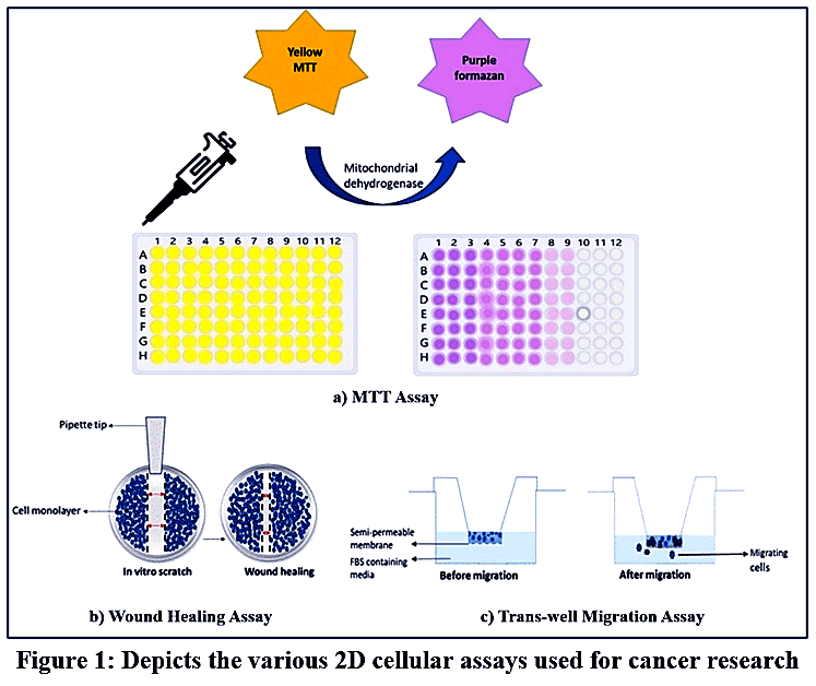 |
Figure 1: Depicts the various 2D cellular assays used for cancer research.
|
Advanced Three-Dimensional Culture Models
Spheroids
Spheroids are cell or tumor aggregates that are grown in a 3D space. These spheroids mimic in vivo tumor niches, and thereby these models serve as crucial in vitro models for studying tumor initiation and development.35 Figure 2 depicts the establishment of spheroids and organoids. Spheroids are widely employed as in vitro models in various research domains such as tumor biology, cancer biology, and immunology.36 Distinct cellular populations are generated during spheroid formation, with a clear distinction between the outer layer consisting of actively proliferating cells, quiescent cells that dominate the middle intermediate layer, and the core of spheroids populated by necrotic or hypoxic cells.8
The hanging drop method is a technique that is used to generate spheroids where small droplets of cell suspension are introduced onto a non-adhesive flat surface that stimulates cells to aggregate towards the bottom of the droplet, forming spheroids.37 A three-dimensional spheroid of size 1.5 mm from human colorectal carcinoma, HCT116, was developed using the flipped well plate hanging drop technique, and cell-cell and cell-extracellular matrix interaction studies were performed.38 A further improved version of the hanging drop method, where a methyl cellulose polymer is introduced into the hanging drop culture medium, and a pancreatic 3D spheroid model was developed. The modified hanging drop method offers several advantages over the classical hanging drop method. In this pancreatic 3D spheroid model, the molecular aggregates were tightly packed, structurally stable, and resistant to mechanical stress.39 Another approach is the spinner cell culture techniques that foster spheroid formation by employing the usage of certain equipment such as spinner flasks and rotating bioreactors, where the cells are subjected to constant agitation. The constant agitation favours the initiation of cell-to-cell contact and self-assembly of cells in the absence of a cell attachment surface.40 A multicellular tumor spheroid model using SK-Hep-1 (Sloan-Kettering Hepatoma-1), human hepatoma cells, was developed by culturing SK-Hep cells in a siliconized cell spin flask. Cellular aggregates were observed during the first 48 hours, and over the following two weeks, the cell aggregates attained a distinct spheroid shape. The model served as an ideal in vitro model for nutrients, specifically the glutamine transport rate.41 A multicellular tumor spheroid of human adenocarcinoma cells, the HT29-based 3D spheroid model was developed using spinner culture technology. The in vitro tumor model served as a reliable model for investigating the effects of anticancer drugs and evaluating the efficacy of different drug delivery systems.42 In some cases, a scaffold system is utilized for spheroid development that supports cell-to-cell and cell-matrix interactions, guiding tumor formation.28These scaffolds mimic the extracellular matrix (ECM), rendering physical support, thereby enhancing the growth of the cell. Various factors such as biocompatibility, biodegradability, surface attachment, bioactivity, and their ability to transport oxygen and nutrients should be considered. Hydrogel-based scaffolds are networks of hydrophilic polymers that are derived from natural components such as alginate, gelatin, and chitosan, as well as prepared from synthetic polymers like polylactide (PLA), polyglycolide (PGA), polycaprolactone (PCL) etc.43Hydrogels such as Matrigel, with extracellular matrix components such as collagen, are widely employed as a 3D matrix.10 A 3D bioengineered hydrogel culture was constructed to investigate the interaction between patient-derived glioblastoma cells and a hydrogel with extracellular matrix containing hyaluronic acid. This interaction was crucial in conferring chemotherapeutic resistance in glioblastoma. This model renders valuable insights about apoptosis mechanisms, DNA damage, and proliferation rates after being subjected to chemotherapeutic drugs.44 Advanced spheroid development techniques include microfluidics-based cell culture systems wherein the cells are nurtured in chambers with interconnected microchannels. This facilitates oxygen and nutrient transfer and also facilitates precise control of in vitro tumor development.45 Using microfluidics technology, a tumor microenvironment was constructed by a blend of pancreatic tumor spheroids with stellate cells, which were nourished in interconnected microchannels of the microfluidics device. The model successfully elucidated the paracrine- and juxtacrine-driven cell-cell interactions, the epithelial-to-mesenchymal transition, and the drug resistance mechanism.46
Spheroids offer a wide range of applications, especially in cancer research for studying various signaling pathways, metastatic properties of tumors, drug delivery systems, analysing drug efficacy, and elucidating the underlying mechanism of drug resistance.47 The well-differentiated layers in spheroids provide more insights into cell signal transduction and cell-to-cell communication, thereby providing a better understanding of tumor progression and tumor metastasis. Spheroids can be used to evaluate the efficacy of anticancer drugs, their mode of action and toxicity, and the data generated are more reliable compared to traditional 2D cultures.
However, certain limitations are associated with spheroid cultures. One of them is the significant increase in dimensionality of the spheroid culture system that may lead to variability in the distribution of oxygen, nutrients, and waste products.8 This heterogeneity leads to misinterpretation and limits reproducibility and reliability of results.48 There is also a requirement for high-resolution imaging facilities for constant spheroid monitoring.49 To address these limitations associated with spheroid culture, various advancements in biomaterials, microfluidics, and imaging are employed to overcome these constraints of spheroids.
Organoids
Organoids are 3D culture models that are grown in laboratory culture conditions using different cell sources, including adult stem cells (ASCs), embryonic stem cells (ESCs), induced pluripotent stem cells (iPSCs), and patient-derived tumour cells or tissues.50 Unlike conventional 2D cell cultures, which grow as flat monolayers, organoids are able to form 3D structures that mimic the real architecture of organs and tissues in the body. So, this potential makes organoids a powerful tool in research field.51 There are human organoids successfully developed, such as the brain, gut, liver, kidney, thyroid gland, and heart. These developed organoids resemble the structure and functional aspects of the corresponding organ in the body.52 ASCs are established by culturing stem or progenitor cells isolated from adult tissue under 3D culture conditions. These organoids are more resemblance to adult stem cells and are useful for studying disease mechanisms, tissue biology, and regenerative medicine. Tumor derived organoids also known as tumoroids are derived from tumor tissue obtained from procedures such as biopsy or surgical resection and it retains the histological, molecular, and genetic characteristics of original tumor. Therefore, tumoroids are valuable models for investigating the tumor biology and discovering potential therapeutic drug response.50
Organoids have wide application in developmental biology, tissue engineering, cancer research, and drug discovery. The process of development of organoids involves a stepwise differentiation process. The selection of appropriate stem cells is the initial process. Next, these cells are embedded in a supportive scaffold or matrix, which helps them to grow in three dimensions. Along with these, growth factors and nutrient-rich media will provide the differentiation and maturation of cells into a complete organoid structure.53
Initially, the stem cells are directed to form three germ layers, such as ectoderm, endoderm, and mesoderm. These germ layers are fundamental in embryonic development and give rise to different organs and tissues in the body. The ectoderm is responsible for forming the nervous system and skin, the endoderm develops into the internal organs such as the liver, pancreas, stomach, lung, thyroid, and intestine, and the mesoderm gives rise to the heart and kidney.54 Researchers can generate the organoids by mimicking the natural development process in laboratory conditions. Organoids can be used as valuable tools in research fields, including drug screening. Since organoids can be developed from patient-specific cells, they can be used to predict patient-specific drug responses.55 Organoids can help to understand an individual’s cells’ response to different treatments, so it is often employed in studies associated with personalized medicine. This approach will improve patient outcomes due to disease conditions.56 Organoid technology faces a major restriction because it lacks functional blood vessels, which results in tissue death and restricts organoid development to small sizes. All tissues require nutrient and oxygen delivery and waste product removal through complex branching vascular networks.56
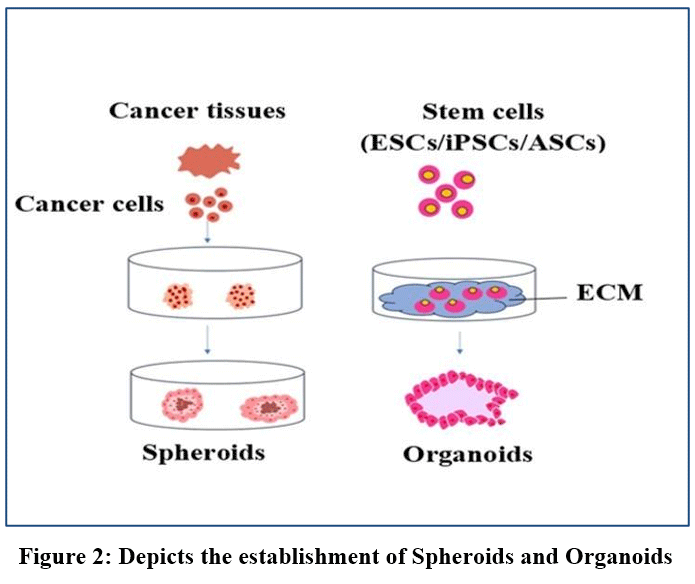 |
Figure 2: Depicts the establishment of Spheroids and Organoids
|
Enhanced recapitulation of Tumor microenvironment complexity in 3D culture systems
The tumor microenvironment (TME) consists of a group of malignant cells, non-malignant cells, blood cells, immune cells, along with the ECM secreted by the specific cell type. The cellular interaction among these cells mediates the complex cell signaling pathway, permitting cancer metastasis and tumor progression.57Monolayer cultures fail to display cell-cell interaction and cell-extracellular matrix interaction, signalling pathways, and unequal oxygen distribution, leading to tumor hypoxic regions along with the ability to initiate tumor heterogeneity.58The 3D culture models such as spheroids and organoids mimic the hallmarks of TME, the oxygen gradient, vascularization, and cellular heterogeneity.59Spheroids precisely establish the oxygen gradient and ECM architecture, while the organoids recapitulate the cellular heterogeneity.59Vascularization is essential for oxygen and nutrient delivery and the removal of waste in cultures. The integration of vasculature could increase the size of the spheroids and organoids. Vascularization strategies are classified as internal or external. Internal induction relies on cell-driven differentiation and self-organization, while external induction uses growth factors, medium composition, and hydrogel properties. Both methods will enhance vascularization and tissue integration.60,61 The absence of blood vessels prevents organoids from growing beyond three millimetres because their central areas become oxygen deprived and die from lack of oxygen. The development of 3D-bioprinted tumor models using patient-derived tumor cells and microenvironmental and immune cells has solved the essential problem of tissue vasculature.60,62
The 3D bioprinting technology enables the layer-by-layer fabrication of complex tissues and organs with precise structural organization.63,64 3D bioprinters are classified into three main categories: inkjet-based, laser-based, and extrusion-based bioprinting. Inkjet-based bioprinting is a cost-effective and accurate method. It deposits bioink as droplets or microspheres but is limited by nozzle clogging when printing dense bioinks. Laser-based bioprinting uses a nozzle-free approach, which deposits bioink droplets without contact. This avoids clogging and improves reproducibility. It is not suitable for complex structures. Extrusion-based bioprinting deposits continuous filaments of bioink to form structures. It is the common method and has more advantages, as it is versatile and compatible with many bioinks. Lower resolution and shear force can damage cells, which is one of the disadvantages.64–66
The combination of 3D-assembled structures, including organoids and spheroids, and cell-laden microgels shows great potential for creating vascularized tissues through bioprinting.67 The construction of tissue through bottom-up methods involves uniting living cells into structures that resemble natural tissues. Bioinks need to fulfil four essential conditions to be effective: they should maintain their shape during printing while having enough viscosity, they should gel quickly to support printed structure stability, they should match the stiffness of natural tissues, and they should be non-toxic for cell survival and operation.62 The biocompatibility of natural hydrogels such as collagen, fibrin, and hyaluronic acid is high, but their mechanical properties are insufficient. The mechanical strength of synthetic hydrogels exceeds that of natural hydrogels, but they need chemical treatment to enable cell attachment. Multiple successful bioink formulations use a combination of natural and synthetic materials.
Multi-Barrel Bioprinting and Tumor-on-Chip Integration
A multi-barrel 3D bioprinting method allows researchers to build human glioblastoma cells in the core and endothelial cells in the peripheral layer, which creates an oxygen gradient.68 The system places glioblastoma cells in the core area, while endothelial cells receive placement in the outer layer. The specific arrangement of cells in this system produces oxygen, and nutrient levels decrease from the centre to the edges, which matches the hypoxic conditions found in glioblastoma tumors. The construct development leads endothelial cells to form vessel-like structures through vasculogenesis, while the established oxygen gradient causes tumor cells to express genes that match patient tumor hypoxia-adapted phenotypes.68
The decellularized stroma method provides distinct benefits because it removes cells from tumor tissue while maintaining the original biochemical elements and structural arrangement of the tumor environment. The combination of tumor-on-a-chip technology with 3D bioprinting allows scientists to create models that duplicate blood vessel perfusion and microcirculation systems.69 The microfluidic-assisted bioprinting technique enables researchers to create functional blood and lymphatic hollow tubes within organ-on-a-chip systems.70 The perfusion system performs three essential tasks by delivering nutrients and oxygen to the construct, removing metabolic waste products, and generating shear stress that enhances endothelial cell development and vascular barrier function. The system allows scientists to study cancer cell behaviour during blood vessel entry and exit processes, which require tumor cells to interact with functioning blood vessels.
Spheroids and organoid 3D culture systems serve as excellent platforms to conduct multiomic approaches, as they are able to recapitulate the tumor under in vitro conditions.71 Multiomics is an interdisciplinary approach that provides high-throughput data on gene expression, proteins, and biological interactions.72 Various genomic profiling techniques, such as Next Generation Sequencing (NGS), are used to gain relevant information about the mutational status and differential gene expression patterns of patient-derived 3D spheroids and organoid models. Single-cell sequencing technologies include single-nucleus RNA sequencing (snRNA-seq), a sequencing technique that quantifies transcriptomes of the whole cell genome of a single cell, whereas advanced techniques such as spatial transcriptomics analyze and quantify gene expression within tissues.73
Spatial transcriptomics meets Organoid technology: Mapping cancer ecosystems in 3D
The Spatial transcriptomic technology connects individual cell analysis to tissue structure assessment to show how spatial arrangements affect cellular activities. snRNA-seq delivers detailed molecular information but fails to show where each cell exists within tissue structures.74 The RNA is mapped in accordance with specific location and is further sequenced to identify various cell types, generate differential gene expression data, and study intertumoral and intratumor heterogeneity.
Spatial transcriptomics maintains essential location data, which allows scientists to study gene expression patterns and cellular positions and their interactions with surrounding cells. The combination of high-resolution spatial techniques with bioinformatic tools allows researchers to build 3D spatial transcriptomics atlases from multiple adjacent tissue slices for studying human heart tissue and cardiac organoids and mouse brain samples.75 The different spatial transcriptomics methods, including 10x Genomics Visium and Slide-seq and Stereo-seq and MERFISH, offer distinct combinations of spatial detail, gene detection capabilities, and processing speed.76 The 10x Genomics Visium platform delivers complete transcriptome analysis through 55-micrometer spots, which can contain multiple cells. The MERFISH technique achieves high-resolution subcellular imaging through sequential fluorescence hybridization while measuring thousands of specific genes. The Slide-seq platform uses 10-micrometer bead-based capture to achieve single-cell spatial resolution while maintaining complete genome-wide coverage.76
Integrating Spatial transcriptomics with patient-derived Organoids
Organoid models serve as a platform for conducting spatial transcriptomic experiments.77 Organoids constructed of placenta tissue generate scalable and reproducible architectural information of transcriptomes, devoid of ethical constraints.78 Organoids from gall bladder epithelia were also developed, and by spatial transcriptomics, the gene expression profile was mapped to understand the major stages in cancer progression.79 Advanced organoids, such as laminar organoid models, were developed by integrating engineering technology such as lamination-based organoid spatially resolved transcriptomics, and a spatial transcriptomic study was carried out in laminated mouse lung and liver organoids.80
The lamination-based organoid spatially resolved transcriptomics method enables automated characterization of primary lung and liver organoid profiles through integrated spatial transcriptomic analysis.80 The technique solves a critical problem because previous spatial transcriptomics methods needed to handle small organoid samples through time-consuming sectioning procedures, which damaged the spatial organization of the samples. The lamination method produces flat cell layers from three-dimensional organoids, which allows researchers to use spatial transcriptomics tools directly without needing complicated sectioning methods that could harm sensitive organoid structures.
The 1 cm × 1 cm chip supports four organoids at present, but researchers can expand it to process more than ten organoids at once, which results in decreased sequencing expenses by more than ten times while needing minimal initial material.80 The combination of reduced costs and minimal sample requirements through this method makes spatial transcriptomics available to all researchers who study organoids.
Genetic Engineering Tools in Cancer Research
CRISPR-Cas9
CRISPR-Cas9, or Clustered Regularly Interspaced Short Palindromic Repeats and CRISPR- associated protein9, is a genome editing tool obtained from the adaptive immune systems of bacteria and archaea.81,82 The representation of CRISPR-Cas9 is shown in Figure 3. The CRISPR-Cas9 system lies in its ability to target and modify specific DNA sequences within a genome.83 This is achieved through the use of a guide RNA (gRNA) that directs the Cas9 nuclease to a complementary DNA sequence.84 CRISPR-Cas9 technology has revolutionized modern research by offering precision and versatility in genome editing across diverse organisms and cell types to understand, diagnose, and treat various complex diseases, mainly cancer.84 CRISPR-Cas9’s ability to induce targeted breaks in DNA has a wide range of applications, including gene knockout, knock-in, gene regulation, epigenome editing, and high-throughput screening.85 The mechanism of action is initiated with the creation of a single gRNA, a synthetic RNA molecule comprising a CRISPR RNA (crRNA) sequence complementary to the target DNA sequence and a trans-activating crRNA (tracrRNA) scaffold required for Cas9 binding.86 The gRNA, along with Cas9, forms a complex and directs the nuclease to a specific locus by base pairing between crRNA and target DNA.87 Upon binding to the target DNA, the Cas9 nuclease induces a double-strand break approximately three base pairs upstream of the protospacer adjacent motif, a short DNA sequence that is essential for Cas9 recognition and binding.88 The system’s flexibility is further enhanced by the constant development of novel Cas9 variants and delivery methods, extending its applicability in diverse cancer research settings.89,90 The different types of CRISPR-Cas9 variants are depicted in Table 1. CRISPR-Cas9 can be used to inactivate oncogenes, such as KRAS (Kirsten rat sarcoma virus), which are often mutated in various cancers, including lung and pancreatic cancer.91 Loss-of-function screens using CRISPR-Cas9 libraries have identified novel tumor suppressor genes and therapeutic targets.89 It is also employed to study the role of non-coding RNAs in cancer development. Cancer dependency genes, which are essential for cancer cell survival but dispensable in normal cells, can be identified through CRISPR-based screens, offering potential targets for cancer-specific therapies. In total, CRISPR-Cas9 serves as a molecular tool, enabling the precise dissection of cancer genomes and the functional characterization of individual genes and regulatory elements. CRISPR-Cas9 has been used in cancer treatments to precisely modify tumor suppressor and oncogene genes, allowing for targeted gene deletion and functional confirmation of cancer drivers. Additionally, it is being investigated in immunotherapy, where CRISPR-engineered T cells (such PD-1 knockdown CAR-T cells) improve treatment efficacy and boost anti-tumor immune responses.92Furthermore, new drug resistance mechanisms and cancer relying genes have been found using CRISPR-based high-throughput screening techniques, which has made it easier to build personalised cancer treatments.93,94
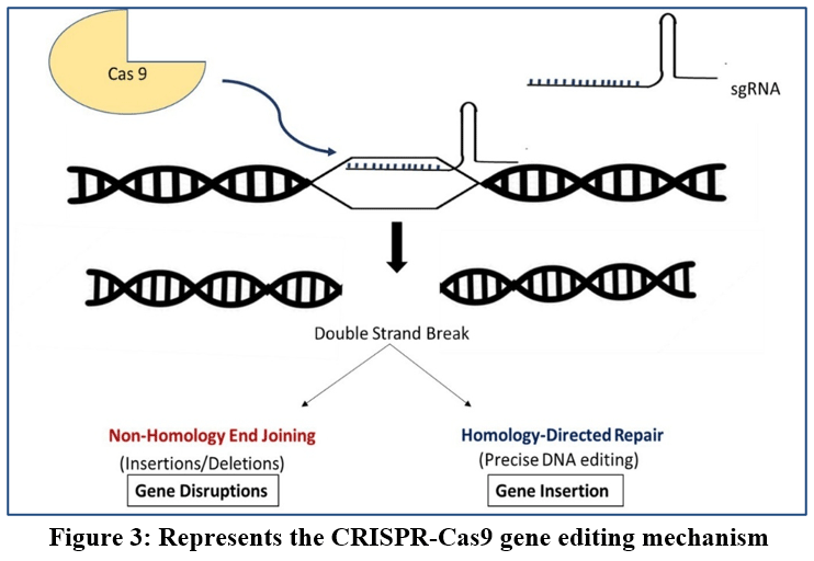 |
Figure 3: Represents the CRISPR-Cas9 gene editing mechanism
|
Table 1: Comparison of engineered Cas9 variants and their functional features
| Variant | Key feature |
| SpCas9 | ● Wild-type Cas9 with high on-target editing efficiency but relatively higher off-target activity.
● Cleavage specificity can be affected by single or double mismatches between the gRNA and target DNA.115 |
| eSpCas9 | ● Engineered high-fidelity Cas9 variant with reduced nonspecific interactions between Cas9 and the DNA backbone.
● Retains efficient on-target nuclease activity.116 |
| SpCas9 HF1 | ● High fidelity variant with modifications intended to decrease non-specific DNA interactions.117 |
| HypaCas9 | ● Exhibit significant genome-wide specificity without sacrificing on-target activity.118 |
| xCas9 | ● Can identify a wide variety of PAM sequences, such as NG, GAA, and GAT.119 |
| nCas9 | ● Cas9 nickase variant with a mutation in one nuclease domain, resulting in single-strand DNA nicking rather than double-strand cleavage.
● Provides increased targeting precision and is useful for applications such as base editing and paired-nickase genome engineering.95 |
| Sniper-Cas9 | ● High specificities in human cells without causing on-target cell death.
● This enables DNA-free genome editing in a preassembled ribonucleoprotein (RNP) structure.120
|
Advanced CRISPR Technologies: Precision Genome Editing Beyond Cas9
Cas12a Systems for Multiplexed Editing with Reduced Off-Targets
The genome editing capabilities of traditional CRISPR-Cas9 systems remain limited because they produce off-target effects.95 The CRISPR effector Cas12a operates as a separate class 2 system, which provides better performance than Cas9 through its ability to detect thymine-rich PAM (Protospacer Adjacent Motif) sequences (TTTV, where V represents A, C, or G), its enhanced guide RNA-target DNA mismatch sensitivity and reduced off-target activity, and its ability to perform multiplex gene editing with one crRNA array.96 The ability to perform multiplex gene editing with Cas12a proves essential because it allows researchers to use one CRISPR array that produces multiple guide RNAs from a single transcript, which Cas12a cuts into separate functional guides.
The Cas12a-knock-in mouse system allows scientists to perform efficient multiplexed genome editing.97 The enAsCas12a variant of Acidaminococcus sp. Cas12a shows a strong ability to produce compound genotypes, which include different cancer types that result from tumor suppressor gene inactivation of three separate genes.98 The ability of Cas12a-based systems to modify multiple genes at once produces models that better represent natural biological processes.
Base Editing: Single-Nucleotide Precision without DNA breaks
Base editors perform target nucleotide conversions through direct modification without causing DNA double-strand breaks.99 Base editors represent a new generation of genome editing tools because they perform single-nucleotide changes without inflicting DNA double-strand breaks. The CRISPR-Cas9 system generates double-strand breaks, which cells repair through faulty mechanisms that produce insertions and deletions together with the desired modifications. The base editors solve these issues through chemical nucleotide modification of individual bases while keeping DNA structures whole.
The three operational sections of CRISPR base editing include a non-active Cas protein, which attaches to DNA targets without cutting them; a cytidine deaminase enzyme that transforms cytosine into uracil; and a uracil glycosylase inhibitor, which blocks cellular editing systems from reversing the modification. The cell system recognizes uracil as thymine during DNA replication and repair processes, which results in the completion of C-to-T base substitution. The adenine base editors perform adenine-to-inosine conversions through adenosine deaminases, which cells recognize as guanine, to achieve A-to-G base substitutions.99 The enzymatic method allows researchers to make exact single-base changes without the DNA damage that occurs when cells repair double-strand breaks.
The Cas12a base editing system allows processing of multiple gRNAs from one transcript, which enables simultaneous base editing at 15 different sites across various human cell types.100 The combination of Cas homologs with base editors enables researchers to target both cytosine and adenine bases, which could restore about 95% of disease-causing transition mutations.101The majority of disease-causing single nucleotide variants exist as transition mutations, which involve purine-purine exchanges (A↔G) and pyrimidine-pyrimidine exchanges (C↔T). The clinical development of Beam-101 continues to progress well in Phase I/II clinical trials.102
Applications of CRISPR-Cas9 in identifying and validating oncogenic targets
Recent research demonstrates that CRISPR-Cas9 has been widely utilized to find and validate oncogenic targets through genome-wide loss-of-function screens that reveal cancer dependence genes necessary for tumor survival.103 Critical vulnerabilities in oncogenic pathways, such as dependencies associated with KRAS-mutant malignancies, have been identified by CRISPR screens. These findings have been confirmed by targeted gene knockout experiments showing decreased tumor growth and proliferation.104 The therapeutic significance of DNA repair pathways has been confirmed by the identification of effective targets in cancers with BRCA1 and BRCA2 abnormalities using CRISPR-based synthetic lethality techniques.105Additionally, by introducing or rectifying mutations and monitoring their direct influence on tumor initiation and progression, CRISPR-engineered tumor models have made it possible to functionally validate oncogenes and tumor suppressors in vivo.92
RNA Interference in cancer research
The process in which specific genes are silenced by small RNA molecules is called RNA interference (RNAi) or gene silencing. RNAi molecules degrade and inhibit the translation of target mRNA. There are different types of RNAi molecules involved in gene silencing, such as small interfering RNAs (siRNAs), short hairpin RNA (shRNAs), microRNAs (miRNAs), and piwi-interacting RNA (piRNAs). siRNAs and miRNAs are vastly studied small RNAs that play a critical role in cell proliferation, cell death, and cancer.106,107 siRNAs are synthetic, double-stranded RNAs with a nucleotide length of about 20-25 base pairs, whereas miRNAs are small, endogenous, non-coding single-stranded RNAs naturally present in the genomes of animal cells. The shRNA is artificially synthesized by the vector, which is then processed to form active siRNA. Later, active siRNA and the RNA-induced silencing complex (RISC) together silence the targeted mRNA.108
siRNA- and miRNA-mediated RNAi
Figure 4 demonstrates the gene silencing pathway of siRNA and miRNA. The double-stranded RNA is processed by the enzyme Dicer into small RNA molecules such as siRNAs, which are then incorporated into RISC, leading to the separation of the passenger strand (sense) and guide strand (antisense). The guide strand then guides the RISC complex to the complementary mRNA for its degradation or blocking of translation. Thereby, it silences the target gene expression.109
miRNAs are first transcribed as pri-miRNAs in the nucleus. This pri-miRNA is then processed by a microprocessor complex, including Drosha and Digeorge syndrome Critical Region 8 (DGCR8), into pre-miRNAs. The pre-miRNAs translocate from the nucleus to the cytoplasm, where they are converted into mature miRNAs by Dicer and incorporated into RISC, leading to the separation of the passenger strand (sense) and guide strand (antisense). The guide strand then guides the RISC complex to the complementary mRNA for its degradation or blocking of translation. Thereby, it silences the target gene expression.110,111
siRNAs have become attractive agents in cancer therapy because they allow for the sequence-specific silencing of oncogenes implicated in tumor growth, metastasis, and drug resistance. The stability and targeted distribution of siRNAs to tumor cells have been enhanced by developments in nanoparticle-based delivery systems, increasing therapeutic efficacy while reducing off-target effects.112 Additionally, siRNA-based treatments are being investigated in conjunction with chemotherapy and immunotherapy to produce synergistic anti-cancer effects.113 RNAi therapy can specifically target and silence the gene expression of multiple genes involved in tumorigenesis. RNAi-based therapy has fewer side effects compared to conventional chemotherapy. It can be used to develop personalized treatment, thus increasing the effectiveness of the therapy.114 Table 2 summarises the relevant techniques used in cancer research as discussed in the manuscript.
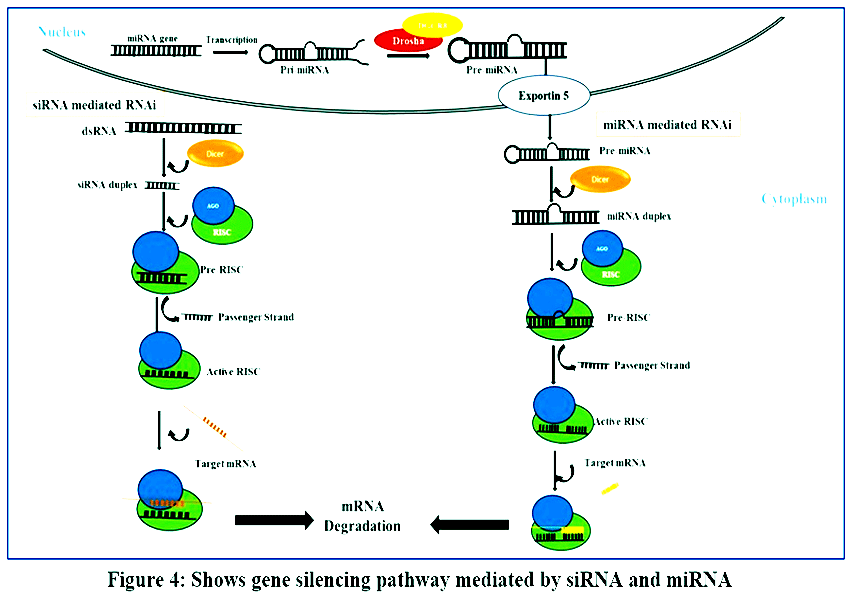 |
Figure 4: Shows gene silencing pathway mediated by siRNA and miRNA
|
Table 2: Summary of Key Experimental tools and their limitations in Cancer Research
| Category | Tools/Techniques | Application | Advantages | Limitations |
| 2D Culture | Monolayer cell lines | Drug screening, functional assays | Easy, quick growth, reproducible, low cost | Less accuracy, Poor physiological relevance3,8 |
| Cellular assays | MTT, Brdu, wound healing | Analyze cell metabolic viability, proliferation and migration. | Functional characterization | Prone to experimental errors12 |
| 3D Culture | Spheroids, organoids | Tumor modeling, drug response, monitoring signalling pathways. | Mimics TME, heterogeneity | Limited functional blood vessels and oxygen deprivation35,46,47,50,56 |
| Gene Editing | CRISPR-Cas9 | Gene knockout/knock-in | High precision, scalable | Off-target effects81,84 |
| RNAi | siRNA/shRNA | Gene silencing using Dicer, RISC mechanisms to degrade mRNA. | Transient modulation | Delivery challenges101,104,111,113 |
Application of genetic tools in organoid systems
Organoids are 3D in vitro cultures made from stem or progenitorcells that have an inherent property to replicate a wide range of cell types.Spheroids and organoids are used by researchers to more closely mimic the in vivo condition, going beyond conventional flat cultures. By integrating organoids with cutting-edge genetic engineering techniques, researchers can replicate and analyse complex human diseases.121,122 This process requires consideration of the method of delivering these genetic tools into the target cells. They could be delivered with a non-viral approach (lipofection or electroporation), or with a viral approach (retrovirus, lentivirus, or adenovirus). Genetic engineering tools such as CRISPR/Cas, RNAi or transposase have been implemented to organoid thereby enhancing disease modelling and enabling developmental research. In organoid genetics, these various methods of genetic engineering are used in organoids to impart required modification on coding regions of genomic sequence.123 Most of the research on organoid genetics has concentrated on improving the knowledge of brain disorders. CRISPR-Cas9-mediated editing can replicate the phenotypes of major neurodegenerative disorders, such as Alzheimer’s and Parkinson’s, in a three-dimensional organoid environment.121
Conclusion
The landscape of oncology research has undergone an integrative transformation driven by the cross-disciplinary fusion of advanced in vitro culture platforms and next-generation genetic engineering technologies. While traditional 2D culture environments and their associated functional assays remain widely utilized due to their high throughput, reproducibility, and simplicity, they fail to replicate the complex architectural, mechanical, and cellular heterogeneities of human solid tumors. The transition to 3D platforms, specifically multicellular spheroids and patient-derived organoids, successfully addresses these shortcomings by capturing the physical oxygen gradients and true tissue architectures characteristic of the tumor microenvironment. Furthermore, the integration of cutting-edge multi-barrel 3D bioprinting and organ-on-a-chip technologies has paved the way for functional tumor vascularization and micro perfusion modeling. These specialized 3D structures, along with advanced spatial transcriptomics techniques, enable high-resolution mapping of the structural organization and transcriptomic landscapes of evolving cancer ecosystems. In parallel, genome editing has expanded far beyond standard wild-type Cas9 protocols to provide unmatched multi-locus manipulation with minimized off-target effects, and the implementation of specialized Cas12a arrays facilitates highly efficient engineering, while cytidine and adenine base editors enable single-nucleotide precision changes without the cytotoxicity of double-strand DNA breaks. Concurrently, RNA interference strategies via optimized delivery of siRNAs and miRNAs remain indispensable tools for target validation and the sequence-specific silencing of oncogenic pathways. Thus, as discussed in this review, different models offer advantages for cancer research. The 2D culture model is simple, reproducible, and cost-effective, whereas 3D culture models can better mimic the tumour microenvironment. The CRISPR-Cas9 and RNAi provide the researcher with the ability to investigate the gene function. Therefore, combining these approaches with appropriate validation can provide more understanding of cancer biology. Ultimately, these basic as well as advanced techniques act as platforms that accelerate the development of clinically viable, personalized cancer therapeutics and robust drug-screening pipelines.
Acknowledgement
The authors gratefully acknowledge the support provided by the Principal, faculty members, and students of Government Arts College, Trivandrum, and the Executive Director, staff, and students of CEPCI, Kollam. DP gratefully acknowledges Karpagam Academy of Higher Education for the facilities.
Funding Sources
The author(s) received no financial support for the research, authorship, and/or publication of this article.
Conflict of interest
The authors do not have any conflict of interest.
Data Availability Statement
This statement does not apply to this article.
Ethics Statement
This research did not involve human participants, animal subjects, or any material that requires ethical approval.
Informed Consent Statement
This study did not involve human participants, and therefore, informed consent was not required.
Clinical Trial Registration
This research does not involve any clinical trials.
Permission to reproduce material from other sources
Not Applicable.
Author Contributions:
- Dhanya Krishnan: Writing- Original Draft, Review & Editing
- Gowri Priya Jayasree: Writing- Original Draft, Review & Editing
- Sneha Ajilal: Writing- Original Draft, Review & Editing
- Anusha Baby Satravada: Writing- Original Draft, Review & Editing
- Dhamodharan Prabhu: Writing- Original Draft, Supervision, Review & Editing
- Ramya Ramachandran Prabhu: Conceptualization, Supervision, Review and Editing
References
- Bray F, Laversanne M, Sung H, et al. Global cancer statistics 2022: GLOBOCAN estimates of incidence and mortality worldwide for 36 cancers in 185 countries. CA A Cancer J Clinicians. 2024;74(3):229-263. https://doi.org/10.3322/caac.21834
CrossRef - Sung H, Ferlay J, Siegel RL, et al. Global Cancer Statistics 2020: GLOBOCAN Estimates of Incidence and Mortality Worldwide for 36 Cancers in 185 Countries. CA A Cancer J Clin. 2021;71(3):209-249. https://doi.org/10.3322/caac.21660
CrossRef - Zhou Y. Understanding the cancer/tumour biology from 2D to 3D. J Thorac Dis. 2016;8(11):E1484-E1486. https://doi.org/10.21037/jtd.2016.11.54
CrossRef - Gunti S, Hoke ATK, Vu KP, London NR. Organoid and Spheroid Tumour Models: Techniques and Applications. Cancers. 2021;13(4):874. https://doi.org/10.3390/cancers13040874
CrossRef - Kopeina GS, Senichkin VV, Zhivotovsky B. Caloric restriction – A promising anti-cancer approach: From molecular mechanisms to clinical trials. Biochimica et Biophysica Acta (BBA) – Reviews on Cancer. 2017;1867(1):29-41. https://doi.org/10.1016/j.bbcan.2016. 11.002
CrossRef - Sharma AK, Giri AK. Engineering CRISPR/Cas9 therapeutics for cancer precision medicine. Front Genet. 2024;15:1309175. https://doi.org/10.3389/fgene.2024.1309175
CrossRef - Rehman SU, Ullah N, Zhang Z, et al. Recent insights into the functions and mechanisms of antisense RNA: emerging applications in cancer therapy and precision medicine. Front Chem. 2024;11:1335330. https://doi.org/10.3389/fchem.2023.1335330
CrossRef - Chaicharoenaudomrung N, Kunhorm P, Noisa P. Three-dimensional cell culture systems as an in vitro platform for cancer and stem cell modeling. WJSC. 2019;11(12):1065-1083. http://dx.doi.org/10.4252/wjsc.v11.i12.1065
CrossRef - Alberts B, ed. Molecular Biology of the Cell. 4th ed. Garland Science; 2002.
- Duval K, Grover H, Han LH, et al. Modeling Physiological Events in 2D vs. 3D Cell Culture. Physiology. 2017;32(4):266-277. https://doi.org/10.1152/physiol.00036.2016
CrossRef - Yao T, Asayama Y. Animal‐cell culture media: History, characteristics, and current issues. Reprod Medicine & Biology. 2017;16(2):99-117. https://doi.org/10.1002/rmb2.12024
CrossRef - Ciapetti G, Cenni E, Pratelli L, Pizzoferrato A. In vitro evaluation of cell/biomaterial interaction by MTT assay. Biomaterials. 1993;14(5):359-364.https://doi.org/10.1016/0142-9612(93)90055-7
CrossRef - Bahuguna A, Khan I, Bajpai VK, Kang SC. MTT assay to evaluate the cytotoxic potential of a drug. Bangladesh J Pharmacol. 2017;12(2).https://doi.org/10.3329/bjp.v12i2.30892
CrossRef - Ahmadian S, Barar J, Saei AA, Fakhree MAA, Omidi Y. Cellular Toxicity of Nanogenomedicine in MCF-7 Cell Line: MTT assay. JoVE. 2009;(26):1191.https://dx.doi.org/10.3791/1191
CrossRef - Stockert JC, Horobin RW, Colombo LL, Blázquez-Castro A. Tetrazolium salts and formazan products in Cell Biology: Viability assessment, fluorescence imaging, and labeling perspectives. Acta Histochemica. 2018;120(3):159-167. https://doi.org/10.1016/ j.acthis.2018.02.005
CrossRef - Zitko J, Mindlová A, Valášek O, et al. Design, Synthesis and Evaluation of N-pyrazinylbenzamides as Potential Antimycobacterial Agents. Molecules. 2018;23(9):2390.https://doi.org/10.3390/molecules23092390
CrossRef - Patel N, Oudemans PV, Hillman BI, Kobayashi DY. Use of the tetrazolium salt MTT to measure cell viability effects of the bacterial antagonist Lysobacter enzymogenes on the filamentous fungus Cryphonectria parasitica. Antonie van Leeuwenhoek. 2013;103(6):1271-1280. https://doi.org/10.1007/s10482-013-9907-3
CrossRef - Adan A, Kiraz Y, Baran Y. Cell Proliferation and Cytotoxicity Assays. CPB. 2016;17(14):1213-1221. https://doi.org/10.2174/1389201017 666160808160513
CrossRef - Pijuan J, Barceló C, Moreno DF, et al. In vitro Cell Migration, Invasion, and Adhesion Assays: From Cell Imaging to Data Analysis. Front Cell Dev Biol. 2019;7:107. https://doi.org/10.3389/fcell.2019.00107
CrossRef - Justus CR, Leffler N, Ruiz-Echevarria M, Yang LV. In vitro Cell Migration and Invasion Assays. JoVE. 2014;(88):51046. https://dx.doi.org/10.3791/51046
CrossRef - Conboy JP, Istúriz Petitjean I, Van Der Net A, Koenderink GH. How cytoskeletal crosstalk makes cells move: Bridging cell-free and cell studies. Biophysics Reviews. 2024;5(2):021307. https://doi.org/10.1063/5.0198119
CrossRef - Bucher N, Britten CD. G2 checkpoint abrogation and checkpoint kinase-1 targeting in the treatment of cancer. Br J Cancer. 2008;98(3):523-528. https://doi.org/10.1038/sj.bjc.6604208
CrossRef - Rothaeusler K, Baumgarth N. Assessment of Cell Proliferation by 5‐Bromodeoxyuridine (BrdU) Labeling for Multicolor Flow Cytometry. CP Cytometry. 2007;40(1). https://doi.org/10.1002/0471142956.cy0731s40
CrossRef - Pozarowski P, Darzynkiewicz Z. Analysis of Cell Cycle by Flow Cytometry. In: Checkpoint Controls and Cancer. Vol 281. Humana Press; 2004:301-312. https://doi.org/10.1385/1-59259-811-0:301
CrossRef - Filograna R, Civiero L, Ferrari V, et al. Analysis of the Catecholaminergic Phenotype in Human SH-SY5Y and BE(2)-M17 Neuroblastoma Cell Lines upon Differentiation. Castresana JS, ed. PLoS ONE. 2015;10(8):e0136769. https://doi.org/10.1371/ journal. pone.0136769
CrossRef - Galati DF, Asai DJ. Immunofluorescence Microscopy. Current Protocols. 2023;3(8):e842. https://doi.org/10.1002/cpz1.842
CrossRef - Im K, Mareninov S, Diaz MFP, Yong WH. An Introduction to Performing Immunofluorescence Staining. In: Yong WH, ed. Biobanking. Vol 1897. Methods in Molecular Biology. Springer New York; 2019:299-311.https://doi.org/10.1007/978-1-4939-8935-5_26
CrossRef - Vermeulen K, Van Bockstaele DR, Berneman ZN. The cell cycle: a review of regulation, deregulation and therapeutic targets in cancer. Cell Proliferation. 2003;36(3):131-149.https://doi.org/10.1046/j.1365-2184.2003.00266.x
CrossRef - Jan R, Chaudhry G e S. Understanding Apoptosis and Apoptotic Pathways Targeted Cancer Therapeutics. Adv Pharm Bull. 2019;9(2):205-218. https://doi.org/10.15171/apb.2019.024
CrossRef - Luo T, Yu S, Ouyang J, et al. Identification of a apoptosis-related LncRNA signature to improve prognosis prediction and immunotherapy response in lung adenocarcinoma patients. Front Genet. 2022;13:946939. https://doi.org/10.3389/fgene.2022. 946939
CrossRef - Gousias K, Theocharous T, Simon M. Mechanisms of Cell Cycle Arrest and Apoptosis in Glioblastoma. Biomedicines. 2022;10(3):564. https://doi.org/10.3390/biomedicines10030564
CrossRef - Zhra M, Qasem RJ, Aldossari F, Saleem R, Aljada A. A Comprehensive Exploration of Caspase Detection Methods: From Classical Approaches to Cutting-Edge Innovations. IJMS. 2024;25(10):5460.https://doi.org/10.3390/ijms25105460
CrossRef - Oosterhuis GJE, Mulder AB, Kalsbeek-Batenburg E, Lambalk CB, Schoemaker J, Vermes I. Measuring apoptosis in human spermatozoa: a biological assay for semen quality? Fertility and Sterility. 2000;74(2):245-250. https://doi.org/10.1016/S0015-0282(00)00623-3
CrossRef - Liu K, Liu P cheng, Liu R, Wu X. Dual AO/EB Staining to Detect Apoptosis in Osteosarcoma Cells Compared with Flow Cytometry. Med Sci Monit Basic Res. 2015;21:15-20.1.https://doi.org/10.12659/MSMBR.89332
CrossRef - Zhang L, Liao W, Chen S, et al. Towards a New 3Rs Era in the construction of 3D cell culture models simulating tumor microenvironment. Front Oncol. 2023;13:1146477.https://doi.org/10.3389/fonc.2023.1146477
CrossRef - Nunes AS, Barros AS, Costa EC, Moreira AF, Correia IJ. 3D tumor spheroids as in vitro models to mimic in vivo human solid tumors resistance to therapeutic drugs. Biotech & Bioengineering. 2019;116(1):206-226.https://doi.org/10.1002/bit.26845
CrossRef - Braccini S, Tacchini C, Chiellini F, Puppi D. Polymeric Hydrogels for In Vitro 3D Ovarian Cancer Modeling. IJMS. 2022;23(6):3265.https://doi.org/10.3390/ijms23063265
CrossRef - Jeong SH, Hong HS, Lee EH, Kwak JJ, Lee JY. Analysis of RAS mutation in thyroid nodular hyperplasia and follicular neoplasm in a Korean population. Endocrino Diabet & Metabol. 2018;1(4):e00040.https://doi.org/10.1002/edm2.40
CrossRef - Ware MJ, Colbert K, Keshishian V, et al. Generation of Homogenous Three-Dimensional Pancreatic Cancer Cell Spheroids Using an Improved Hanging Drop Technique. Tissue Engineering Part C: Methods. 2016;22(4):312-321. https://doi.org/10.1089/ten.tec. 2015.0280
CrossRef - Sart S, Tomasi RFX, Amselem G, Baroud CN. Multiscale cytometry and regulation of 3D cell cultures on a chip. Nat Commun. 2017;8(1):469.https://doi.org/10.1038/s41467-017-00475-x
CrossRef - Pawlik TM, Souba WW, Sweeney TJ, Bode BP. Amino Acid Uptake and Regulation in Multicellular Hepatoma Spheroids. Journal of Surgical Research. 2000;91(1):15-25.https://doi.org/10.1006/jsre.2000.5888
CrossRef - Yakavets I, Yankovsky I, Millard M, et al. The alteration of temoporfin distribution in multicellular tumor spheroids by β-cyclodextrins. International Journal of Pharmaceutics. 2017;529(1-2):568-575. https://doi.org/10.1016/j.ijpharm.2017.07.037
CrossRef - Mandeda Madaiah P, Ghosh RN, Namboothiri PK, Peter M. Advancement in Scaffold-Based 3D Cell Culture Models for Osteosarcoma Drug Screening. ACS Biomater Sci Eng. 2025;11(11):6426-6442. https://doi.org/10.1021/acsbiomaterials.5c01174
CrossRef - Xiao W, Wang S, Zhang R, et al. Bioengineered scaffolds for 3D culture demonstrate extracellular matrix-mediated mechanisms of chemotherapy resistance in glioblastoma. Matrix Biology. 2020;85-86:128-146. https://doi.org/10.1016/j.matbio.2019.04.003
CrossRef - Kalla J, Pfneissl J, Mair T, Tran L, Egger G. A systematic review on the culture methods and applications of 3D tumoroids for cancer research and personalized medicine. Cell Oncol. 2025;48(1):1-26.https://doi.org/10.1007/s13402-024-00960-8
CrossRef - Lee JH, Kim SK, Khawar IA, Jeong SY, Chung S, Kuh HJ. Microfluidic co-culture of pancreatic tumor spheroids with stellate cells as a novel 3D model for investigation of stroma-mediated cell motility and drug resistance. J Exp Clin Cancer Res. 2018;37(1):4. https://doi.org/10.1186/s13046-017-0654-6
CrossRef - Achilli TM, Meyer J, Morgan JR. Advances in the formation, use and understanding of multi-cellular spheroids. Expert Opinion on Biological Therapy. 2012;12(10):1347-1360. https://doi.org/10.1517/14712598.2012.707181
CrossRef - Bassi G, Grimaudo MA, Panseri S, Montesi M. Advanced Multi-Dimensional Cellular Models as Emerging Reality to Reproduce In Vitro the Human Body Complexity. IJMS. 2021;22(3):1195. https://doi.org/10.3390/ijms22031195
CrossRef - Bicer M, Cottrell GS, Widera D. Impact of 3D cell culture on bone regeneration potential of mesenchymal stromal cells. Stem Cell Res Ther. 2021;12(1):31. https://doi.org/10.1186/s13287-020-02094-8
CrossRef - Yang S, Hu H, Kung H, et al. Organoids: The current status and biomedical applications. MedComm. 2023;4(3):e274. https://doi.org/10.1002/mco2.274
CrossRef - Jubelin C, Muñoz-Garcia J, Griscom L, et al. Three-dimensional in vitro culture models in oncology research. Cell Biosci. 2022;12(1):155. https://doi.org/10.1186/s13578-022-00887-3
CrossRef - De Jongh D, Massey EK, the VANGUARD consortium, et al. Organoids: a systematic review of ethical issues. Stem Cell Res Ther. 2022;13(1):337. https://doi.org/10.1186/s13287-022-02950-9
CrossRef - Salinas-Vera YM, Valdés J, Pérez-Navarro Y, et al. Three-Dimensional 3D Culture Models in Gynecological and Breast Cancer Research. Front Oncol. 2022;12:826113. https://doi.org/10.3389/fonc.2022.826113
CrossRef - Shankaran A, Prasad K, Chaudhari S, Brand A, Satyamoorthy K. Advances in development and application of human organoids. 3 Biotech. 2021;11(6):257. https://doi.org/10.1007/s13205-021-02815-7
CrossRef - Lehmann R, Lee CM, Shugart EC, et al. Human organoids: a new dimension in cell biology. Drubin DG, ed. MBoC. 2019;30(10):1129-1137. https://doi.org/10.1091/mbc.E19-03-0135
CrossRef - Drost J, Clevers H. Organoids in cancer research. Nat Rev Cancer. 2018;18(7):407-418. https://doi.org/10.1038/s41568-018-0007-6
CrossRef - Plava J, Cehakova M, Kuniakova M, et al. The third dimension of tumor microenvironment—The importance of tumor stroma in 3D cancer models. Exp Biol Med (Maywood). 2023;248(15):1347-1358. https://doi.org/10.1177/15353702231198050
CrossRef - Panez-Toro I, Mountford J, Muñoz-Garcia J, Heymann D. Advancing cancer research through 3D cell culture models. EXCLI J. 2025;24:1061-1099. Published 2025 Aug 28https://doi.org/10.17179/EXCLI2025-8563
- Aggarwal V, Miranda O, Johnston PA, Sant S. Three dimensional engineered models to study hypoxia biology in breast cancer. Cancer Letters. 2020;490:124-142. https://doi.org/10.1016/j.canlet.2020.05.030
CrossRef - Revokatova D, Bikmulina P, Heydari Z, et al. Getting Blood out of a Stone: Vascularization via Spheroids and Organoids in 3D Bioprinting. Cells. 2025;14(9):665. https://doi.org/10.3390/cells14090665
CrossRef - Zhang J, Wehrle E, Rubert M, Müller R. 3D Bioprinting of Human Tissues: Biofabrication, Bioinks, and Bioreactors. IJMS. 2021;22(8):3971. https://doi.org/10.3390/ijms22083971
CrossRef - Blandino G, Satchi-Fainaro R, Tinhofer I, et al. Cancer Organoids as reliable disease models to drive clinical development of novel therapies. J Exp Clin Cancer Res. 2024;43(1):334. https://doi.org/10.1186/s13046-024-03258-7
CrossRef - Lee JM, Sing SL, Zhou M, Yeong WY. 3D bioprinting processes: A perspective on classification and terminology. IJB. 2024;4(2):151. https://doi.org/10.18063/ijb.v4i2.151
CrossRef - Mallya D, Gadre MA, Varadharajan S, Vasanthan KS. 3D bioprinting for the construction of drug testing models-development strategies and regulatory concerns. Front Bioeng Biotechnol. 2025;13:1457872. https://doi.org/10.3389/fbioe.2025.1457872
CrossRef - Dababneh AB, Ozbolat IT. Bioprinting Technology: A Current State-of-the-Art Review. Journal of Manufacturing Science and Engineering. 2014;136(6):061016. https://doi.org/10.1115/1.4028512
CrossRef - He Y, Gu Z, Xie M, Fu J, Lin H. Why choose 3D bioprinting? Part II: methods and bioprinters. Bio-des Manuf. 2020;3(1):1-4. https://doi.org/10.1007/s42242-020-00064-w
CrossRef - Shirure VS, George SC. Design considerations to minimize the impact of drug absorption in polymer-based organ-on-a-chip platforms. Lab Chip. 2017;17(4):681-690. https://doi.org/10.1039/C6LC01401A
CrossRef - Yi HG, Jeong YH, Kim Y, et al. A bioprinted human-glioblastoma-on-a-chip for the identification of patient-specific responses to chemoradiotherapy. Nat Biomed Eng. 2019;3(7):509-519. https://doi.org/10.1038/s41551-019-0363-x
CrossRef - Wang X, Luo Y, Ma Y, Wang P, Yao R. Converging bioprinting and organoids to better recapitulate the tumor microenvironment. Trends Biotechnol. 2024;42(5):648-663. https://doi.org/10.1016/j.tibtech.2023.11.006
CrossRef - Hwangbo H, Chae S, Kim W, Jo S, Kim GH. Tumor-on-a-chip models combined with mini-tissues or organoids for engineering tumor tissues. Theranostics. 2024;14(1):33-55. https://doi.org/10.7150/thno.90093
CrossRef - Nielsen BS, Madsen NH, Larsen J, Skandorff I, Gad M, Holmstrøm K. Architectural organization and molecular profiling of 3D cancer heterospheroids and their application in drug testing. Front Oncol. 2024;14:1386097. https://doi.org/10.3389/fonc.2024.1386097
CrossRef - Heo YJ, Hwa C, Lee GH, Park JM, An JY. Integrative Multi-Omics Approaches in Cancer Research: From Biological Networks to Clinical Subtypes. Molecules and Cells. 2021;44(7):433-443. https://doi.org/10.14348/molcells.2021.0042
CrossRef - Smirnov A, Melino G, Candi E. Gene expression in organoids: an expanding horizon. Biol Direct. 2023;18(1):11. https://doi.org/10.1186/s13062-023-00360-2
CrossRef - Asp M, Bergenstråhle J, Lundeberg J. Spatially Resolved Transcriptomes—Next Generation Tools for Tissue Exploration. BioEssays. 2020;42(10):1900221. https://doi.org/10.1002/bies.201900221
CrossRef - Rao A, Barkley D, França GS, Yanai I. Exploring tissue architecture using spatial transcriptomics. Nature. 2021;596(7871):211-220. https://doi.org/10.1038/s41586-021-03634-9
CrossRef - Moses L, Pachter L. Museum of spatial transcriptomics. Nat Methods. 2022;19(5):534-546.https://doi.org/10.1038/s41592-022-01409-2
CrossRef - Cao J, Li C, Cui Z, et al. Spatial Transcriptomics: A Powerful Tool in Disease Understanding and Drug Discovery. Theranostics. 2024;14(7):2946-2968. https://doi.org/10.7150/thno.95908
CrossRef - Soncin F. Advancing comparative placentation through spatial transcriptomics and organoid models. PLoS Biol. 2025;23(8):e3003346. https://doi.org/10.1371/journal.pbio.3003346
CrossRef - Pirenne S, Manzano-Núñez F, Loriot A, et al. Spatial transcriptomics profiling of gallbladder adenocarcinoma: a detailed two-case study of progression from precursor lesions to cancer. BMC Cancer. 2024;24(1):1025. https://doi.org/10.1186/s12885-024-12770-0
CrossRef - Ma S, Wang W, Zhou J, et al. Lamination-based organoid spatially resolved transcriptomics technique for primary lung and liver organoid characterization. Proceedings of the National Academy of Sciences. 2024;121(46):e2408939121. https://doi.org/10.1073/pnas.2408939121
CrossRef - Doudna JA, Charpentier E. The new frontier of genome engineering with CRISPR-Cas9. Science. 2014;346(6213):1258096. https://doi.org/10.1126/science.1258096
CrossRef - Zhang F, Wen Y, Guo X. CRISPR/Cas9 for genome editing: progress, implications and challenges. Human Molecular Genetics. 2014;23(R1):R40-R46. https://doi.org/10.1093/hmg/ddu125
CrossRef - Redman M, King A, Watson C, King D. What is CRISPR/Cas9? Arch Dis Child Educ Pract Ed. 2016;101(4):213-215. https://doi.org/10.1136/archdischild-2016-310459
CrossRef - Ansori ANm, Antonius Y, Susilo RJk, et al. Application of CRISPR-Cas9 genome editing technology in various fields: A review. Narra J. 2023;3(2):e184. https://doi.org/10.52225/narra.v3i2.184
CrossRef - Martinez-Lage M, Torres-Ruiz R, Rodriguez-Perales S. CRISPR/Cas9 Technology: Applications and Human Disease Modeling. In: Progress in Molecular Biology and Translational Science. Vol 152. Elsevier; 2017:23-48.https://doi.org/10.1093/bfgp/elw025
CrossRef - Rodríguez-Rodríguez DR, Ramírez-Solís R, Garza-Elizondo MA, Garza-Rodríguez ML, Barrera-Saldaña HA. Genome editing: A perspective on the application of CRISPR/Cas9 to study human diseases (Review). Int J Mol Med. 2019;43(4):1559-1574. https://doi.org/10.3892/ijmm.2019.4112
CrossRef - Ding D, Chen K, Chen Y, Li H, Xie K. Engineering Introns to Express RNA Guides for Cas9- and Cpf1-Mediated Multiplex Genome Editing. Molecular Plant. 2018;11(4):542-552.https://doi.org/10.1016/j.molp.2018.02.005
CrossRef - Mengstie MA, Wondimu BZ. Mechanism and Applications of CRISPR/Cas-9-Mediated Genome Editing. BTT. 2021;Volume 15:353-361.https://doi.org/10.2147/BTT.S326422
CrossRef - Huang CH, Lee KC, Doudna JA. Applications of CRISPR-Cas Enzymes in Cancer Therapeutics and Detection. Trends in Cancer. 2018;4(7):499-512.https://doi.org/10.1016/j.trecan.2018.05.006
CrossRef - Huang YY, Zhang XY, Zhu P, Ji L. Development of clustered regularly interspaced short palindromic repeats/CRISPR-associated technology for potential clinical applications. WJCC. 2022;10(18):5934-5945. https://doi.org/10.12998/wjcc.v10.i18.5934
CrossRef - Davis DJ, Yeddula SGR. CRISPR Advancements for Human Health. Mo Med. 2024;121(2):170-176.
- Stadtmauer EA, Fraietta JA, Davis MM, et al. CRISPR-engineered T cells in patients with refractory cancer. Science. 2020;367(6481):eaba7365. https://doi.org/10.1126/science.aba7365
CrossRef - Wang S, Xiong Y, Luo Y, et al. Genome-wide CRISPR screens identify PKMYT1 as a therapeutic target in pancreatic ductal adenocarcinoma. EMBO Mol Med. 2024;16(5):1115-1142. https://doi.org/10.1038/s44321-024-00060-y
CrossRef - Zhang B, Chen Y, Chen X, et al. Genome-wide CRISPR screen identifies ESPL1 limits the response of gastric cancer cells to apatinib. Cancer Cell Int. 2024;24(1):83.https://doi.org/10.1186/s12935-024-03233-4
CrossRef - Ran FA, Hsu PD, Wright J, Agarwala V, Scott DA, Zhang F. Genome engineering using the CRISPR-Cas9 system. Nat Protoc. 2013;8(11):2281-2308. https://doi.org/10.1038/nprot.2013.143
CrossRef - Zetsche B, Gootenberg JS, Abudayyeh OO, et al. Cpf1 is a single RNA-guided endonuclease of a class 2 CRISPR-Cas system. Cell. 2015;163(3):759-771.https://doi.org/10.1016/j.cell.2015.09.038
CrossRef - Tang K, Zhou L, Tian X, et al. Cas12a-knock-in mice for multiplexed genome editing, disease modelling and immune-cell engineering. Nat Biomed Eng. 2025;9(8):1290-1308. https://doi.org/10.1038/s41551-025-01371-2
CrossRef - Hebert JD, Xu H, Tang YJ, et al. Efficient and multiplexed somatic genome editing with Cas12a mice. Nat Biomed Eng. 2025;9(11):1982-1997.https://doi.org/10.1038/s41551-025-01407-7
CrossRef - Komor AC, Kim YB, Packer MS, Zuris JA, Liu DR. Programmable editing of a target base in genomic DNA without double-stranded DNA cleavage. Nature. 2016;533(7603):420-424. https://doi.org/10.1038/nature17946
CrossRef - Schweitzer AY, Adams EW, Nguyen MTA, Lek M, Isaacs FJ. Precision multiplexed base editing in human cells using Cas12a-derived base editors. Nat Commun. 2025;16(1):5061. https://doi.org/10.1038/s41467-025-59653-x
CrossRef - Cabré-Romans JJ, Cuella-Martin R. CRISPR-dependent base editing as a therapeutic strategy for rare monogenic disorders. Front Genome Ed. 2025;7:1553590. https://doi.org/10.3389/fgeed.2025.1553590
CrossRef - Cetin B, Erendor F, Eksi YE, Sanlioglu AD, Sanlioglu S. Advancing CRISPR genome editing into gene therapy clinical trials: progress and future prospects. Expert Rev Mol Med. 2025;27:e16.https://doi.org/10.1017/erm.2025.10
CrossRef - Wang Y, Jiang H, Li M, et al. Delivery of CRISPR/Cas9 system by AAV as vectors for gene therapy. Gene. 2024;927:148733. https://doi.org/10.1016/j.gene.2024.148733
CrossRef - Álvarez-Pérez JC, Sanjuán-Hidalgo J, Arenas AM, et al. High-fidelity Cas9-mediated targeting of KRAS driver mutations restrains lung cancer in preclinical models. Nat Commun. 2025;16(1):7080. https://doi.org/10.1038/s41467-025-62350-4
CrossRef - Li X, Zou L. BRCAness, DNA gaps, and gain and loss of PARP inhibitor–induced synthetic lethality. J Clin Invest. 2024;134(14). https://doi.org/10.1172/JCI181062
CrossRef - Cuciniello R, Filosa S, Crispi S. Novel approaches in cancer treatment: preclinical and clinical development of small non-coding RNA therapeutics. J Exp Clin Cancer Res. 2021;40(1):383. https://doi.org/10.1186/s13046-021-02193-1
CrossRef - Mansoori B, Sandoghchian Shotorbani S, Baradaran B. RNA interference and its role in cancer therapy. Adv Pharm Bull. 2014;4(4):313-321. https://doi.org/10.5681/APB.2014.046
- Xin Y, Huang M, Guo WW, Huang Q, Zhang LZ, Jiang G. Nano-based delivery of RNAi in cancer therapy. Mol Cancer. 2017;16(1):134. https://doi.org/10.1186/s12943-017-0683-y
CrossRef - Kang H, Ga YJ, Kim SH, et al. Small interfering RNA (siRNA)-based therapeutic applications against viruses: principles, potential, and challenges. J Biomed Sci. 2023;30(1):88. https://doi.org/10.1186/s12929-023-00981-9
CrossRef - Manasa V, Kannan S. Impact of microRNA dynamics on cancer hallmarks: An oral cancer scenario. Tumour Biol. 2017;39(3):101042831769592. https://doi.org/10.1177/1010428317695920
CrossRef - O’Brien J, Hayder H, Zayed Y, Peng C. Overview of MicroRNA Biogenesis, Mechanisms of Actions, and Circulation. Front Endocrinol. 2018;9:402. https://doi.org/10.3389/fendo.2018.00402
CrossRef - Yan C, Zhang J, Huang M, et al. Design, strategies, and therapeutics in nanoparticle-based siRNA delivery systems for breast cancer. J Mater Chem B. 2023;11(34):8096-8116. https://doi.org/10.1039/D3TB00278K
CrossRef - Bavishi L, Butani SB, Patel SS. Small interfering RNA (siRNA) based therapies for cancer treatment: A special focus on cervical cancer. European Journal of Pharmacology. 2025;1007:178198. https://doi.org/10.1016/j.ejphar.2025.178198
CrossRef - Tian Z, Liang G, Cui K, et al. Insight Into the Prospects for RNAi Therapy of Cancer. Front Pharmacol. 2021;12:644718. https://doi.org/10.3389/fphar.2021.644718
CrossRef - Sung K, Jung Y, Kim N, et al. A rational engineering strategy for structural dynamics modulation enables target specificity enhancement of the Cas9 nuclease. Nucleic Acids Research. 2025;53(12):gkaf535. https://doi.org/10.1093/nar/gkaf535
CrossRef - Slaymaker IM, Gao L, Zetsche B, Scott DA, Yan WX, Zhang F. Rationally engineered Cas9 nucleases with improved specificity. Science. 2016;351(6268):84-88.https://doi.org/10.1126/science.aad5227
CrossRef - Kleinstiver BP, Pattanayak V, Prew MS, et al. High-fidelity CRISPR–Cas9 nucleases with no detectable genome-wide off-target effects. Nature. 2016;529(7587):490-495. https://doi.org/10.1038/nature16526
CrossRef - Skeens E, Sinha S, Ahsan M, et al. High-fidelity, hyper-accurate, and evolved mutants rewire atomic-level communication in CRISPR-Cas9. Sci Adv. 2024;10(10):eadl1045.https://doi.org/10.1126/sciadv.adl1045
CrossRef - Hu JH, Miller SM, Geurts MH, et al. Evolved Cas9 variants with broad PAM compatibility and high DNA specificity. Nature. 18;556(7699):57-63. https://doi.org/10.1038/nature26155
CrossRef - Lee JK, Jeong E, Lee J, et al. Directed evolution of CRISPR-Cas9 to increase its specificity. Nat Commun. 2018;9(1):3048. https://doi.org/10.1038/s41467-018-05477-x
CrossRef - Gopal S, Rodrigues AL, Dordick JS. Exploiting CRISPR Cas9 in Three-Dimensional Stem Cell Cultures to Model Disease. Front Bioeng Biotechnol. 2020;8:69 2.https://doi.org/10.3389/fbioe.2020.00692
CrossRef - Klompstra TM, Yoon KJ, Koo BK. Evolution of organoid genetics. European Journal of Cell Biology. 2025;104(2):151481.https://doi.org/10.1016/j.ejcb.2025.151481
CrossRef - Teriyapirom I, Batista-Rocha AS, Koo BK. Genetic engineering in organoids. J Mol Med. 2021;99(4):555-568.https://doi.org/10.1007/s00109-020-02029-z
CrossRef
Abbreviations
2D – Two-dimensional
3D – Three-dimensional
CRISPR – Clustered Regularly Interspaced Palindromic Repeats
siRNA – Small interfering RNAs
MTT – (3-(4,5-dimethylthiazol-2-yl)-2,5-diphenyltetrazolium bromide)
BrdU – 5-bromo-2′-deoxyuridine
FUCCI – Fluorescent Ubiquitination-based Cell Cycle Indicator
PI – Propidium Iodide
AO/EtBr – Acridine orange/Ethidium bromide
SK-HEP-1 – Sloan-Kettering Hepatoma-1
PLA – Polylactide
PGA – Polyglycolide
PCL – Polycaprolactone
TME – Tumour Microenvironment
ECM – Extra Cellular Matrix
snRNAseq – Single – nucleus RNA sequencing
NGS – Next Generation Sequencing
gRNA – Guide RNA
crRNA – CRISPR RNA
tracrRNA – Trans-activating CRISPR RNA
KRAS – Kirsten rat sarcoma virus
PAM – Protospacer Adjacent Motif
shRNA – Short hairpin RNA
miRNA – Micro RNA
piRNA – Piwi-interacting RNA
RISC – RNA-Induced Silencing Complex
DGCR8 – Drosha and Digeorge syndrome Critical Region 8
ABEs – Adenine Base Editors
Accepted on: 21-08-2026
Second Review by: Dr. Akshaya Arva
Final Approval by: Dr. Hifzur R. Siddique