Phytochemicals Targeting Inflammatory Signaling Pathways to Combat Gastric Cancer
 , Priti Sharad Khilare, Kishor Vasant Otariand Ajay Yashwant Kale
 Department of Pharmacology, Navsahyadri Institute of Pharmacy, Pune, India
Corresponding Author’s E-mail:aparnabura2000@gmail.com
DOI : http://dx.doi.org/10.13005/bbra/3553
Download this article as:

Gastric cancer remains one of the leading causes of cancer-related mortality worldwide, largely due to delayed diagnosis, complex molecular mechanisms, and resistance to existing therapies. Although treatment options such as surgery, chemotherapy, and targeted therapy are available, their effectiveness is often limited by toxicity, adverse effects, and the development of drug resistance. Therefore, there is a growing need for safer and more effective therapeutic strategies. In recent years, plant-derived phytochemicals have gained considerable attention due to their relatively low toxicity and ability to target multiple molecular pathways. Compounds such as curcumin, resveratrol, epigallocatechin-3-gallate (EGCG), quercetin, berberine, luteolin, apigenin, sulforaphane, and ginsenosides have demonstrated notable anticancer potential, with promising results in preclinical studies and increasing evaluation in clinical settings. These bioactive molecules modulate key signaling pathways, including NF-κB, PI3K/Akt/mTOR, JAK/STAT3, MAPK, and Wnt/β-catenin, thereby suppressing inflammation and regulating oxidative stress, cell proliferation, apoptosis, angiogenesis, and metastasis. Furthermore, these phytochemicals have been reported to enhance the efficacy of conventional chemotherapeutic agents and reduce drug resistance, supporting their role in combination therapy. However, their clinical application remains limited due to poor bioavailability, rapid metabolism, lack of standardized formulations, and insufficient clinical evidence. Advances in nanotechnology-based delivery systems may help overcome these limitations and improve therapeutic outcomes. In conclusion, this review highlights the potential of phytochemicals as multi-targeted, low-toxicity agents in gastric cancer management and emphasizes their promise as adjuncts to current therapies. However, further well-designed clinical studies are essential to establish their efficacy and safety.
KEYWORDS:Apoptosis; Chemosensitization; Gastric cancer; Herbal phytochemicals; Molecular signaling pathways; Oxidative stress; Targeted therapy
Introduction
Gastric cancer is still ranked amongst those cancers that have higher prevalence and are also hazardous globally, making significant contributions to both incidence and mortality associated with cancer, especially in East Asia and developing countries.1 Also, most incidences are diagnosed at an advanced level; therefore, the five-year survival rate is very low, indicating that despite the decrease in stomach cancer prevalence in some countries, its outlook is still poor.2
Gastric cancer grows in stages and is caused by a multitude of factors that include infections, environmental, nutritional, and inherited components. The chief cause of gastric cancer concerns a Helicobacter pylori infection in early stages that triggers a cascade of gastric inflammation, atrophy, intestinal metaplasia, dysplasia, and cancer in a result.3 High dietary intake of sodium, dietary intake of processed and smoked foods, nicotine use, alcohol use, and inherited biological factors also carry secondary roles in pathogenesis.4 Symptoms in the early stages of gastric cancer remain vague or non-specific. But in the secondary stage weight loss, anemia, abdominal pain, and gastrointestinal bleeding are commonly observed in patients with gastric cancer through endoscopy biopsy and TNM criteria used for determination in patients.5
The treatments that are being practiced for gastric cancer are surgical removal, chemotherapy, radiation therapy, target therapy, and immunotherapy. These treatments can be administered individually or in combination, as they are known to confer greater benefits to the patient, though they are toxic and can lead to recurrence and genetic heterogeneity, though it is understood that they are toxic and result in recurrence and genetic heterogeneity.6 Therefore, safer and more effective treatment strategies are increasingly being explored to target multiple oncogenic processes in one treatment.7
Their capability to influence multiple molecular pathways involved in stomach carcinogenesis with very low toxicity has made herbal phytochemicals promising multi-target therapies. Inflammation, oxidative stress, apoptosis, cell cycle progression, angiogenesis, and metastasis are the processes regulated by these bioactive compounds which have both therapeutic and chemopreventive effects.8 Very importantly, it has been documented that in the case of gastric cancer, phytochemicals can influence several dysregulated signaling pathways such as p53-dependent cell cycle control, JAK/STAT3, Wnt/β-catenin, MAPK cascades, NF-κB-mediated inflammatory signaling, and PI3K/Akt/mTOR.9
Furthermore, phytochemicals induce autophagy via Beclin-1 and LC3, activate apoptotic proteins such as Bax and caspases, and promote programmed cell death via novel pathways, including ferroptosis.10 Further, they inhibit angiogenesis by downregulating HIF-1α and VEGF signaling, and they inhibit the epithelial-mesenchymal transition and metastasis by regulating E-cadherin, matrix metalloproteinases, and EMT-associated transcription factors.11 In addition, the influence of the Nrf2/Keap1 pathway on modifying redox homeostasis and regulating epigenetic processes, including microRNAs and long non-coding RNAs, reveals the wide molecular influence of phytochemicals in the biology of gastric cancer.12
The scientific evidence suggests that numerous phytochemicals, including curcumin, resveratrol, EGCG, quercetin, berberine, luteolin, apigenin, kaempferol, sulforaphane, thymoquinone, ginsenosides, honokiol, emodin, and either ursolic or oleanolic acid, have the potential to inhibit gastric cancer by simultaneously targeting multiple signal transduction pathways.13 This provides a scientific basis to assess the use of herbal-derived phytochemicals for their effects on gastric cancer through the molecular mechanisms of action as alternate/adjunctive therapies.14
Methodology: Search Strategy Used to Extract Data for this Review
The present review was conducted using a structured literature search approach to collect relevant scientific evidence regarding herbal phytochemicals targeting inflammatory and oncogenic molecular pathways involved in gastric cancer progression. Electronic databases including PubMed, Scopus, Web of Science, and Google Scholar were systematically searched for articles published up to 2026. The search strategy was developed using combinations of keywords and Boolean operators such as:
“gastric cancer” OR “stomach cancer”
“phytochemicals” OR “plant-derived compounds” OR “natural products”
“inflammatory signaling pathways” OR “molecular pathways” OR “signaling pathways”
“PI3K/Akt/mTOR” OR “NF-κB” OR “MAPK” OR “JAK/STAT3” OR “Wnt/β-catenin”
“apoptosis” OR “autophagy” OR “ferroptosis” OR “oxidative stress” OR “angiogenesis”
“epithelial-mesenchymal transition” OR “EMT” OR “epigenetics”
These terms were combined using AND/OR Boolean operators to retrieve the most relevant studies related to phytochemical-mediated modulation of signaling pathways in gastric cancer.
Inclusion Criteria
- Peer-reviewed research articles and review papers
- Studies focusing on gastric cancer and phytochemicals
- Articles describing molecular mechanisms or signaling pathways
- In vitro, in vivo, and clinical studies
- Articles published in English
Exclusion Criteria
- Non-English publications
- Conference abstracts, editorials, and unpublished data
- Studies not related to gastric cancer or lacking mechanistic insights
Study Selection and Data Extraction
Relevant articles were screened based on title, abstract, and full text. Data were extracted focusing on:
- Type of phytochemical
- Targeted molecular pathways
- Mechanism of action
- Experimental model (in vitro/in vivo/clinical)
Duplicate studies were removed, and only the most relevant and recent studies were included to ensure scientific accuracy and clarity.
Type of Review
This study is a narrative review with a structured literature search approach, summarizing current evidence on phytochemicals targeting molecular pathways in gastric cancer.
Objectives
The purpose of this narrative review is to evaluate the evidence on the chemical constituents of plants/herbs that may regulate biological mechanisms involved in the initiation and progression of gastric cancer. Based upon our findings, we will also be able to identify voids for future clinical research. Our data will provide a clear definition and classification of various types of biological chemicals; an explanation of the molecular mechanisms by which phytochemicals affect biological processes; an evaluation of the possible additive or synergistic effects of phytochemicals and sundry cancer treatments; and identification of any knowledge gaps concerning phytochemicals during the investigation and treatment of cancer.15
Molecular Pathways Associated with Gastric Cancer
The development of gastric cancer is a complex, multistep process driven by the gradual accumulation of genetic and epigenetic alterations that ultimately result in the malignant transformation of gastric epithelial cells.16 In most cases, the disease originates from chronic inflammation, commonly associated with Helicobacter pylori infection, which initiates a well-recognized sequence of pathological changes, including chronic gastritis, mucosal atrophy, intestinal metaplasia, dysplasia, and eventually carcinoma. In addition to infection, environmental and lifestyle factors such as high dietary salt intake, smoking, alcohol consumption, and genetic susceptibility further influence disease progression.17
At the molecular level, gastric cancer involves the dysregulation of several interconnected signaling pathways that collectively contribute to uncontrolled cell proliferation, survival, invasion, and resistance to therapy. Among these, the NF-κB signaling pathway is particularly important in sustaining a chronic inflammatory microenvironment. Its persistent activation promotes the expression of pro-inflammatory mediators such as TNF-α, IL-6, COX-2, and iNOS, which in turn support tumor growth and immune evasion.18
The PI3K/Akt/mTOR pathway is another key regulator frequently activated in gastric cancer. It plays a central role in controlling cellular metabolism, protein synthesis, and survival. When aberrantly activated, it enhances tumor growth, suppresses apoptosis, and contributes to therapeutic resistance.19
In addition, the JAK/STAT3 signaling pathway remains constitutively active in many gastric tumors and promotes the transcription of genes associated with proliferation, angiogenesis, and immune modulation. The Wnt/β-catenin pathway further contributes to epithelial-mesenchymal transition (EMT), stemness, and metastatic behavior. Similarly, MAPK signaling (including ERK, JNK, and p38) regulates cellular responses to growth signals and stress, often interacting with other pathways such as PI3K/Akt to reinforce tumor progression.20
Alterations in tumor suppressor pathways also play a critical role. Loss or mutation of p53 disrupts normal cell cycle control and apoptotic mechanisms, allowing abnormal cells to survive and proliferate. Furthermore, emerging pathways such as Nrf2/Keap1 are involved in maintaining redox balance, enabling cancer cells to adapt to oxidative stress and develop resistance to therapy.21
It is important to note that these pathways do not function independently but are interconnected through complex crosstalk, which amplifies oncogenic signaling and makes therapeutic targeting more challenging. Therefore, strategies that simultaneously target multiple pathways may offer greater therapeutic benefit. In this context, herbal phytochemicals have attracted attention due to their ability to modulate several signaling networks at once. Among the various pathways involved, PI3K/Akt/mTOR and NF-κB appear to play particularly dominant roles, as they regulate key processes such as proliferation, survival, and inflammation in gastric cancer. The interconnected oncogenic signaling pathways involved in gastric cancer progression and their multi-target modulation by herbal phytochemicals are illustrated in Figure 1.
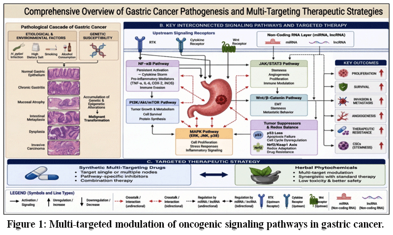 |
Figure 1: Multi-targeted modulation of oncogenic signaling pathways in gastric cancer.
|
Herbal Phytochemicals Targeting the Signaling Pathways
Pi3k/Akt/Mtor Pathway
The phosphoinositide 3-kinase (PI3K)/Akt/mTOR pathway plays a central role in the progression of gastric cancer by regulating key cellular processes such as proliferation, metabolism, survival, and resistance to apoptosis. In many cases, this pathway becomes persistently activated due to upstream receptor tyrosine kinase signaling, mutations in PI3K, or loss of tumor suppressors like PTEN. As a result, Akt remains continuously phosphorylated, leading to sustained activation of mTOR and downstream signaling events that support tumor growth and survival. Akt-mediated activation of Ras homolog enriched in brain (Rheb), a critical upstream regulator of mTOR, further enhances downstream signaling. Activated mTOR subsequently regulates effectors such as ribosomal protein S6 kinase (S6K) and eukaryotic translation initiation factor 4E-binding protein 1 (4E-BP1), thereby promoting protein synthesis, metabolic activity, and gastric cancer cell survival. These alterations not only drive uncontrolled cellular proliferation but also allow cancer cells to adapt to metabolic stress and evade therapeutic interventions.22 (As illustrated in Figure 2.)
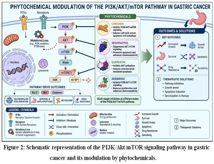 |
Figure 2: Schematic representation of the PI3K/Akt/mTOR signaling pathway in gastric cancer and its modulation by phytochemicals.
|
In this context, natural phytochemicals have attracted increasing attention because of their ability to interfere with multiple components of this pathway rather than acting on a single molecular target. A substantial portion of the available evidence comes from in vitro studies using gastric cancer cell lines and in vivo experiments in animal or xenograft models, where these compounds consistently reduce PI3K/Akt signaling activity. This multi-target behavior is particularly relevant in gastric cancer, where signaling redundancy often limits the effectiveness of conventional therapies. However, despite encouraging results from preclinical studies, translation into clinical evidence in human subjects remains limited, and this gap continues to be a major challenge.22-26
Curcumin is one of the most extensively studied phytochemicals in this context. Findings from in vitro studies indicate that curcumin can reduce Akt phosphorylation and suppress mTOR activity, ultimately leading to cell cycle arrest and apoptosis in gastric cancer cells. These effects are further supported by in vivo studies, where curcumin has been shown to reduce tumor growth in experimental models. Interestingly, beyond inducing apoptosis, curcumin also promotes autophagy through mTOR inhibition, suggesting that it influences multiple forms of programmed cell death simultaneously. This dual action may partly explain its broad anticancer potential.23,24 Resveratrol shows a similar pattern of activity, although its effects are somewhat more context-dependent. Evidence from both in vitro and in vivo studies suggests that resveratrol suppresses Akt/mTOR signaling while promoting apoptosis through modulation of the Bax/Bcl-2 balance and activation of caspases. In addition, several studies have reported that resveratrol enhances the responsiveness of gastric cancer cells to chemotherapeutic agents, indicating a possible role as a chemosensitizer. Nevertheless, the lack of well-controlled clinical studies means that its therapeutic value in patients is still not fully established.25,26
Quercetin, a widely distributed dietary flavonoid, has been primarily investigated in in vitro models, where it interferes with Akt activation and promotes mitochondrial-mediated apoptosis. By shifting the balance toward pro-apoptotic signaling, it increases the susceptibility of cancer cells to cell death. In contrast, berberine has been evaluated in both in vitro and in vivo systems, where it not only suppresses PI3K/Akt/mTOR signaling but also reduces tumor proliferation and enhances sensitivity to chemotherapeutic agents such as cisplatin. These findings suggest that berberine may have broader therapeutic applicability compared to compounds supported solely by in vitro evidence.27,28
Despite the consistency of these preclinical observations, several limitations continue to hinder clinical translation. Many phytochemicals suffer from poor oral bioavailability, rapid metabolism, and limited systemic stability, which reduce their effectiveness in vivo. In addition, differences in formulation, dosing, and experimental design across studies make it difficult to directly compare outcomes or establish standardized therapeutic protocols.41,42 Another important consideration is that the PI3K/Akt/mTOR pathway does not function in isolation. It interacts closely with other signaling networks, including NF-κB, MAPK, and p53 pathways, and these interactions can influence how cancer cells respond to treatment in a context-dependent manner.22
Taken together, phytochemicals such as curcumin, resveratrol, quercetin, and berberine offer promising multi-target strategies for modulating the PI3K/Akt/mTOR pathway in gastric cancer. Their ability to simultaneously influence proliferation, apoptosis, autophagy, and drug sensitivity highlights their potential as adjunct therapeutic agents. However, moving from experimental models to clinical application will require carefully designed clinical trials, improved delivery systems, and a better understanding of how these compounds behave within complex signaling networks.43,44 A summary of key phytochemicals targeting the PI3K/Akt/mTOR pathway, along with their mechanisms and evidence types, is presented in Table 1.
Table 1: Phytochemicals targeting the PI3K/Akt/mTOR pathway in gastric cancer.
| Phytochemicals | Molecular Target / Action | Biological Effect | Evidence Type |
| Curcumin | Inhibits PI3K activation; reduces Akt phosphorylation; suppresses mTOR signaling; induces autophagy via mTOR inhibition | Cell cycle arrest, apoptosis, autophagy induction, reduced tumor growth | In vitro (cell lines); In vivo (xenograft models) |
| Resveratrol | Suppresses Akt/mTOR signaling; modulates Bax/Bcl-2 balance; activates caspases | Apoptosis, enhanced chemosensitivity | In vitro; In vivo |
| Quercetin | Inhibits Akt activation; regulates mitochondrial apoptotic pathways (↑Bax, ↓Bcl-2) | Apoptosis induction, reduced cell survival | In vitro |
| Berberine | Inhibits PI3K/Akt/mTOR signaling; enhances response to chemotherapeutic agents | Reduced proliferation, apoptosis, chemosensitization | In vitro; In vivo |
Inflammatory Pathways and NF-κB in Gastric Cancer
The nuclear factor-κB (NF-κB) signaling pathway plays a central role in linking chronic inflammation to the initiation and progression of gastric cancer. NF-κB comprises a family of transcription factors (p65/RelA, RelB, c-Rel, p50, and p52) that regulate genes involved in immune responses, cell survival, and proliferation. Under physiological conditions, NF-κB remains inactive in the cytoplasm through its association with inhibitor proteins (IκBs). However, in gastric cancer, persistent stimuli such as Helicobacter pylori infection, inflammatory cytokines, and oxidative stress activate IκB kinase (IKK), leading to IκB degradation and subsequent nuclear translocation of NF-κB. NF-κB signaling occurs through both canonical (p65/p50-mediated) and non-canonical (p52/RelB-mediated) pathways, which contribute to the regulation of inflammatory and survival-related genes in gastric cancer. This activation subsequently drives the expression of pro-inflammatory cytokines (TNF-α, IL-6), chemokines, and enzymes such as COX-2 and iNOS, thereby establishing a tumor-promoting microenvironment that supports proliferation, angiogenesis, invasion, and resistance to apoptosis.9,29-31 The molecular mechanisms and therapeutic targeting of NF-κB signaling in gastric cancer are illustrated in Figure 3.
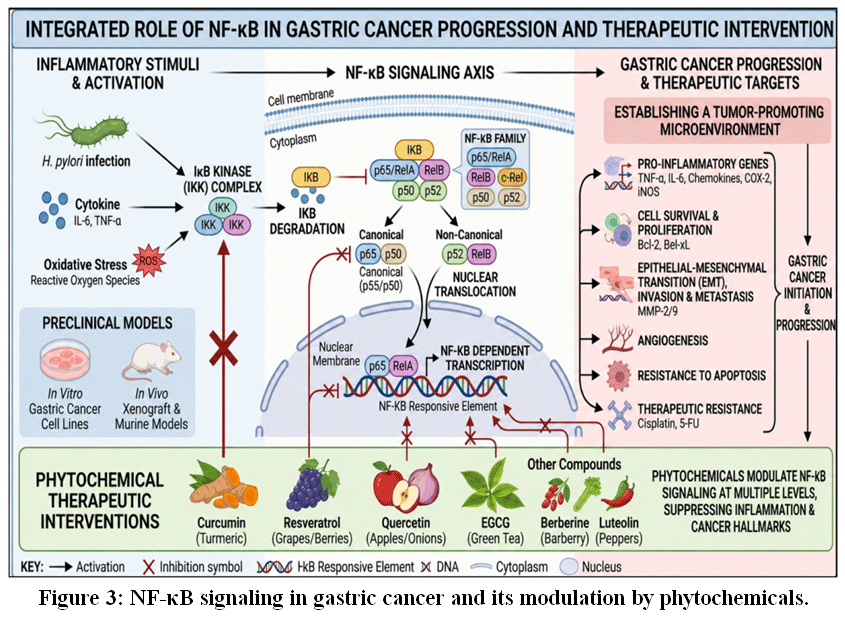 |
Figure 3: NF-κB signaling in gastric cancer and its modulation by phytochemicals.
|
A substantial body of preclinical evidence highlights the contribution of aberrant NF-κB signaling to gastric cancer progression. In vitro studies using gastric cancer cell lines consistently show that constitutive activation of this pathway enhances cell survival, promotes epithelial-mesenchymal transition (EMT), and reduces sensitivity to chemotherapeutic agents, including cisplatin and 5-fluorouracil. Complementary in vivo studies using xenograft and murine models further demonstrate that inhibition of NF-κB signaling suppresses tumor growth, reduces angiogenesis, and promotes apoptotic cell death.9,31 These findings collectively establish NF-κB as a key driver of tumor aggressiveness and therapeutic resistance.
Targeting NF-κB has therefore emerged as a promising therapeutic strategy. Inhibition of this pathway leads to reduced expression of anti-apoptotic proteins (Bcl-2, Bcl-xL), decreased activity of matrix metalloproteinases (MMP-2/9), and attenuation of inflammatory signaling, ultimately limiting tumor invasion and enhancing apoptosis. Several dietary phytochemicals have shown the ability to modulate NF-κB at multiple regulatory levels. Curcumin suppresses IKK activity and stabilizes IκB, thereby preventing nuclear translocation of NF-κB. Resveratrol interferes with upstream signaling cascades and enhances chemosensitivity. Similarly, quercetin and epigallocatechin-3-gallate (EGCG) inhibit NF-κB-dependent transcription and reduce the expression of genes associated with inflammation and survival. In addition to these compounds, other phytochemicals such as berberine, apigenin, and luteolin have also been reported to inhibit NF-κB signaling through suppression of inflammatory mediators and downstream gene expression, contributing to reduced tumor growth and metastasis in preclinical studies.13,45
These effects are consistently observed across experimental systems. In vitro, these compounds inhibit proliferation, induce apoptosis, and reduce migratory potential of gastric cancer cells. In vivo, they decrease tumor burden and improve response to chemotherapy when used alone or in combination. Emerging translational and early clinical evidence also suggests that polyphenols targeting NF-κB may enhance therapeutic efficacy while reducing treatment-related toxicity. However, clinical validation in gastric cancer remains limited, and most findings are derived from preclinical models.13
Despite these promising outcomes, several challenges limit clinical translation. Many phytochemicals exhibit poor bioavailability, rapid metabolism, and variability in formulation, which can affect therapeutic efficacy. In addition, NF-κB signaling has context-dependent functions, including roles in normal immune regulation, raising concerns about unintended systemic effects. Therefore, future research should focus on well-designed clinical trials, improved delivery systems, and combination strategies to optimize NF-κB-targeted therapies. Collectively, current evidence supports NF-κB as both a critical mediator of inflammation-driven gastric carcinogenesis and a viable therapeutic target, while underscoring the need for rigorous clinical investigation.9,29 The key phytochemicals targeting NF-κB signaling and their biological effects are summarized in Table 2.
Table 2: Phytochemicals Targeting NF-κB Signaling in Gastric Cancer.
| Phytochemicals | Molecular Target/Mechanism | Biological Effects | Evidence Type |
| Curcumin | Inhibits IKK activation; prevents IκB degradation; blocks NF-κB nuclear translocation | Reduced proliferation; increased apoptosis; decreased inflammation | In vitro, In vivo |
| Resveratrol | Inhibits upstream kinases regulating NF-κB signaling | Enhanced chemosensitivity; reduced survival signaling | In vitro, In vivo, Preclinical |
| Quercetin | Suppresses NF-κB-dependent transcription | Reduced invasion and inflammation; increased apoptosis | In vitro, Preclinical |
| EGCG | Inhibits NF-κB activation and downstream cytokine expression | Reduced angiogenesis and proliferation | In vitro, In vivo |
| Berberine | Suppresses NF-κB signaling pathway | Reduced tumor growth; increased apoptosis | In vitro, In vivo |
| Apigenin | Inhibits NF-κB activation and inflammatory mediators | Reduced inflammation and metastasis | Preclinical |
| Luteolin | Modulates NF-κB signaling and cytokine production | Reduced proliferation and inflammation | Preclinical |
MAPK Signaling In Gastric Cancer (ERK/JNK/P38)
The mitogen-activated protein kinase (MAPK) signaling cascade, comprising extracellular signal-regulated kinase (ERK), c-Jun N-terminal kinase (JNK), and p38 MAPK modules, plays a pivotal role in regulating proliferation, survival, inflammation, and cellular stress responses in gastric cancer. Dysregulation of this cascade is a key driver of tumor initiation and progression, largely through sustained activation of downstream transcription factors and oncogenic signaling networks.32,33 (as illustrated in Figure 4).
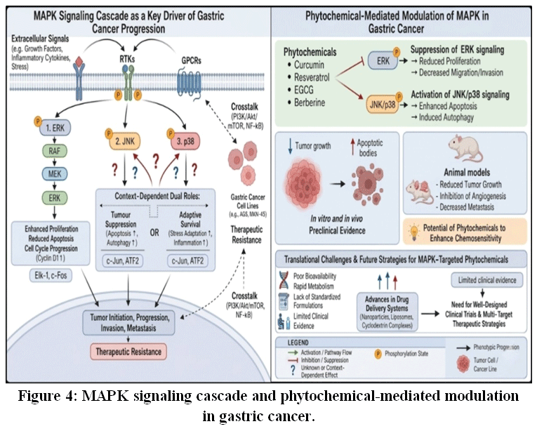 |
Figure 4: MAPK signaling cascade and phytochemical-mediated modulation in gastric cancer.
|
Among these components, ERK1/2 activation is consistently associated with enhanced cell cycle progression, increased proliferation, and resistance to apoptosis. In contrast, JNK and p38 exhibit context-dependent and sometimes opposing roles, acting either as tumor suppressors by promoting apoptosis or as adaptive stress mediators that support tumor survival under unfavorable microenvironmental conditions.33 This dual behavior reflects the complexity of MAPK signaling and highlights the importance of context-specific therapeutic targeting.
At the transcriptional level, ERK signaling activates downstream factors such as Elk-1 and c-Fos, while JNK and p38 regulate c-Jun and ATF2, thereby influencing gene expression involved in proliferation, apoptosis, and stress responses.32,33
In gastric cancer, MAPK signaling contributes not only to tumor growth but also to invasion and metastasis. ERK-driven signaling enhances cyclin expression and promotes cell cycle progression, whereas JNK and p38 regulate apoptosis, autophagy, and inflammatory responses. Importantly, this cascade is tightly interconnected with other oncogenic networks, particularly PI3K/Akt/mTOR and NF-κB pathways, forming an integrated signaling system that amplifies tumor-promoting processes and contributes to therapeutic resistance.22
Studies in gastric cancer cell lines such as AGS and MKN-45 demonstrate that phytochemicals including curcumin, resveratrol, epigallocatechin-3-gallate (EGCG), and berberine modulate MAPK activity in a coordinated manner. These compounds suppress ERK phosphorylation while activating JNK and p38, thereby shifting cellular signaling toward apoptosis and reducing proliferation, migration, and invasion.34,35
Animal models further support these findings, where phytochemical-mediated modulation of MAPK signaling leads to reduced tumor growth, inhibition of angiogenesis, and decreased metastatic potential. These effects are frequently associated with enhanced apoptotic signaling and suppression of inflammatory mediators, reinforcing the therapeutic relevance of targeting this pathway.34
Despite compelling preclinical evidence, clinical validation remains limited. Although compounds such as curcumin and resveratrol have been explored in clinical settings for various malignancies, there is insufficient evidence specifically demonstrating MAPK-targeted effects in gastric cancer patients.34 This gap underscores the need for well-designed clinical trials to establish translational relevance.
An additional therapeutic advantage of MAPK-targeting phytochemicals lies in their ability to enhance chemosensitivity. By modulating apoptosis-related pathways and interfering with resistance mechanisms, these compounds can improve responses to conventional therapies. Moreover, their simultaneous influence on interconnected signaling networks, including PI3K/Akt and NF-κB, supports a multi-target therapeutic strategy.25 However, this broad activity may also result in variable outcomes depending on tumor heterogeneity and experimental conditions.
Nevertheless, several challenges limit clinical application. Poor bioavailability, rapid metabolism, lack of standardized formulations, and variability across experimental models remain significant barriers.42 Furthermore, the predominance of in vitro and in vivo studies, with limited clinical corroboration, restricts definitive conclusions regarding therapeutic efficacy.34 Advances in drug delivery systems, including nanoparticle-based carriers and cyclodextrin complexes, offer promising approaches to overcome these limitations and improve pharmacokinetic profiles.42
In summary, MAPK signaling represents a critical regulatory axis in gastric cancer, influencing proliferation, survival, and stress adaptation. Phytochemicals targeting this cascade demonstrate significant potential in preclinical models; however, their clinical utility requires further validation through rigorous studies, improved formulation strategies, and integrated therapeutic approaches.Key phytochemicals targeting MAPK signaling, along with their molecular mechanisms, biological effects, and levels of evidence, are summarized in Table 3.
Table 3: Phytochemicals targeting MAPK signaling in gastric cancer: molecular mechanisms, biological effects, and evidence type.
| Phytochemical | Molecular Target / Mechanism of Action | Biological Effects in Gastric Cancer | Evidence Type |
| Curcumin | Inhibits ERK1/2 phosphorylation; activates JNK and p38 MAPK; modulates cross-talk with PI3K/Akt and NF-κB pathways | Induces apoptosis; suppresses proliferation; inhibits invasion and metastasis; enhances chemosensitivity | In vitro (AGS, MKN-45); In vivo (xenograft models); Limited clinical evidence |
| Resveratrol | Suppresses ERK signaling; activates JNK/p38 pathways; regulates apoptosis-related signaling and MAPK cross-talk | Promotes apoptosis; inhibits tumor growth and metastasis; improves chemotherapeutic response | In vitro; In vivo; Limited clinical evidence |
| Epigallocatechin-3-gallate (EGCG) | Inhibits ERK activation; stimulates JNK/p38 signaling; modulates MAPK-associated inflammatory pathways | Reduces proliferation; induces apoptosis; inhibits migration and angiogenesis | In vitro; In vivo |
| Berberine | Downregulates ERK phosphorylation; activates JNK/p38 signaling; interacts with MAPK-PI3K/Akt signaling axis | Induces apoptosis; inhibits tumor growth and invasion; suppresses metastatic potential | In vitro; In vivo |
Herbal Phytochemicals Target Apoptosis (Bax/Bcl-2, Caspases), Autophagy, and Ferroptosis in Gastric Cancer
Apoptosis (BAX/BCL-2, Caspases)
Evasion of apoptosis is a defining hallmark of cancer, including gastric malignancies, and is largely driven by disruption of mitochondrial apoptotic signaling and impaired caspase activation.36 In gastric cancer, increased expression of anti-apoptotic proteins such as Bcl-2, along with reduced levels of pro-apoptotic proteins like Bax, interferes with mitochondrial outer membrane permeabilization (MOMP), thereby preventing cytochrome-c release and subsequent activation of caspase-9 and caspase-3.37 This imbalance plays a crucial role in tumor progression and resistance to therapy.36,37
A growing body of evidence indicates that herbal phytochemicals can restore apoptotic signaling by modulating the Bax/Bcl-2 ratio, promoting mitochondrial dysfunction, and triggering caspase-dependent cell death. Compounds such as curcumin, berberine, quercetin, resveratrol, and epigallocatechin-3-gallate (EGCG) have demonstrated notable pro-apoptotic activity in gastric cancer models.At the cellular level, in vitro studies (cell line models) show that curcumin induces apoptosis through activation of caspase-3 and caspase-9, along with cleavage of poly (ADP-ribose) polymerase (PARP).38 Similar effects have been observed with other phytochemicals, which enhance apoptotic signaling via mitochondrial pathways and oxidative stress mechanisms.
Evidence from in vivo studies (animal models) further substantiates these findings, where phytochemical treatment has been associated with reduced tumor growth, increased apoptotic index, and enhanced caspase activation in xenograft systems. Despite these promising results, clinical evidence (human trials) evaluating phytochemical-induced apoptosis in gastric cancer remains insufficient, reflecting a gap between experimental findings and clinical application.36,46 In addition, phytochemicals may influence upstream signaling pathways such as PI3K/Akt and NF-κB, thereby increasing tumor cell susceptibility to apoptosis and improving therapeutic responsiveness.46
Autophagy (LC3/Beclin-1 Signaling)
Autophagy is a highly regulated lysosomal degradation pathway that exerts a dual role in gastric cancer, functioning either as a survival mechanism under stress conditions or as a mediator of programmed cell death. Alterations in key autophagy-related proteins, including Beclin-1 and LC3, have been closely linked to tumor progression and therapeutic resistance.10
Recent studies suggest that herbal phytochemicals can effectively regulate autophagic processes by influencing LC3 conversion (LC3-I to LC3-II), enhancing Beclin-1 expression, and suppressing mTOR signaling. Bioactive compounds such as curcumin, ginsenosides (Rg3 and Rh2), oridonin, and oleanolic acid exhibit significant autophagy-modulating potential.39,40
Experimental findings from in vitro models (gastric cancer cell lines) demonstrate that these phytochemicals can induce autophagic flux, which may either promote cell survival or lead to autophagy-mediated cell death depending on the cellular context.39 Consistent with these observations, in vivo studies (animal models) have shown that modulation of autophagy by phytochemicals contributes to tumor suppression, decreased tumor volume, and enhanced sensitivity to chemotherapeutic agents.40,46 Notably, the interplay between autophagy and apoptosis suggests a coordinated mechanism underlying phytochemical-mediated anticancer effects.46
However, clinical evidence (human studies) supporting the role of phytochemicals in regulating autophagy in gastric cancer is still limited, with most insights derived from preclinical research.10,39 Further well-designed clinical investigations are required to establish the therapeutic relevance of targeting autophagy in patients.
Ferroptosis
Ferroptosis is an iron-dependent form of regulated cell death characterized by excessive lipid peroxidation and depletion of intracellular glutathione due to inhibition of glutathione peroxidase 4 (GPX4). Unlike apoptosis, ferroptosis represents an alternative mechanism for eliminating cancer cells, particularly those that exhibit resistance to conventional apoptotic pathways.47,48
Emerging evidence indicates that phytochemicals can trigger ferroptosis in gastric cancer through multiple mechanisms. Compounds such as artemisinin derivatives, quercetin, baicalein, and tanshinone IIA have been shown to enhance reactive oxygen species (ROS) generation, disrupt iron homeostasis, and inhibit GPX4 activity, ultimately leading to ferroptotic cell death. Findings from in vitro studies (cell line models) reveal increased lipid peroxidation, elevated ROS levels, and decreased viability of gastric cancer cells following phytochemical treatment.48
These effects are further supported by in vivo evidence (animal studies), where induction of ferroptosis is associated with suppression of tumor growth and increased oxidative stress within tumor tissues. At present, clinical evidence (human trials) investigating ferroptosis-targeting phytochemicals in gastric cancer is lacking, and available data remain largely confined to experimental models.Nevertheless, ferroptosis continues to gain attention as a promising therapeutic approach, particularly for overcoming apoptosis resistance and enhancing anticancer efficacy.47,48 The molecular mechanisms of phytochemical-induced cell death and the associated phytochemicals with their targets and evidence levels are summarized in Figure 5 and Table 4, respectively.
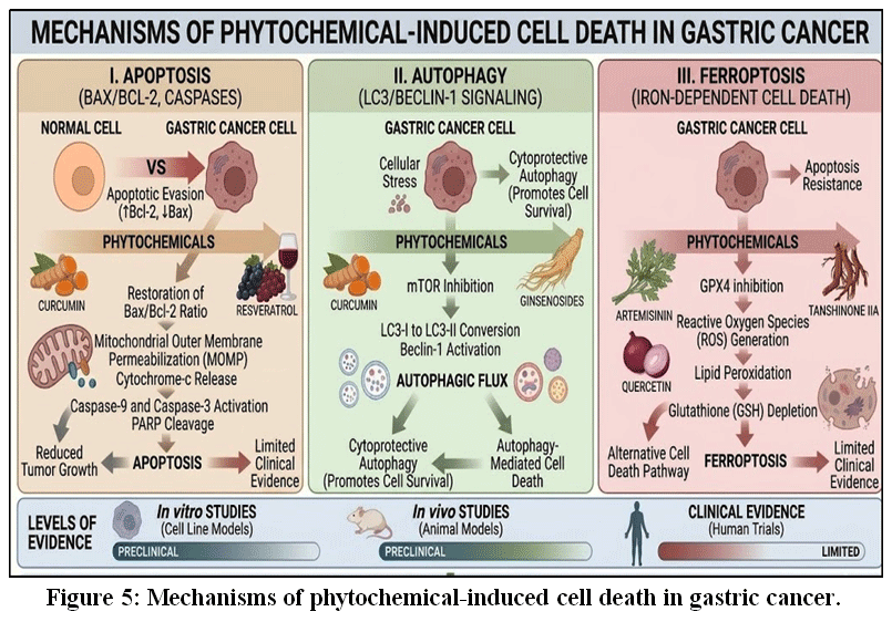 |
Figure 5: Mechanisms of phytochemical-induced cell death in gastric cancer.
|
Table 4; Herbal phytochemicals targeting apoptosis, autophagy, and ferroptosis in gastric cancer.
| Phytochemical | Molecular Target / Mechanism of Action | Biological Effect in Gastric Cancer | Evidence Type |
| Curcumin | Increases Bax/Bcl-2 ratio; activates caspase-3/-9; inhibits PI3K/Akt and NF-κB | Induces apoptosis; enhances chemosensitivity | In vitro; In vivo |
| Berberine | Modulates Bax/Bcl-2; induces mitochondrial dysfunction | Promotes apoptosis; inhibits tumor growth | In vitro; In vivo |
| Quercetin | Induces ROS; activates caspases; disrupts mitochondrial membrane potential | Triggers apoptosis and oxidative stress | In vitro; In vivo |
| Resveratrol | Activates intrinsic apoptosis; modulates NF-κB | Enhances apoptosis; inhibits proliferation | In vitro; In vivo |
| EGCG | Modulates Bcl-2 proteins; activates caspases; inhibits survival pathways | Induces apoptosis; suppresses tumor survival | In vitro; In vivo |
| Ginsenosides (Rg3, Rh2) | Upregulates Beclin-1; promotes LC3 conversion; inhibits mTOR | Induces autophagy; suppresses tumor growth | In vitro; In vivo |
| Oridonin | Activates LC3 and Beclin-1; regulates autophagy | Induces autophagy and apoptosis | In vitro |
| Oleanolic acid | Modulates mTOR; enhances autophagy | Promotes autophagy-mediated tumor suppression | In vitro; In vivo |
| Artemisinin derivatives | Increases ROS; disrupts iron metabolism; inhibits GPX4 | Induces ferroptosis | In vitro; In vivo |
| Baicalein | Promotes lipid peroxidation; inhibits GPX4 | Triggers ferroptosis | In vitro |
| Tanshinone IIA | Enhances ROS; modulates ferroptosis pathways | Induces ferroptosis; suppresses tumor growth | In vitro; In
vivo |
P53-Cell Cycle Dysregulation in Gastric Cancer: Cyclins and CDKS
Loss of functional p53 tumor suppressor activity is a key molecular event in the pathogenesis of gastric cancer, leading to disruption of DNA damage response mechanisms and deregulation of cell cycle checkpoints. Under normal physiological conditions, p53 plays a central role in maintaining genomic stability by regulating transcription of genes involved in cell cycle arrest, apoptosis, and DNA repair. In normal cells, p53 activity is further stabilized through acetylation by p300/CBP-associated factor (PCAF), which enhances p21-mediated cell cycle arrest and DNA repair. However, in gastric cancer, mutation or inactivation of p53 results in aberrant activation of cyclins (Cyclin D1, Cyclin E, and Cyclin B1) and cyclin-dependent kinases (CDK4/6, CDK2, and CDK1), which drive uncontrolled progression through the G1/S and G2/M phases of the cell cycle and promote tumor growth.49 Large-scale molecular analyses have further demonstrated that alterations in p53 signaling are closely associated with tumor heterogeneity, disease progression, and therapeutic resistance in gastric cancer.16
Most mechanistic insights are derived from gastric cancer cell line models. Curcumin has been shown to enhance p53 stability and transcriptional activity, leading to increased expression of p21 and subsequent inhibition of Cyclin D1/CDK4 complexes, thereby inducing G1 phase arrest.50 Resveratrol suppresses Cyclin E/CDK2 signaling and activates p53-dependent transcription, limiting S-phase progression and reducing proliferation.51 Epigallocatechin-3-gallate (EGCG), a major dietary polyphenol, further enhances p53-mediated growth arrest while downregulating Cyclin D1 expression.52 In addition, berberine and quercetin modulate cell cycle progression through p53-dependent mechanisms, with quercetin specifically inducing G2/M arrest via inhibition of Cyclin B1/CDK1 complexes. Compounds such as genistein and apigenin similarly suppress CDK activity and restore regulatory control of the cell cycle. Recent studies also indicate that phytochemicals influence upstream signaling pathways, including MAPK cascades, thereby enhancing p53-mediated anticancer effects through pathway cross-talk.20
Preclinical animal models provide supportive evidence for these mechanisms. Polyphenols such as resveratrol and curcumin have demonstrated tumor growth inhibition in gastric cancer xenograft models, partly through restoration of p53 signaling and suppression of cyclin-CDK activity.51 Furthermore, recent studies highlight that combinations of phytochemicals can enhance anticancer efficacy by synergistically activating apoptotic pathways and improving chemosensitivity, suggesting an important role for combination strategies in targeting cell cycle dysregulation.53 However, these findings should be interpreted cautiously due to variability in experimental conditions, dosing, and bioavailability.
Clinical translation remains limited, which represents a critical gap in current research. Although phytochemicals such as curcumin and resveratrol have been evaluated in early-phase clinical studies across various cancers, robust clinical evidence specifically in gastric cancer is still lacking. Available data suggest modest improvements in biomarkers related to oxidative stress and proliferation, but direct validation of p53 reactivation and cell cycle regulation in human patients remains insufficient. Recent studies emphasize the need for improved delivery systems, enhanced bioavailability, and well-designed clinical trials to establish therapeutic efficacy.54 Mechanistically, phytochemicals exert multi-dimensional effects beyond direct modulation of p53. Polyphenols are known to regulate intracellular redox balance and signaling pathways that influence p53 stability and function, demonstrating actions beyond their classical antioxidant properties.52 This integrated mode of action supports their role as network-targeting agents capable of modulating multiple oncogenic pathways simultaneously.
In summary, herbal phytochemicals demonstrate significant potential in regulating the p53-cyclin-CDK axis, thereby restoring cell cycle control and inhibiting gastric cancer progression. However, consistent with reviewer recommendations, it is important to recognize that current evidence is predominantly derived from in vitro studies, with supportive but limited in vivo validation and insufficient clinical confirmation. Therefore, further well-designed clinical investigations are required to validate their therapeutic potential and facilitate translation into clinical practice.49-54 The dysregulation of the p53-cyclin-CDK axis and its modulation by phytochemicals are illustrated in Figure 6. and summarized in Table 5.
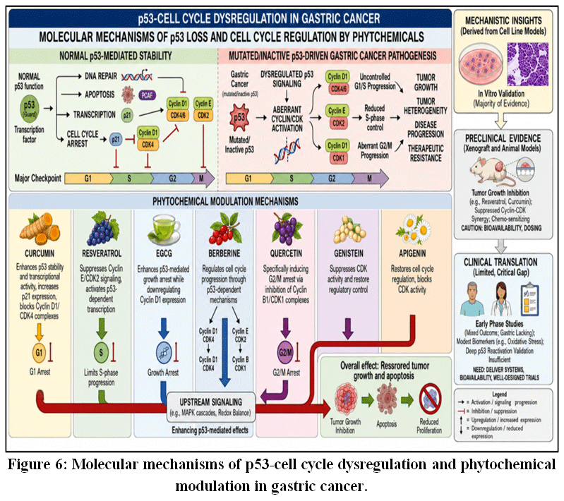 |
Figure 6: Molecular mechanisms of p53-cell cycle dysregulation and phytochemical modulation in gastric cancer.
|
Table 5: Herbal Phytochemicals Targeting p53-Cyclin-CDK Axis in Gastric Cancer.
| Phytochemical | Molecular Target / Mechanism of Action | Biological Effect in Gastric Cancer | Evidence Type |
| Curcumin | Enhances p53 stability; upregulates p21; inhibits Cyclin D1/CDK4 | G1 arrest; anti-proliferative; tumor suppression | In vitro; In vivo; Limited clinical |
| Resveratrol | Activates p53; inhibits Cyclin E/CDK2 | Reduces S-phase progression; induces apoptosis | In vitro; In vivo; Limited clinical |
| EGCG | Enhances p53 signaling; downregulates Cyclin D1 | Growth arrest; anti-proliferative | In vitro |
| Berberine | Activates p53 checkpoints; modulates CDKs | Cell cycle arrest; reduced proliferation | In vitro |
| Quercetin | Inhibits Cyclin B1/CDK1 | G2/M arrest; anti-proliferative | In vitro |
| Genistein | Suppresses CDK activity; restores p53 regulation | Inhibits proliferation; cell cycle control | In vitro |
| Apigenin | Inhibits CDKs; enhances p53 signaling | Cell cycle arrest; anti-proliferative | In vitro |
EMT and Metastasis in Gastric Cancer
Epithelial-mesenchymal transition (EMT) is a critical biological process that drives gastric cancer progression, particularly in tumor invasion, dissemination, and metastasis. During EMT, epithelial cells lose polarity and intercellular adhesion, primarily due to downregulation of E-cadherin, and acquire mesenchymal features such as increased N-cadherin and vimentin expression, which enhance motility and invasiveness. This transition is regulated by transcription factors including Snail, Slug, and Twist, along with matrix metalloproteinases (MMP-2 and MMP-9), which facilitate extracellular matrix degradation and tumor cell migration.56,61,62
EMT is tightly controlled by interconnected oncogenic signaling pathways, including PI3K/Akt, NF-κB, Wnt/β-catenin, and TGF-β cascades, which integrate inflammatory, metabolic, and microenvironmental signals to promote metastatic behavior.13,14,61 Importantly, EMT exists along a dynamic spectrum of intermediate states rather than as a binary process, contributing to tumor plasticity, immune evasion, and resistance to therapy.62
Evidence from gastric cancer cell lines such as AGS, MKN-45, and SGC-7901 demonstrates that phytochemicals can effectively suppress EMT by restoring epithelial characteristics and inhibiting mesenchymal transition. Curcumin enhances E-cadherin expression while reducing Snail and MMP-9 levels, resulting in decreased migration and invasion of cancer cells.13,56 Resveratrol similarly attenuates EMT by modulating transcriptional regulators and reducing MMP-2/9 activity, thereby limiting invasive potential.55 Berberine also suppresses EMT by regulating adhesion molecules and extracellular matrix-degrading enzymes, ultimately reducing tumor cell motility.57 These findings indicate that phytochemicals target multiple regulatory nodes involved in EMT progression.
Preclinical animal studies further support the anti-metastatic effects of phytochemicals. In gastric cancer xenograft models, curcumin reduces tumor growth and metastatic spread through modulation of EMT markers and inhibition of MMP activity.55 Berberine exhibits similar effects by suppressing tumor invasion and metastasis via regulation of EMT-associated signaling pathways and tumor microenvironment interactions.57 Additionally, resveratrol has been shown to reduce metastatic burden and restore epithelial features in vivo, reinforcing its role in inhibiting EMT-driven tumor progression.14 These findings highlight the translational relevance of targeting EMT using phytochemicals.
Direct clinical evidence linking phytochemicals to EMT inhibition in gastric cancer remains limited; however, available human studies provide supportive insights into their therapeutic potential. Curcumin-based interventions have demonstrated improved treatment response, reduced inflammatory markers, and enhanced tolerability when used alongside conventional chemotherapy.58,60 Resveratrol has also been evaluated clinically, showing favorable safety, pharmacokinetics, and biological activity in patients with gastrointestinal malignancies.59 Although these findings are not EMT-specific, they suggest that phytochemicals may influence metastatic and inflammatory pathways in human cancers.14
Despite encouraging evidence, several limitations restrict the clinical translation of phytochemicals targeting EMT. These include poor bioavailability, rapid metabolism, and lack of standardized formulations, which can affect therapeutic efficacy. Additionally, EMT exhibits context-dependent behavior, where partial EMT states may enhance tumor adaptability, stemness, and drug resistance rather than fully suppress metastasis.62 Therefore, therapeutic strategies should focus on controlled modulation of EMT rather than complete inhibition. Future research should prioritize improved delivery systems, standardized formulations, and validation of EMT-related biomarkers in well-designed clinical trials.
In summary, phytochemicals such as curcumin, resveratrol, and berberine demonstrate significant potential to inhibit EMT and metastasis in gastric cancer through multi-targeted molecular mechanisms. While strong in vitro and in vivo evidence supports their efficacy, further well-designed clinical studies are required to confirm their role in translational and clinical oncology.13,14,55 The molecular mechanisms and pathway interactions involved in EMT regulation by phytochemicals are illustrated in Figure 7 and A summary of phytochemical targets, mechanisms, and supporting evidence is provided in Table 6.
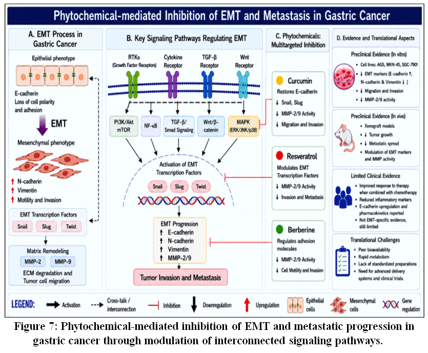 |
Figure 7: Phytochemical-mediated inhibition of EMT and metastatic progression in gastric cancer through modulation of interconnected signaling pathways.
|
Table 6: Herbal phytochemicals targeting epithelial-mesenchymal transition (EMT) and metastasis in gastric cancer.
| Phytochemicals | Molecular Target/ Mechanism of Action | Biological Effect | Evidence Type |
| Curcumin | Upregulates E-cadherin expression; downregulates N-cadherin, Snail, and MMP-9; inhibits PI3K/Akt and NF-κB signaling pathways | Suppresses EMT, reduces cell migration and invasion, inhibits metastatic progression, and restores epithelial phenotype | In vitro (AGS, MKN-45, SGC-7901); In vivo (xenograft models); Clinical (adjunct evidence) |
| Resveratrol | Inhibits Snail-mediated EMT; downregulates MMP-2 and MMP-9; modulates transcriptional regulators and inflammatory pathways | Attenuates EMT, decreases invasive potential, reduces metastatic burden, and promotes epithelial marker expression | In vitro; In vivo; Clinical (supportive evidence) |
| Berberine | Upregulates E-cadherin expression; downregulates N-cadherin and MMP-2/9; modulates EMT-associated signaling pathways | Reverses EMT, suppresses tumor cell motility, and inhibits invasion and metastasis | In vitro; In vivo |
Angiogenesis (HIF-1Α/VEGF) as a Therapeutic Target in Gastric Cancer
Angiogenesis is a critical hallmark of gastric cancer progression and is primarily governed by the hypoxia-inducible factor-1 alpha (HIF-1α)/vascular endothelial growth factor (VEGF) signaling axis. Under hypoxic conditions within the tumor microenvironment, HIF-1α escapes oxygen-dependent degradation, accumulates intracellularly, and translocates to the nucleus, where it activates transcription of VEGF and other pro-angiogenic genes. This cascade drives endothelial cell proliferation, migration, and neovascularization, thereby promoting tumor growth, invasion, and metastasis. Sustained activation of this pathway is closely associated with aggressive disease behavior and poor clinical outcomes in gastric cancer.63 In addition, HIF-1α interacts with multiple oncogenic signaling networks, including PI3K/Akt and MAPK pathways, highlighting significant pathway cross-talk that enhances tumor survival, metabolic adaptation, and therapeutic resistance.64,55 The mechanistic role of phytochemicals targeting the HIF-1α/VEGF signaling axis in gastric cancer is illustrated in Figure 8.
Experimental studies in gastric cancer cell lines (such as AGS and MKN-45) demonstrate that several phytochemicals exert potent anti-angiogenic effects at the molecular level. Epigallocatechin-3-gallate (EGCG) inhibits hypoxia-induced stabilization of HIF-1α and reduces VEGF expression by modulating upstream signaling pathways, including PI3K/Akt and ERK.64,65 Similarly, apigenin suppresses angiogenesis by downregulating HIF-1α activity and VEGF production, thereby impairing endothelial cell function and angiogenic signaling.67 In addition, these compounds influence oxidative stress and intracellular signaling cascades, contributing to reduced angiogenic potential.55 Collectively, in vitro findings support the role of phytochemicals as multi-target modulators that interfere with both transcriptional regulation and signaling pathways involved in angiogenesis. A summary of key phytochemicals, their molecular targets, mechanisms of action, and evidence types is presented in Table 7.
Preclinical animal models provide more robust evidence for the anti-angiogenic activity of phytochemicals. Genistein has been shown to significantly reduce tumor growth and microvessel density (MVD) by inhibiting VEGF-mediated endothelial signaling.64,66 Likewise, silibinin suppresses tumor vascularization by targeting VEGF signaling and interfering with key hallmarks of cancer, including angiogenesis and proliferation.68 Baicalein and related compounds further contribute to inhibition of tumor progression through modulation of oxidative stress, apoptosis, and angiogenic pathways.69 These findings indicate that phytochemicals exert anti-angiogenic effects not only through direct inhibition of VEGF signaling but also by influencing tumor microenvironment components such as inflammation and redox balance.55
Despite encouraging preclinical results, clinical evidence remains insufficient. Certain phytochemicals, including EGCG and genistein, have been evaluated in early-phase clinical or translational studies, where they demonstrated favorable safety profiles and modest reductions in circulating VEGF levels. However, large-scale, well-controlled clinical trials specifically targeting gastric cancer are still lacking, and current evidence is insufficient to confirm their therapeutic efficacy in patients. Furthermore, pharmacokinetic limitations-such as poor bioavailability, rapid metabolism, and lack of standardized formulations-continue to hinder clinical translation. Combination strategies integrating phytochemicals with conventional chemotherapeutic agents are currently being explored to enhance therapeutic outcomes and overcome drug resistance.53,55
Importantly, the anti-angiogenic effects of phytochemicals are context-dependent and may vary according to tumor stage, dosage, and microenvironmental conditions. This variability is largely influenced by tumor heterogeneity and dynamic interactions within the tumor microenvironment, including hypoxia, oxidative stress, and inflammatory signaling.55 Moreover, HIF-1α exhibits a dual role, functioning as a survival factor under hypoxic stress while simultaneously promoting angiogenesis and tumor progression, which necessitates careful interpretation of therapeutic targeting strategies.63
Beyond direct inhibition of VEGF signaling, these compounds also regulate key components of the tumor microenvironment, including oxidative stress and inflammatory mediators. By modulating redox balance, cytokine signaling, and pathways such as NF-κB and PI3K/Akt, phytochemicals indirectly influence angiogenesis through interconnected molecular networks. This multi-targeted mode of action underscores their potential advantage over single-target therapies in complex diseases such as gastric cancer.55,64
In conclusion, targeting the HIF-1α/VEGF signaling axis using herbal phytochemicals represents a promising strategy for inhibiting angiogenesis in gastric cancer. Nevertheless, the current body of evidence is largely derived from in vitro and in vivo studies, with limited clinical validation, emphasizing the need for well-designed translational and clinical investigations to establish their efficacy, safety, and therapeutic applicability in human patients.53,55,63-69
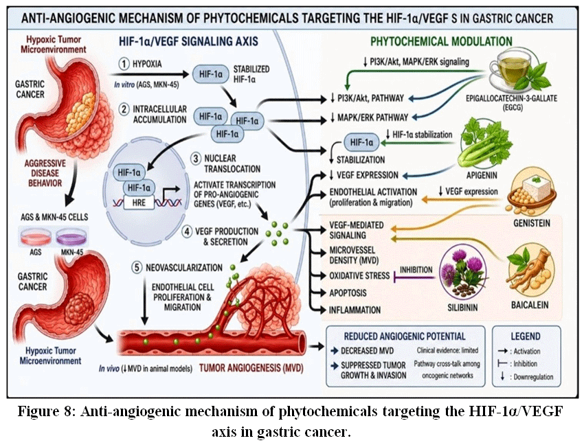 |
Figure 8: Anti-angiogenic mechanism of phytochemicals targeting the HIF-1α/VEGF axis in gastric cancer.
|
Table 7: Herbal phytochemicals targeting HIF-1α/VEGF-mediated angiogenesis in gastric cancer.
| Phytochemicals | Molecular Target / Mechanism of Action | Biological Effect | Evidence Type |
| Epigallocatechin-3-gallate (EGCG) | Inhibits HIF-1α stabilization; downregulates VEGF expression; modulates PI3K/Akt and ERK signaling | Reduces endothelial proliferation, angiogenesis, and tumor vascularization | In vitro, Clinical |
| Apigenin | Suppresses HIF-1α activity and VEGF production; interferes with hypoxia-induced signaling | Inhibits angiogenesis and endothelial cell function | In vitro |
| Genistein | Inhibits VEGF-mediated endothelial signaling; reduces microvessel density (MVD) | Suppresses tumor growth and angiogenesis | In vivo, Clinical |
| Silibinin | Targets VEGF signaling pathways; interferes with angiogenesis-related cancer hallmarks | Reduces tumor vascularization and proliferation | In vivo |
| Baicalein | Modulates oxidative stress, apoptosis, and angiogenic signaling pathways | Inhibits tumor progression and angiogenesis | In vivo |
NRF2/KEAP1 Redox Signaling in Gastric Cancer
The nuclear factor erythroid 2-related factor 2 (Nrf2)/Kelch-like ECH-associated protein 1 (Keap1) signaling pathway plays a pivotal role in maintaining cellular redox homeostasis and regulating antioxidant defense under oxidative stress. Activation of Nrf2 induces transcription of antioxidant response element (ARE)-driven genes, including heme oxygenase-1 (HO-1), NAD(P)H quinone oxidoreductase 1 (NQO1), and glutamate-cysteine ligase catalytic subunit (GCLC), thereby enhancing detoxification and cytoprotective responses.21,70
In gastric cancer, persistent oxidative stress, chronic inflammation, and oncogenic signaling frequently lead to aberrant and sustained activation of Nrf2, which contributes to tumor progression. Constitutive Nrf2 activation enhances cancer cell survival, promotes metabolic reprogramming, and increases resistance to chemotherapy. Importantly, emerging evidence highlights the dual and context-dependent role of Nrf2, where transient activation protects normal cells, whereas prolonged activation supports tumor growth, chemoresistance, and aggressive phenotypes.71-73 Furthermore, Nrf2 signaling has been specifically implicated in gastric cancer progression, where it regulates oxidative stress adaptation and tumor cell survival.74
In gastric cancer cell lines, several phytochemicals regulate Nrf2 signaling and intracellular redox balance. Sulforaphane activates Nrf2 through modification of Keap1 cysteine residues, leading to enhanced antioxidant gene expression. Curcumin activates Nrf2-dependent antioxidant pathways while simultaneously elevating reactive oxygen species (ROS) levels, resulting in apoptosis. Resveratrol exhibits a biphasic effect, where low concentrations promote cytoprotection via Nrf2 activation, whereas higher concentrations induce oxidative stress-mediated cell death. Flavonoids such as quercetin, luteolin, and apigenin also modulate Nrf2/Keap1 signaling, suppress redox-driven proliferation, and enhance chemosensitivity in gastric cancer cells.75,54 The mechanistic role of Nrf2/Keap1 signaling and its modulation by phytochemicals in gastric cancer is illustrated in Figure 9, while a concise summary of phytochemical mechanisms, biological effects, and supporting evidence is presented in Table 8.
Preclinical studies using gastric cancer xenograft models further support the regulatory role of phytochemicals on Nrf2 signaling. Compounds such as curcumin have been shown to modulate Nrf2 activity, reduce tumor growth, and improve therapeutic efficacy. Notably, recent findings indicate that Nrf2 depletion or inhibition can enhance the anticancer effects of phytochemicals, suggesting its involvement in chemoresistance mechanisms.76
Despite strong preclinical findings, clinical evidence remains limited. While phytochemicals such as curcumin and resveratrol have been evaluated in broader oncology settings, there is a lack of well-designed clinical trials specifically targeting Nrf2 signaling in gastric cancer patients. Current evidence suggests potential benefits as adjunct therapies; however, variability in response, poor pharmacokinetics, and limited bioavailability significantly hinder clinical translation.73
Critically, targeting the Nrf2/Keap1 pathway presents both opportunities and challenges. While inhibition of aberrant Nrf2 activation may help overcome drug resistance, complete suppression could impair physiological antioxidant defense mechanisms. Therefore, a balanced and context-specific modulation strategy is required. Moreover, extensive cross-talk between Nrf2 and other oncogenic pathways, including PI3K/Akt, NF-κB, and MAPK signaling, further complicates its therapeutic targeting.72
In addition, the clinical application of phytochemicals is constrained by poor solubility, rapid metabolism, and low systemic bioavailability, which limit their therapeutic efficacy. Recent advances in nanotechnology-based delivery systems, including nanoparticles, liposomes, and phytosome formulations, have shown promise in improving stability, targeted delivery, and bioavailability of phytochemicals, thereby enhancing their anticancer potential.54
In summary, phytochemicals represent multi-target redox modulators with significant translational potential, supported by substantial in vitro and in vivo evidence demonstrating their ability to regulate oxidative stress, apoptosis, and chemosensitivity in gastric cancer. However, the lack of robust clinical validation, along with the context-dependent dual role of Nrf2 and pharmacokinetic limitations, remains a major challenge. Future research should focus on well-designed clinical trials, advanced delivery systems, and pathway-specific targeting strategies to enable effective clinical translation.71-76
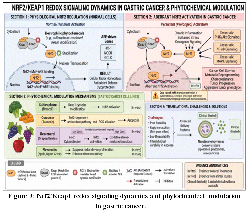 |
Figure 9: Nrf2/Keap1 redox signaling dynamics and phytochemical modulation in gastric cancer.
|
Table 8: Phytochemical modulation of Nrf2/Keap1 signaling in gastric cancer.
| Sulforaphane | Modifies Keap1 cysteine residues → disrupts Keap1-Nrf2 interaction → Nrf2 activation and ARE gene transcription (HO-1, NQO1, GCLC) | Enhances antioxidant defense, increases cytoprotection, sensitizes cancer cells to oxidative stress | In vitro (gastric cancer cell lines) |
| Curcumin | Activates Nrf2-dependent antioxidant pathway; simultaneously induces ROS generation at higher doses | Dual effect: antioxidant cytoprotection (low dose) and ROS-mediated apoptosis (high dose); improves therapeutic response | In vitro; In vivo (xenograft models) |
| Resveratrol | Modulates Nrf2 signaling in a dose-dependent (biphasic/hormetic) manner | Low dose: cytoprotection via Nrf2 activation; High dose: oxidative stress-induced apoptosis; enhances chemosensitivity | In vitro |
| Flavonoids | Regulate Nrf2/Keap1 signaling; modulate redox balance and protein interactions | Suppress redox-driven proliferation; enhance chemosensitivity; inhibit tumor growth pathways | In vitro; In vivo |
Epigenetics and Non-Coding Rnas (Mirnas andnd Lncrnas) in Gastric Cancer
Epigenetic dysregulation represents a fundamental mechanism underlying gastric cancer progression, particularly through altered expression of non-coding RNAs such as microRNAs (miRNAs) and long non-coding RNAs (lncRNAs), which regulate gene expression without modifying the DNA sequence.77,78 These regulatory RNAs are critically involved in multiple oncogenic processes, including cell proliferation, apoptosis, epithelial-mesenchymal transition (EMT), invasion, metastasis, and resistance to therapy. Importantly, ncRNAs interact with key signaling pathways such as PI3K/Akt, NF-κB, and Wnt/β-catenin, thereby contributing to pathway cross-talk and tumor heterogeneity.78
In gastric cancer, several tumor-suppressive miRNAs, including miR-34a and members of the miR-200 family, are frequently downregulated. This loss facilitates EMT, enhances cellular invasiveness, and is associated with poor prognosis. Mechanistically, reduced expression of these miRNAs promotes activation of EMT-inducing transcription factors such as ZEB1/2 and disrupts apoptotic signaling pathways.78 In contrast, oncogenic miRNAs such as miR-21 and miR-155 are commonly upregulated and contribute to tumor progression by suppressing tumor suppressor genes and activating inflammatory and survival signaling cascades.77 Alongside miRNAs, lncRNAs such as HOTAIR and MALAT1 play a crucial role in epigenetic regulation by interacting with chromatin-modifying complexes, thereby promoting transcriptional reprogramming, metastasis, and drug resistance.79 Evidence supporting these mechanisms is largely derived from in vitro studies (gastric cancer cell lines such as AGS and MKN-45)andin vivo models (xenograft mouse systems), where modulation of ncRNA expression significantly alters tumor growth and metastatic potential.78
Notably, herbal phytochemicals have emerged as promising epigenetic modulators capable of restoring ncRNA balance. Curcumin, for example, influences DNA methylation and histone acetylation, leading to reactivation of tumor-suppressive miRNAs and suppression of oncogenic miRNAs. These effects have been consistently demonstrated in cell-based experimental systems, where curcumin induces apoptosis and inhibits proliferation, as well as in animal models, where it reduces tumor burden.77 Similarly, resveratrol has been shown to reshape miRNA expression profiles associated with cell cycle arrest and apoptosis, thereby improving chemosensitivity; these findings are supported by preclinical experimental evidence.80 Epigallocatechin-3-gallate (EGCG) exerts epigenetic effects through inhibition of DNA methyltransferases (DNMTs), resulting in reactivation of silenced tumor-suppressor genes and miRNAs. This mechanism has been validated in in vitro models and in vivo studies, where EGCG suppresses proliferation and tumor progression.81,82
In addition, other phytochemicals such as genistein, sulforaphane, quercetin, and berberine further contribute to epigenetic regulation by targeting chromatin-modifying enzymes and ncRNA networks. Through these mechanisms, they inhibit proliferation, induce apoptosis, and reverse EMT, highlighting the interconnected nature of epigenetic and signaling pathways in gastric cancer.77,53
Despite compelling preclinical evidence (in vitro and in vivo), translation into clinical practice remains limited. Clinical evidence (human trials) evaluating the epigenetic effects of phytochemicals in gastric cancer is still scarce, with most available data derived from other cancer types.53 This represents a significant gap in current research. Moreover, ncRNA-mediated regulation is highly context-dependent, and certain miRNAs may exhibit dual roles depending on tumor stage and microenvironment, which necessitates cautious interpretation of experimental findings.78
In summary, ncRNA-driven epigenetic alterations play a central role in gastric cancer progression and therapeutic resistance. Herbal phytochemicals function as multi-target epigenetic modulators capable of restoring ncRNA homeostasis and suppressing tumor-promoting pathways. While strong evidence from in vitro and in vivo studies supports their therapeutic potential, the lack of robust clinical validation remains a major limitation. Future research should therefore focus on well-designed clinical trials and advanced delivery strategies to facilitate the successful translation of these agents into clinical applications.77,78,53 Epigenetic dysregulation, ncRNA signaling, and oncogenic pathways in gastric cancer are illustrated in Figure 10, while key phytochemicals and their mechanisms are summarized in Table 9.
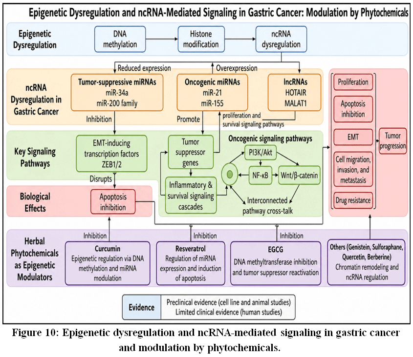 |
Figure 10: Epigenetic dysregulation and ncRNA-mediated signaling in gastric cancer and modulation by phytochemicals.
|
Table 9: Phytochemicals Targeting Epigenetic ncRNA Regulation in Gastric Cancer.
| Phytochemical | Molecular Target / Mechanism of Action | Biological Effect | Evidence Type |
| Curcumin | Regulates DNA methylation and histone acetylation; enhances expression of tumor-suppressive miRNAs such as miR-34a and suppresses oncogenic miRNAs | Promotes apoptosis, inhibits cell proliferation, and suppresses epithelial-mesenchymal transition and metastasis | In vitro (AGS, MKN-45 cell lines); In vivo (xenograft models) |
| Resveratrol | Modulates miRNA expression profiles and influences TGF-β-related signaling pathways | Induces cell cycle arrest and apoptosis; improves chemosensitivity | In vitro; In vivo |
| EGCG (Epigallocatechin-3-gallate) | Inhibits DNA methyltransferase activity, leading to reactivation of silenced tumor-suppressor genes and miRNAs | Reduces cell proliferation, induces apoptosis, and inhibits tumor growth | In vitro; In vivo |
| Genistein | Regulates histone modification and miRNA expression through modulation of epigenetic enzymes | Inhibits proliferation, induces apoptosis, and reduces angiogenesis | In vitro; In vivo |
| Sulforaphane | Inhibits histone deacetylases and modulates miRNA expression along with Nrf2 signaling pathways | Induces apoptosis, enhances antioxidant responses, and suppresses tumor progression | In vitro; In vivo |
| Quercetin | Regulates miRNA networks and influences chromatin remodeling and PI3K/Akt signaling pathways | Promotes apoptosis and inhibits proliferation and epithelial-mesenchymal transition | In vitro; In vivo |
| Berberine | Modulates ncRNA expression and targets chromatin-modifying enzymes and signaling pathways | Suppresses cell proliferation, induces apoptosis, and reverses epithelial-mesenchymal transition and drug resistance | In vitro; In vivo |
Combination Therapy and Chemosensitization
Phytochemicals have emerged as promising adjunctive agents in combination cancer therapy owing to their ability to modulate multiple oncogenic signaling networks and overcome chemoresistance in gastric cancer. Resistance to widely used chemotherapeutic agents such as doxorubicin, cisplatin, and 5-fluorouracil is frequently associated with aberrant activation of survival pathways, including PI3K/Akt, NF-κB, STAT3, and epithelial-mesenchymal transition (EMT). Accumulating evidence indicates that phytochemicals act as effective chemosensitizers, enhancing therapeutic efficacy by targeting these interconnected molecular pathways and restoring drug responsiveness.53-55
Experimental studies in gastric cancer cell lines (e.g., AGS, MKN-45, SGC-7901) consistently demonstrate that bioactive compounds such as curcumin, resveratrol, quercetin, berberine, and epigallocatechin-3-gallate (EGCG) potentiate the cytotoxic effects of standard chemotherapeutics. These compounds suppress key pro-survival signaling cascades, leading to inhibition of proliferation and induction of apoptosis. Mechanistically, they downregulate anti-apoptotic proteins (Bcl-2, survivin) while upregulating pro-apoptotic mediators such as Bax, thereby increasing the Bax/Bcl-2 ratio and promoting mitochondrial dysfunction. This triggers cytochrome-c release and subsequent activation of caspase-9 and caspase-3, culminating in programmed cell death. In parallel, inhibition of STAT3 signaling and EMT-associated transcription factors (Snail, Twist, ZEB1) reduces invasive potential and reverses drug-resistant phenotypes. Importantly, phytochemicals also attenuate the expression of multidrug resistance (MDR) proteins, including P-glycoprotein, thereby enhancing intracellular drug accumulation and efficacy.46,53
The chemosensitizing potential of phytochemicals is further supported by preclinical in vivo studies. In gastric cancer xenograft models, combination treatment with phytochemicals and conventional chemotherapeutic agents results in significant tumor growth inhibition, reduced metastatic spread, and improved survival compared to monotherapy. These effects are mediated through coordinated suppression of oncogenic signaling, inhibition of angiogenesis, and modulation of EMT processes. Additionally, phytochemicals exhibit protective effects against chemotherapy-induced systemic toxicity by mitigating oxidative stress and inflammatory responses in normal tissues, thereby improving the overall therapeutic index. Nevertheless, heterogeneity in experimental design, dosage, and formulation remains a limitation for direct clinical translation.54,55
Despite compelling preclinical evidence, clinical validation of phytochemicals as chemosensitizers in gastric cancer remains limited. Preliminary clinical studies and observational data suggest that compounds such as curcumin and resveratrol may enhance treatment response and reduce adverse effects when used alongside chemotherapy. However, the lack of well-designed, large-scale randomized controlled trials specifically targeting gastric cancer patients represents a significant gap. Furthermore, challenges such as poor bioavailability, rapid metabolism, lack of standardized formulations, and inter-individual variability continue to hinder clinical applicability. Addressing these limitations through advanced delivery systems and rigorous clinical evaluation is essential for successful translation.53,54
The therapeutic advantage of phytochemicals lies in their ability to simultaneously target multiple signaling pathways and disrupt the complex cross-talk that underlies cancer progression and chemoresistance. However, this multi-target nature also introduces challenges, as certain pathways (e.g., autophagy and Nrf2 signaling) exhibit context-dependent dual roles that may influence treatment outcomes. Therefore, a more nuanced understanding of pathway interactions, tumor heterogeneity, and dose-dependent effects is crucial for optimizing combination strategies and minimizing unintended effects.
In summary, phytochemicals represent a promising class of multi-target chemosensitizers capable of enhancing chemotherapy efficacy, reversing drug resistance, and promoting apoptosis in gastric cancer. While robust in vitro and in vivo evidence supports their potential, the lack ofsubstantial clinical validation remains a critical limitation. Future research should prioritize standardized formulations, improved bioavailability, and well-designed clinical trials to establish their role in evidence-based gastric cancer therapy.46,53-55
Pharmacokinetics, Bioavailability, and Delivery Systems
The therapeutic use of phytochemicals in gastric cancer is often limited by poor pharmacokinetic properties such as rapid metabolism, low aqueous solubility, poor membrane permeability, and reduced oral bioavailability. These factors lower systemic drug exposure and ultimately reduce anticancer efficacy, despite the strong multitargeted potential of many phytochemicals. As a result, improving the pharmacokinetic profile of phytochemicals through advanced delivery systems and bioavailability-enhancing approaches has become an important area of current cancer research.83,84,86
Several in vitro studies using gastric cancer cell lines have shown that advanced delivery systems can significantly improve the stability, solubility, and intracellular uptake of phytochemicals. Nanotechnology-based formulations, including polymeric nanoparticles, liposomes, and inclusion complexes, help protect phytochemicals from premature degradation while also improving drug solubility.83 Curcumin nanoformulations, for example, demonstrate greater cellular uptake and stronger cytotoxic effects than free curcumin, leading to enhanced apoptosis and suppression of proliferative signaling pathways in gastric cancer cells.84 Similarly, cyclodextrin-based inclusion complexes improve the dissolution and solubility of poorly water-soluble compounds, allowing more effective interaction with intracellular molecular targets.85 In addition, these systems support sustained drug release and prolonged intracellular retention, which may contribute to improved pharmacodynamic activity in cancer cells.83
Findings from animal studies further support the pharmacokinetic benefits of advanced phytochemical delivery systems. Nanoparticle-mediated formulations improve circulation time, tissue distribution, and tumor accumulation, thereby enhancing overall bioavailability and therapeutic efficacy.83 In murine models, cyclodextrin inclusion complexes have demonstrated improved oral absorption and better pharmacokinetic parameters, including prolonged half-life and increased peak plasma concentration.85 Phytosome technology, which involves complexing phytochemicals with phospholipids, has also shown improved gastrointestinal absorption and membrane permeability, resulting in enhanced therapeutic responses in preclinical cancer models.86 Besides improving pharmacokinetics, these delivery systems may also reduce systemic toxicity and facilitate selective accumulation within tumor tissues through passive targeting mechanisms.83
Although preclinical findings are encouraging, clinical translation remains limited and should be interpreted cautiously. Current clinical evidence suggests that nanoformulations may improve pharmacokinetic properties such as absorption and systemic exposure; however, consistent therapeutic efficacy in gastric cancer patients has not yet been clearly established.83 Resveratrol, for instance, has demonstrated considerable anticancer activity through multiple molecular mechanisms, but its clinical application remains limited because of rapid metabolism and poor bioavailability.26 In addition, differences in formulation methods, dosing regimens, and patient populations contribute to variability in clinical outcomes, highlighting the need for standardized delivery strategies and well-designed clinical trials.83,26
Another promising strategy for overcoming pharmacokinetic limitations involves the use of bioenhancers and specialized delivery technologies that reduce metabolic degradation and improve absorption. Nanoparticle systems and phytosome-based carriers enhance membrane permeability and protect phytochemicals from enzymatic breakdown, thereby increasing systemic availability and therapeutic effectiveness.83,86 These approaches may also improve therapeutic selectivity while minimizing off-target adverse effects.
Despite these advances, several important limitations still remain. Many advanced delivery systems lack standardized manufacturing methods, large-scale reproducibility, and long-term safety evaluation, which restricts their broader clinical application. Furthermore, although improved pharmacokinetic performance has been reported in several studies, enhanced bioavailability does not always translate directly into superior clinical efficacy.83,86 Notably, increased systemic exposure alone may not guarantee proportional therapeutic benefit because of the complex and heterogeneous tumor microenvironment associated with gastric cancer. Therefore, further studies involving robust clinical validation, standardized formulations, and comparative pharmacokinetic-pharmacodynamic analyses are needed to bridge the gap between promising preclinical findings and successful clinical translation.83,26
In summary, advanced delivery systems such as nanoparticles, cyclodextrin inclusion complexes, and phytosomes represent promising strategies for overcoming the pharmacokinetic limitations associated with phytochemicals in gastric cancer therapy. Although significant improvements have been demonstrated in both in vitro and in vivo studies, additional clinical investigations are still necessary before these approaches can be effectively translated into routine therapeutic applications for gastric cancer patients.83-86
Safety, Toxicity, and Limitations of Current Evidence
Although dietary phytochemicals have shown considerable promise in gastric cancer therapy, several concerns related to safety, toxicity, and clinical applicability still remain unresolved. Most available evidence suggests that these natural compounds possess relatively low toxicity and favorable safety profiles when compared with conventional chemotherapeutic agents. Findings from in vitro cell line studies using AGS, MKN-45, SGC-7901, and NCI-N87 gastric cancer cells have demonstrated that phytochemicals such as curcumin, quercetin, apigenin, luteolin, epigallocatechin gallate (EGCG), and resveratrol can effectively suppress cancer cell proliferation, induce apoptosis, and regulate oxidative stress and inflammatory signaling pathways.87-89 In several studies, these compounds exhibited selective cytotoxicity toward cancer cells while causing comparatively less damage to normal gastric epithelial cells. However, despite these encouraging observations, many in vitro experiments are conducted under highly controlled laboratory conditions using concentrations that may not be achievable in the human body. Furthermore, the biological activity of many phytochemicals appears to be dose dependent. Lower concentrations may exert antioxidant and cytoprotective effects, whereas higher concentrations can promote excessive reactive oxygen species generation, mitochondrial dysfunction, and nonspecific cellular toxicity.87,88 This dual behavior complicates the determination of safe and therapeutically effective dosage ranges.
Evidence from in vivo animal studies has also provided substantial support for the anticancer potential of phytochemicals in gastric cancer. Experimental studies using xenograft and chemically induced gastric cancer models in mice and rats have demonstrated reduced tumor growth, inhibition of angiogenesis, modulation of inflammatory mediators, and enhancement of apoptosis following treatment with phytochemical-rich extracts or purified bioactive compounds.88-90 Several in vivo investigations additionally reported minimal histopathological damage in major organs such as the liver, kidney, and heart, suggesting relatively acceptable systemic safety at therapeutic doses. Nevertheless, important limitations remain within these animal studies. Many investigations involve small sample sizes, short treatment periods, inconsistent dosing protocols, and differences in extraction methods, making comparison between studies difficult. In addition, the phytochemical composition of herbal extracts can vary significantly depending on plant species, geographical origin, cultivation conditions, harvesting season, extraction procedures, and storage conditions. Such variability may alter pharmacological activity, bioavailability, and toxicity profiles, thereby affecting reproducibility and standardization of results.89,90
Another important issue is the possibility of herb-drug interactions during combination therapy. Several phytochemicals are known to influence cytochrome P450 enzymes, ATP-binding cassette transporters, and other drug-metabolizing pathways, which may alter the pharmacokinetics and therapeutic efficacy of conventional anticancer agents such as cisplatin, doxorubicin, and 5-fluorouracil.87,89 Although some interactions may improve chemosensitivity and reduce drug resistance, others could potentially increase adverse effects or interfere with treatment outcomes. Therefore, careful pharmacokinetic and toxicological evaluation remains essential before phytochemicals can be safely integrated into routine clinical practice.
Despite extensive preclinical evidence from in vitro and in vivo studies, strong clinicalevidence in gastric cancer patients is still limited. Most currently available clinical studies are small-scale pilot investigations or early-phase trials involving heterogeneous patient populations and limited follow-up periods. In many cases, the lack of standardized formulations, validated pharmacodynamic biomarkers, and clearly defined therapeutic endpoints further reduces study reproducibility and reliability. Poor oral bioavailability, low aqueous solubility, rapid metabolism, and inadequate tissue distribution also remain major barriers for the clinical translation of several phytochemicals, particularly curcumin and resveratrol.To overcome these limitations, advanced drug delivery systems including nanoparticles, liposomes, phytosomes, polymeric carriers, and micellar formulations are being explored to improve stability, absorption, and targeted delivery. Although these approaches have shown promising results in preclinical models, their long-term clinical safety and therapeutic efficacy still require confirmation through large, well-designed randomized clinical trials.88-90
Overall, phytochemicals represent promising multi-target therapeutic agents with comparatively favorable safety profiles and broad biological activity against gastric cancer. However, the current evidence is still largely dependent on in vitro cell line experiments and in vivo animal studies, while robust evidence from human clinical trials remains insufficient. Future research should therefore focus on standardized extract preparation, detailed toxicological assessment, pharmacokinetic optimization, and large multicenter clinical studies to establish the long-term safety, efficacy, and translational potential of phytochemical-based therapies in gastric cancer management.87-90
Future Directions
Future research should emphasize rigorous standardization and chemical characterization of phytochemical extracts to improve reproducibility, therapeutic consistency, and translational reliability in gastric cancer studies. Advanced analytical approaches including metabolomics, transcriptomics, proteomics, and other omics-based platforms may help identify molecular targets, signaling networks, and predictive biomarkers associated with phytochemical responsiveness.43 In addition, integration of network pharmacology and precision medicine frameworks may provide deeper insight into pathway cross-talk among PI3K/Akt/mTOR, NF-κB, MAPK, JAK/STAT3, and Wnt/β-catenin signaling, thereby facilitating the identification of multi-target phytochemical interventions tailored to specific molecular subtypes of gastric cancer.92
Most currently available evidence is still predominantly derived from in vitro cell line studies, particularly using AGS, MKN-45, SGC-7901, and NCI-N87 gastric cancer cells, where phytochemicals such as curcumin, resveratrol, quercetin, EGCG, and berberine have demonstrated anti-inflammatory, antiproliferative, pro-apoptotic, and anti-metastatic activities through modulation of multiple oncogenic pathways. Although these findings provide important mechanistic insights, conventional monolayer cell cultures cannot fully reproduce tumor heterogeneity, stromal interactions, immune regulation, or pharmacokinetic complexity observed in human gastric cancer.54
To improve translational relevance, future investigations should increasingly incorporate clinically representative in vivo animal studies, including orthotopic tumor models, genetically engineered mouse models, and patient-derived xenograft (PDX) systems. PDX models preserve tumor architecture, molecular heterogeneity, and therapeutic response patterns more effectively than conventional xenografts, thereby providing a more reliable platform for evaluating phytochemical efficacy, toxicity, resistance mechanisms, and combination therapies.91 Furthermore, patient-derived organoids (PDOs) have emerged as promising ex vivo models capable of recapitulating patient-specific genomic and phenotypic characteristics, allowing more accurate assessment of individualized phytochemical responses.91,92
Despite encouraging preclinical findings, robust clinical evidence from human trials remains limited. Most clinical investigations involving phytochemicals have primarily focused on safety, pharmacokinetics, tolerability, or adjunctive therapeutic potential rather than definitive clinical efficacy in gastric cancer patients.54 Therefore, large-scale, multicenter, randomized clinical trials are urgently required to evaluate optimal dosing strategies, long-term safety, pharmacodynamic biomarkers, survival outcomes, and potential herb-drug interactions. Future clinical studies should also adopt molecular stratification and precision oncology approaches to identify patient populations most likely to benefit from phytochemical-based interventions.92
Another important direction involves addressing pharmacokinetic limitations such as poor solubility, rapid metabolism, and low oral bioavailability. Nanotechnology-based delivery systems, including nanoparticles, liposomes, phytosomes, micelles, and hydrogel formulations, may significantly improve targeted delivery, systemic stability, and intracellular accumulation of phytochemicals while minimizing toxicity.54 Such advances may enhance therapeutic efficacy and facilitate clinical translation.
Although phytochemicals have demonstrated significant anticancer activity in gastric cancer through the modulation of multiple signaling pathways, the majority of available evidence is derived from in vitro and in vivo studies. While these preclinical findings provide valuable mechanistic insights, their translation into clinical practice remains challenging. Factors such as poor bioavailability, rapid metabolism, variability in formulations, and tumor heterogeneity may influence therapeutic outcomes in patients. Furthermore, experimental conditions used in laboratory studies do not fully reflect the complexity of human gastric cancer. Future research should focus on the development of standardized formulations, advanced drug delivery systems, and well-designed clinical trials to evaluate the efficacy, safety, optimal dosage, and long-term therapeutic potential of phytochemicals in gastric cancer management.41-44,93
Overall, future research should integrate standardized phytochemical preparations, omics-driven target validation, advanced in vitro cell line studies, clinically relevant in vivo animalmodels, and well-designed clinical human trials to strengthen the scientific and translational potential of phytochemical-based therapies for gastric cancer.43,54
Conclusion
In conclusion, herbal phytochemicals have emerged as promising multi-target therapeutic agents capable of modulating several oncogenic signaling pathways involved in gastric cancer progression, including PI3K/Akt/mTOR, NF-κB, MAPK, STAT3, EMT, angiogenesis, oxidative stress, and apoptosis pathways. Recent evidence from in vitro studies using gastric cancer cell lines such as AGS, MKN-45, SGC-7901, and NCI-N87 has demonstrated that phytochemicals including curcumin, resveratrol, quercetin, berberine, EGCG, and apigenin can inhibit proliferation, induce apoptosis, suppress epithelial-mesenchymal transition, and enhance chemosensitivity through regulation of multiple molecular targets. These findings highlight the ability of phytochemicals to simultaneously interfere with tumor-promoting pathways while reducing inflammatory and oxidative stress responses associated with gastric carcinogenesis.
Furthermore, several in vivo animal studies have confirmed the anticancer potential of phytochemicals in gastric cancer xenograft and chemically induced tumor models. Experimental evidence indicates that compounds such as curcumin, ginsenosides, berberine, and resveratrol can reduce tumor growth, inhibit angiogenesis, suppress metastasis, and improve responsiveness to conventional chemotherapeutic agents with comparatively lower systemic toxicity. Advanced nanoformulations, phytosomes, liposomes, and cyclodextrin-based delivery systems have also demonstrated improved pharmacokinetic behavior, stability, and bioavailability of phytochemicals in preclinical models, thereby enhancing therapeutic efficacy. However, despite encouraging preclinical outcomes, limitations including poor solubility, rapid metabolism, inconsistent extract standardization, and variability in biological responses continue to hinder successful clinical translation.
Importantly, available clinical evidence in humans remains limited and insufficient for definitive therapeutic recommendations in gastric cancer patients. Although clinical trials involving curcumin, resveratrol, and curcuminoid-based adjunct therapies in other gastrointestinal malignancies have shown acceptable safety profiles, anti-inflammatory activity, and potential enhancement of chemotherapy response, robust gastric cancer-specific clinical trials are still lacking. Therefore, current evidence supports phytochemicals primarily as potential adjunctive rather than standalone therapeutic agents. Future investigations should focus on large-scale controlled clinical trials, standardized phytochemical formulations, precise dose optimization, and integration of patient-derived organoids and xenograft models to improve translational relevance.Additionally, incorporation of omics technologies, network pharmacology, and precision medicine approaches may facilitate identification of predictive biomarkers and multi-target therapeutic combinations for personalized gastric cancer management.Collectively, these advances suggest that phytochemical-based adjunct therapies may represent a rational and promising strategy for improving gastric cancer treatment outcomes while minimizing toxicity and drug resistance.
Despite encouraging findings from preclinical studies, clinical evidence supporting the therapeutic use of phytochemicals in gastric cancer remains limited. Future research should focus on conducting large-scale clinical studies and developing standardized formulations to facilitate successful translation of promising laboratory findings into clinical applications.
Acknowledgement
The authors would like to acknowledge the Department of Pharmacology, Navsahyadri Institute of Pharmacy, for providing the necessary facilities to conduct this review.
Funding Sources
The author(s) received no financial support for the research, authorship, and/or publication of this article.
Conflict of Interest
The authors do not have any conflict of interest.
Data Availability Statement
This statement does not apply to this article.
Ethics Statement
This research did not involve human participants, animal subjects, or any material that requires ethical approval.
Informed Consent Statement
This study did not involve human participants, and therefore, informed consent was not required.
Clinical Trial Registration
This research does not involve any clinical trials.
Permission to reproduce material from other sources
Not Applicable.
Author Contributions
- Aparna Shrishail Bura: Conceptualization, Literature Search, Methodology, Data Curation, Writing – Original Draft, Writing – Review & Editing.
- Priti Sharad Khilare: Data Curation, Final Approval.
- Kishor Vasant Otari: Manuscript Review, Final Approval.
- Ajay Yashwant Kale: Supervision, Manuscript Review, Final Approval.
References
- Sung H, Ferlay J, Siegel RL, et al. Global Cancer Statistics 2020: GLOBOCAN Estimates of Incidence and Mortality Worldwide for 36 Cancers in 185 Countries. CA Cancer J Clin. 2021;71(3):209-249. doi:10.3322/caac.21660
CrossRef - Smyth EC, Nilsson M, Grabsch HI, van Grieken NC, Lordick F. Gastric cancer. Lancet. 2020;396(10251):635-648. doi:10.1016/S0140-6736(20)31288-5
CrossRef - Correa P, Piazuelo MB. The gastric precancerous cascade. J Dig Dis. 2012;13(1):2-9. doi:10.1111/j.1751-2980.2011.00550.x
CrossRef - Rawla P, Barsouk A. Epidemiology of gastric cancer: global trends, risk factors and prevention. Prz Gastroenterol. 2019;14(1):26-38. doi:10.5114/pg.2018.80001
CrossRef - Ramachandran R, Grantham T, Parvataneni S, et al. Gastric Cancer: Clinical Features, Screening, Diagnosis, Treatment, and Prevention. J Community Hosp Intern Med Perspect. 2024;14(2):49-57. doi:10.55729/2000-9666.1304
CrossRef - Joshi SS, Badgwell BD. Current treatment and recent progress in gastric cancer. CA Cancer J Clin. 2021;71(3):264-279. doi:10.3322/caac.21657
CrossRef - Thrift AP, El-Serag HB. Burden of Gastric Cancer. Clin Gastroenterol Hepatol. 2020;18(3):534-542. doi:10.1016/j.cgh.2019.07.045
CrossRef - Langner E, Rzeski W. Dietary derived compounds in cancer chemoprevention. Contemp Oncol (Pozn). 2012;16(5):394-400. doi:10.5114/wo.2012.31767
CrossRef - Grivennikov SI, Greten FR, Karin M. Immunity, inflammation, and cancer. Cell. 2010;140(6):883-899. doi:10.1016/j.cell.2010.01.025
CrossRef - White E. The role for autophagy in cancer. J Clin Invest. 2015;125(1):42-46. doi:10.1172/JCI73941
CrossRef - Lambert AW, Pattabiraman DR, Weinberg RA. Emerging Biological Principles of Metastasis. Cell. 2017;168(4):670-691. doi:10.1016/j.cell.2016.11.037
CrossRef - Taguchi K, Motohashi H, Yamamoto M. Molecular mechanisms of the Keap1–Nrf2 pathway in stress response and cancer evolution. Genes Cells. 2011;16(2):123-140. doi:10.1111/j.1365-2443.2010.01473.x
CrossRef - Khan H, Ullah H, Castilho PCMF, et al. Targeting NF-κB signaling pathway in cancer by dietary polyphenols. Crit Rev Food Sci Nutr. 2020;60(16):2790-2800. doi:10.1080/10408398.2019.1661827
CrossRef - Rezvan M, Hossini SE, Sarmast K, Khamesipour F. The Efficiency of Plant Extracts in the Prevention of Gastric Cancer: A Systematic Review. Health Sci Rep. 2025;8(9):e71251. doi:10.1002/hsr2.71251
CrossRef - Cragg GM, Newman DJ. Natural products: a continuing source of novel drug leads. Biochim Biophys Acta. 2013;1830(6):3670-3695. doi:10.1016/j.bbagen.2013.02.008
CrossRef - Cancer Genome Atlas Research Network. Comprehensive molecular characterization of gastric adenocarcinoma. Nature. 2014;513(7517):202-209. doi:10.1038/nature13480
CrossRef - Patel AK, Sethi NS, Park H. Gastric Cancer: A Review. JAMA. 2026;335(5):439-450. doi:10.1001/jama.2025.20034
CrossRef - Ashrafizadeh M, Zarrabi A, Orouei S, et al. STAT3 Pathway in Gastric Cancer: Signaling, Therapeutic Targeting and Future Prospects. Biology (Basel). 2020;9(6):126. doi:10.3390/biology9060126
CrossRef - Tewari D, Patni P, Bishayee A, Sah AN, Bishayee A. Natural products targeting the PI3K-Akt-mTOR signaling pathway in cancer: A novel therapeutic strategy. Semin Cancer Biol. 2022;80:1-17. doi:10.1016/j.semcancer.2019.12.008
CrossRef - Shi A, Liu L, Li S, Qi B. Natural products targeting the MAPK-signaling pathway in cancer: overview. J Cancer Res Clin Oncol. 2024;150(1):6. doi:10.1007/s00432-023-05572-7
CrossRef - Rojo de la Vega M, Chapman E, Zhang DD. NRF2 and the Hallmarks of Cancer. Cancer Cell. 2018;34(1):21-43. doi:10.1016/j.ccell.2018.03.022
CrossRef - Morgos DT, Stefani C, Miricescu D, et al. Targeting PI3K/AKT/mTOR and MAPK Signaling Pathways in Gastric Cancer. Int J Mol Sci. 2024;25(3):1848. doi:10.3390/ijms25031848
CrossRef - Zoi V, Kyritsis AP, Galani V, et al. The Role of Curcumin in Cancer: A Focus on the PI3K/Akt Pathway. Cancers (Basel). 2024;16(8):1554. doi:10.3390/cancers16081554
CrossRef - Zheng X, Liu J, Hu W, et al. Curcumin Induces Autophagy-mediated Ferroptosis by Targeting the PI3K/AKT/mTOR Signaling Pathway in Gastric Cancer. Turk J Gastroenterol. 2024;35(8):625-633. doi:10.5152/tjg.2024.23526
CrossRef - McCubrey JA, Lertpiriyapong K, Steelman LS, et al. Effects of resveratrol, curcumin, berberine and other nutraceuticals on aging, cancer development, cancer stem cells and microRNAs. Aging (Albany NY). 2017;9(6):1477-1536. doi:10.18632/aging.101250
CrossRef - Ko JH, Sethi G, Um JY, et al. The Role of Resveratrol in Cancer Therapy. Int J Mol Sci. 2017;18(12):2589. doi:10.3390/ijms18122589
CrossRef - Cheng S, Gao N, Zhang Z, et al. Quercetin induces tumor-selective apoptosis through downregulation of Mcl-1 and activation of Bax. Clin Cancer Res. 2010;16(23):5679-5691. doi:10.1158/1078-0432.CCR-10-1565
CrossRef - Kou Y, Tong B, Wu W, Liao X, Zhao M. Berberine improves chemo-sensitivity to cisplatin by enhancing cell apoptosis and repressing PI3K/AKT/mTOR signaling pathway in gastric cancer. Front Pharmacol. 2020;11:616251. doi:10.3389/fphar.2020.616251
CrossRef - Ben-Neriah Y, Karin M. Inflammation meets cancer, with NF-κB as the matchmaker. Nat Immunol. 2011;12(8):715-723. doi:10.1038/ni.2060
CrossRef - Perkins ND. The diverse and complex roles of NF-κB subunits in cancer. Nat Rev Cancer. 2012;12(2):121-132. doi:10.1038/nrc3204
CrossRef - Gupta SC, Sundaram C, Reuter S, Aggarwal BB. Inhibiting NF-κB activation by small molecules as a therapeutic strategy. Biochim Biophys Acta. 2010;1799(10-12):775-787. doi:10.1016/j.bbagrm.2010.05.004
CrossRef - Dhillon AS, Hagan S, Rath O, Kolch W. MAP kinase signalling pathways in cancer. Oncogene. 2007;26(22):3279-3290. doi:10.1038/sj.onc.1210421.
CrossRef - Wagner EF, Nebreda AR. Signal integration by JNK and p38 MAPK pathways in cancer development. Nat Rev Cancer. 2009;9(8):537-549. doi:10.1038/nrc2694
CrossRef - Liang Z, Xu Y, Zhang Y, et al. Anticancer applications of phytochemicals in gastric cancer: Effects and molecular mechanism. Front Pharmacol. 2023;13:1078090. doi:10.3389/fphar.2022.1078090
CrossRef - Mao QQ, Xu XY, Shang A, et al. Phytochemicals for the Prevention and Treatment of Gastric Cancer: Effects and Mechanisms. Int J Mol Sci. 2020;21(2):570. doi:10.3390/ijms21020570
CrossRef - Hanahan D, Weinberg RA. Hallmarks of cancer: the next generation. Cell. 2011;144(5):646-674. doi:10.1016/j.cell.2011.02.013
CrossRef - Fulda S. Apoptosis pathways and their therapeutic exploitation in pancreatic cancer. J Cell Mol Med. 2009;13(7):1221-1227. doi:10.1111/j.1582-4934.2009.00748.x
CrossRef - Li W, Zhou Y, Yang J, Li H, Zhang H, Zheng P. Curcumin induces apoptotic cell death and protective autophagy in human gastric cancer cells. Oncol Rep. 2017;37(6):3459-3466. doi:10.3892/or.2017.5637
CrossRef - Sun S, Yu W, Zhang G, et al. Potential mechanism of traditional Chinese medicine intervention in gastric cancer: targeted regulation of autophagy. Front Pharmacol. 2025;16:1548672. doi:10.3389/fphar.2025.1548672
CrossRef - Min Z, Er-Xi C, Lin-Juan W, Lin W, Bi-Li L, Qiu-Fang C. Ginsenosides Rh2 and Rg3 exert their anti-cancer effects on non-small cell lung cancer by regulating cell autophagy and choline-phosphatidylcholine metabolism. Front Pharmacol. 2025;16:1507990. doi:10.3389/fphar.2025.1507990
CrossRef - Lagoa R, Silva J, Rodrigues JR, Bishayee A. Advances in phytochemical delivery systems for improved anticancer activity. Biotechnol Adv. 2020;38:107382. doi:10.1016/j.biotechadv.2019.04.004
CrossRef - Suvarna V, Bore B, Bhawar C, Mallya R. Complexation of phytochemicals with cyclodextrins and their derivatives- an update. Biomed Pharmacother. 2022;149:112862. doi:10.1016/j.biopha.2022.112862
CrossRef - Heinrich M, Appendino G, Efferth T, et al. Best practice in research – Overcoming common challenges in phytopharmacological research. J Ethnopharmacol. 2020;246:112230. doi:10.1016/j.jep.2019.112230
CrossRef - Pan MH, Lai CS, Ho CT. Anti-inflammatory activity of natural dietary flavonoids. Food Funct. 2010;1(1):15-31. doi:10.1039/c0fo00103a
CrossRef - Karin M. Nuclear factor-kappaB in cancer development and progression. Nature. 2006;441(7092):431-436. doi:10.1038/nature04870
CrossRef - Rahman MA, Hannan MA, Dash R, et al. Phytochemicals as a Complement to Cancer Chemotherapy: Pharmacological Modulation of the Autophagy-Apoptosis Pathway. Front Pharmacol. 2021;12:639628. doi:10.3389/fphar.2021.639628
CrossRef - Dixon SJ, Lemberg KM, Lamprecht MR, et al. Ferroptosis: an iron-dependent form of nonapoptotic cell death. Cell. 2012;149(5):1060-1072. doi:10.1016/j.cell.2012.03.042
CrossRef - Liang C, Zhang X, Yang M, Dong X. Recent Progress in Ferroptosis Inducers for Cancer Therapy. Adv Mater. 2019;31(51):e1904197. doi:10.1002/adma.201904197
CrossRef - Vousden KH, Lane DP. p53 in health and disease. Nat Rev Mol Cell Biol. 2007;8(4):275-283. doi:10.1038/nrm2147
CrossRef - Shehzad A, Lee J, Lee YS. Curcumin in various cancers. Biofactors. 2013;39(1):56-68. doi:10.1002/biof.1068
CrossRef - Athar M, Back JH, Tang X, et al. Resveratrol: a review of preclinical studies for human cancer prevention. Toxicol Appl Pharmacol. 2007;224(3):274-283. doi:10.1016/j.taap.2006.12.025
CrossRef - Kim HS, Quon MJ, Kim JA. New insights into the mechanisms of polyphenols beyond antioxidant properties; lessons from the green tea polyphenol, epigallocatechin 3-gallate. Redox Biol. 2014;2:187-195. doi:10.1016/j.redox.2013.12.022
CrossRef - Talib WH, Awajan D, Hamed RA, Azzam AO, Mahmod AI, Al-Yasari IH. Combination Anticancer Therapies Using Selected Phytochemicals. Molecules. 2022;27(17):5452. doi:10.3390/molecules27175452
CrossRef - Aljabali AAA, Obeid MA, Bashatwah RM, et al. Phytochemicals in cancer therapy: a structured review of mechanisms, challenges, and progress in personalized treatment. Chem Biodivers. 2025;22:e202402479. doi:10.1002/cbdv.202402479
CrossRef - Liu W, Wang Y, Xia L, Li J. Research Progress of Plant-Derived Natural Products against Drug-Resistant Cancer. Nutrients. 2024;16(6):797. doi:10.3390/nu16060797
CrossRef - Song Y, Ye M, Zhou J, Wang Z, Zhu X. Targeting E-cadherin expression with small molecules for digestive cancer treatment. Am J Transl Res. 2019;11(7):3932-3944.
- Xiong RG, Huang SY, Wu SX, et al. Anticancer Effects and Mechanisms of Berberine from Medicinal Herbs: An Update Review. Molecules. 2022;27(14):4523. doi:10.3390/molecules27144523
CrossRef - Howells LM, Iwuji COO, Irving GRB, et al. Curcumin Combined with FOLFOX Chemotherapy Is Safe and Tolerable in Patients with Metastatic Colorectal Cancer in a Randomized Phase IIa Trial. J Nutr. 2019;149(7):1133-1139. doi:10.1093/jn/nxz029
CrossRef - Patel KR, Brown VA, Jones DJ, et al. Clinical pharmacology of resveratrol and its metabolites in colorectal cancer patients. Cancer Res. 2010;70(19):7392-7399. doi:10.1158/0008-5472.CAN-10-2027
CrossRef - Panahi Y, Saberi-Karimian M, Valizadeh O, et al. Effects of Curcuminoids on Systemic Inflammation and Quality of Life in Patients with Colorectal Cancer Undergoing Chemotherapy: A Randomized Controlled Trial. Adv Exp Med Biol. 2021;1328:1-9. doi:10.1007/978-3-030-73234-9_1
CrossRef - Dongre A, Weinberg RA. New insights into the mechanisms of epithelial-mesenchymal transition and implications for cancer. Nat Rev Mol Cell Biol. 2019;20(2):69-84. doi:10.1038/s41580-018-0080-4
CrossRef - Mittal V. Epithelial Mesenchymal Transition in Tumor Metastasis. Annu Rev Pathol. 2018;13:395-412. doi:10.1146/annurev-pathol-020117-043854
CrossRef - Ucaryilmaz Metin C, Ozcan G. The HIF-1α as a Potent Inducer of the Hallmarks in Gastric Cancer. Cancers (Basel). 2022;14(11):2711. doi:10.3390/cancers14112711
CrossRef - Parveen A, Subedi L, Kim HW, et al. Phytochemicals Targeting VEGF and VEGF-Related Multifactors as Anticancer Therapy. J Clin Med. 2019;8(3):350. doi:10.3390/jcm8030350
CrossRef - Hayakawa S, Ohishi T, Miyoshi N, Oishi Y, Nakamura Y, Isemura M. Anti-Cancer Effects of Green Tea Epigallocatchin-3-Gallate and Coffee Chlorogenic Acid. Molecules. 2020;25(19):4553. doi:10.3390/molecules25194553
CrossRef - Naponelli V, Piscazzi A, Mangieri D. Cellular and Molecular Mechanisms Modulated by Genistein in Cancer. Int J Mol Sci. 2025;26(3):1114. doi:10.3390/ijms26031114
CrossRef - Fang J, Zhou Q, Liu LZ, et al. Apigenin inhibits tumor angiogenesis through decreasing HIF-1alpha and VEGF expression. Carcinogenesis. 2007;28(4):858-864. doi:10.1093/carcin/bgl205
CrossRef - Sameri S, Mohammadi C, Mehrabani M, Najafi R. Targeting the hallmarks of cancer: the effects of silibinin on proliferation, cell death, angiogenesis, and migration in colorectal cancer. BMC Complement Med Ther. 2021;21(1):160. doi:10.1186/s12906-021-03330-1
CrossRef - Rahmani AH, Almatroudi A, Khan AA, Babiker AY, Alanezi M, Allemailem KS. The multifaceted role of baicalein in cancer management through modulation of cell signalling pathways. Molecules. 2022;27(22):8023. doi:10.3390/molecules27228023
CrossRef - Bellezza I, Giambanco I, Minelli A, Donato R. Nrf2-Keap1 signaling in oxidative and reductive stress. Biochim Biophys Acta Mol Cell Res. 2018;1865(5):721-733. doi:10.1016/j.bbamcr.2018.02.010
CrossRef - Liu P, Kerins MJ, Tian W, Neupane D, Zhang DD, Ooi A. Differential and overlapping targets of the transcriptional regulators NRF1, NRF2, and NRF3 in human cells. J Biol Chem. 2019;294(48):18131-18149. doi:10.1074/jbc.RA119.009591
CrossRef - Tonelli C, Chio IIC, Tuveson DA. Transcriptional Regulation by Nrf2. Antioxid Redox Signal. 2018;29(17):1727-1745. doi:10.1089/ars.2017.7342
CrossRef - Lin L, Wu Q, Lu F, et al. Nrf2 signaling pathway: current status and potential therapeutic targetable role in human cancers. Front Oncol. 2023;13:1184079. doi:10.3389/fonc.2023.1184079
CrossRef - Farkhondeh T, Pourbagher-Shahri AM, Azimi-Nezhad M, et al. Roles of Nrf2 in Gastric Cancer: Targeting for Therapeutic Strategies. Molecules. 2021;26(11):3157. doi:10.3390/molecules26113157
CrossRef - Almatroodi SA, Alsahli MA, Almatroudi A, et al. Potential Therapeutic Targets of Quercetin, a Plant Flavonol, and Its Role in the Therapy of Various Types of Cancer through the Modulation of Various Cell Signaling Pathways. Molecules. 2021;26(5):1315. doi:10.3390/molecules26051315
CrossRef - Wang Y, Wang S, Ma C, et al. Nrf2 depletion enhanced curcumin therapy effect in gastric cancer by inducing the excessive accumulation of ROS. Sci Rep. 2024;14(1):30165. doi:10.1038/s41598-024-81375-1
CrossRef - Khan A, Khan A, Khan MA, et al. Phytocompounds targeting epigenetic modulations: an assessment in cancer. Front Pharmacol. 2024;14:1273993. doi:10.3389/fphar.2023.1273993
CrossRef - Han Z, Liu W, Zhu Y, et al. Non-coding RNAs in gastric cancer: mechanisms and therapeutic prospects. Mol Cancer. 2025;24(1):244. doi:10.1186/s12943-025-02467-8
CrossRef - Gutschner T, Diederichs S. The hallmarks of cancer: a long non-coding RNA point of view. RNA Biol. 2012;9(6):703-719. doi:10.4161/rna.20481
CrossRef - Tili E, Michaille JJ, Alder H, et al. Resveratrol modulates the levels of microRNAs targeting genes encoding tumor-suppressors and effectors of TGFβ signaling pathway in SW480 cells. Biochem Pharmacol. 2010;80(12):2057-2065. doi:10.1016/j.bcp.2010.07.003
CrossRef - Fang MZ, Wang Y, Ai N, et al. Tea polyphenol (-)-epigallocatechin-3-gallate inhibits DNA methyltransferase and reactivates methylation-silenced genes in cancer cell lines. Cancer Res. 2003;63(22):7563-7570.
- Liu C, Li P, Qu Z, Xiong W, Liu A, Zhang S. Advances in the Antagonism of Epigallocatechin-3-gallate in the Treatment of Digestive Tract Tumors. Molecules. 2019;24(9):1726. doi:10.3390/molecules24091726
CrossRef - Mostafa MAH, Khojah HMJ. Nanoparticle-based delivery systems for phytochemicals in cancer therapy: molecular mechanisms, clinical evidence, and emerging trends. Drug Dev Ind Pharm. 2025;51(9):1105-1121. doi:10.1080/03639045.2025.2483425
CrossRef - Lee WH, Loo CY, Young PM, Traini D, Mason RS, Rohanizadeh R. Recent advances in curcumin nanoformulation for cancer therapy. Expert Opin Drug Deliv. 2014;11(8):1183-1201. doi:10.1517/17425247.2014.916686
CrossRef - Nakhla DS, Naguib YW, Saha S, et al. Cyclodextrin-Based Inclusion Complexes Improve the In Vitro Solubility and Pharmacokinetics of Ivacaftor Following Oral Administration in Mice. AAPS PharmSciTech. 2025;26(5):135. doi:10.1208/s12249-025-03131-6
CrossRef - P K, R K. Phytosome Technology: A Novel Breakthrough for the Health Challenges. Cureus. 2024;16(8):e68180. doi:10.7759/cureus.68180
CrossRef - Rudzińska A, Juchaniuk P, Oberda J, et al. Phytochemicals in Cancer Treatment and Cancer Prevention-Review on Epidemiological Data and Clinical Trials. Nutrients. 2023;15(8):1896. doi:10.3390/nu15081896
CrossRef - Dimaki A, Lazaridou L, Vakalou K, et al. Natural Compounds in Gastric Cancer Therapy: Molecular Mechanisms and Potential Treatment Options. Int J Mol Sci. 2026;27(2):753. doi:10.3390/ijms27020753
CrossRef - George BP, Chandran R, Abrahamse H. Role of Phytochemicals in Cancer Chemoprevention: Insights. Antioxidants (Basel). 2021;10(9):1455. doi:10.3390/antiox10091455
CrossRef - Chen C-H, Liu Y-W. Dietary Phytochemicals in Cancer Therapy-From Mechanistic Insights to Combination Strategies. Nutrients. 2026; 18(10):1524. doi:10.3390/nu18101524
CrossRef - Hidalgo M, Amant F, Biankin AV, et al. Patient-derived xenograft models: an emerging platform for translational cancer research. Cancer Discov. 2014;4(9):998-1013. doi:10.1158/2159-8290.CD-14-0001
CrossRef - Thombre KR, Gabhane V, Gupta KR, et al. Network pharmacology in the multi-omics era: uncovering novel therapeutic strategies. Discov Chem. 2026;3:111. doi:10.1007/s44371-026-00567-y.
CrossRef - Panneerselvam S, Hussain S, Jeyavelkumaran R. Chrysin as a chemosensitizer: molecular insights into its role in prostate cancer treatment. J Bio-X Res. 2025;8:0060. doi:10.34133/jbioxresearch.0060.
CrossRef
Abbreviations List
Akt: Protein kinase B
ARE: Antioxidant response element
ATF2: Activating transcription factor 2
Bax: Bcl-2-associated X protein
Bcl-2: B-cell lymphoma 2
CBP:CREB-binding protein
CDK: Cyclin-dependent kinase
COX-2: Cyclooxygenase-2
DNMT: DNA methyltransferase
EGCG: Epigallocatechin-3-gallate
EMT: Epithelial–mesenchymal transition
ERK: Extracellular signal-regulated kinase
ERK1/2: Extracellular signal-regulated kinase 1/2
GC: Gastric cancer
GCLC: Glutamate-cysteine ligase catalytic subunit
GPX4: Glutathione peroxidase 4
HIF-1α: Hypoxia-inducible factor 1 alpha
HO-1: Heme oxygenase-1
HOTAIR: HOX transcript antisense RNA
IκB: Inhibitor of nuclear factor kappa B
IKK: IκB kinase
IL: Interleukin
iNOS: Inducible nitric oxide synthase
JAK: Janus kinase
JNK: c-Jun N-terminal kinase
Keap1: Kelch-like ECH-associated protein 1
LC3: Microtubule-associated protein 1A/1B-light chain 3
lncRNA: Long non-coding RNA
MALAT1: Metastasis-associated lung adenocarcinoma transcript 1
MAPK: Mitogen-activated protein kinase
MDR: Multidrug resistance
miRNA: MicroRNA
MMP: Matrix metalloproteinase
MOMP: Mitochondrial outer membrane permeabilization
mTOR: Mechanistic target of rapamycin
MVD: Microvessel density
ncRNA: Non-coding RNA
NF-κB: Nuclear factor kappa B
NQO1: NAD(P)H quinone dehydrogenase 1
Nrf2: Nuclear factor erythroid 2-related factor 2
PARP: Poly(ADP-ribose) polymerase
PCAF: p300/CBP-associated factor
PDO: Patient-derived organoid
PDX: Patient-derived xenograft
PI3K: Phosphoinositide 3-kinase
PTEN: Phosphatase and tensin homolog
Rheb: Ras homolog enriched in brain
ROS: Reactive oxygen species
S6K: Ribosomal protein S6 kinase
STAT3: Signal transducer and activator of transcription 3
TGF-β: Transforming growth factor beta
TNF-α: Tumor necrosis factor alpha
TNM: Tumor-node-metastasis
VEGF: Vascular endothelial growth factor
ZEB1/2: Zinc finger E-box-binding homeobox ½
 PlumX:
 Views: 795 Views
  PDF Downloads:
15
PDF Downloads:
15 Accepted on: 15-07-2026
Second Review by: Dr. Ajay Kumar and Dr. J. Renuka Devi
Final Approval by: Dr. Eugene A. Silow