Development the Exopolysaccharides Production from Ganoderma sichuanense by Submerged Fermentation
 , , 3 , , 3 , , 4 , Luciano Henrique Campestrini5 , Jaqueline Araújo Bezerra6, Larissa Ramos Chevreuil2, Everaldo Silvino dos Santos8*, , , 9
 1Molecular Biology Laboratory, Embrapa Western Amazon, Amazonas, Brazil
2Edible Fungus Cultivation Laboratory, National Institute for Amazonian Research (INPA), Amazonas, Brazil
3Postgraduate Program in Biodiversity and Biotechnology of the BIONORTE network (PPGBIONORTE), School of Health Sciences (ESA), University of Amazonas (UEA), Amazonas, Brazil
4Multi-User Center for Analysis of Biomedical Phenomena (CMABio), State University of Amazonas (UEA), Amazonas, Brazil
5Cellular Zootechnics Laboratory, Agricultural Sciences Sector, Paraná, Brazil
6Coordination of Technology and Innovation (COTEI), National Institute for Amazonian Research (INPA), Amazonas, Brazil
7Department of Chemical Engineering, Federal University of Rio Grande do Norte (UFRN), Rio Grande do Norte, Brazil
8Graduate Program in Biotechnology (PPGBiotec), Institute of Biological Sciences (ICB), Federal University of Amazonas (UFAM), Amazonas, Brazil.
Corresponding Author E-mail: everaldo.santos@ufrn.br
Download this article as:
ABSTRACT:The genus Ganoderma is widely recognized for its therapeutic properties, mainly attributed to bioactive polysaccharides. This study investigated the production and characterization of exopolysaccharides (EPS) from Ganoderma sichuanense (CC22) cultivated by submerged fermentation, evaluating the effects of different carbon sources (glucose and sucrose) at concentrations of 20, 30, and 40 g/L. Glucose at 30 g/L resulted in the highest mycelial growth (8.27 ± 0.01 g/L), while sucrose at 20 g/L showed the lowest performance (2.32 ± 0.01 g/L). The highest EPS yield (4.53 ± 0.01 g/L) was obtained by the freeze-thaw method in a medium with glucose at 40 g/L. Structural analyses of the exopolysaccharide (SEM, FTIR, XRD, UV-Vis, and NMR) confirmed crystalline-amorphous characteristics and the presence of β-glycosidic linkages. The results demonstrate that the choice of carbon source and precipitation method directly influences the quantity and structure of EPS, reinforcing its potential for pharmaceutical and food applications.
KEYWORDS:Basidiomycetes; Bioactive Compounds; Biopolymers; Polysaccharide Production; White-rot Fungi
Introduction
The Ganoderma genus has been widely used in traditional Chinese medicine for over 2000 years.1,2 It is considered by many to be the most extensively studied and applied medicinal mushroom genus, renowned for promoting longevity and valued at approximately 2.5 billion USD in the global Market.3 Among its species, Ganoderma sichuanense has gained increasing attention for its health-promoting properties and has been cultivated in China for over 100 years.4,5
The beneficial effects of macromycetes (Ganoderma lucidum and distinct species) are associated with the presence of bioactive molecules found in their mycelia and spores, such as triterpenoids, lactones, polysaccharides, alkaloids, proteins, fatty acids, amino acids, enzymes, lectins, and steroids.6-9 Among these compounds, polysaccharides are considered some of the main biomolecules responsible for the biological activities of these fungi. They exhibit various health-promoting effects, including anti-inflammatory,10 antioxidant,11 immunomodulatory,12 hypoglycemic,13 antitumor,14 gut-microbiota regulation,15 and hypolipidemic activities,16 among others.
The carbon source is one of the main factors influencing fungal growth, cellular metabolism, and the biosynthesis of metabolites, including extracellular polysaccharides. Among the different substrates available, glucose stands out for being widely used by fungi due to its easy assimilation and its central role in primary metabolic pathways, acting as a precursor of metabolic intermediates such as glucose-6-phosphate and UDP-glucose, which are fundamental for the synthesis of structural and extracellular polysaccharides.17
Recent studies have shown that glucose availability in the culture medium can increase mycelial biomass production and exopolysaccharide yield in Ganoderma species, as well as modulate the expression of enzymes involved in the biosynthetic pathway of activated sugars, which are crucial for the formation of these bioactive macromolecules.18,19 Therefore, the selection and concentration of glucose as a carbon source represent determining variables in the optimization of submerged cultivation and in the production of compounds of biotechnological interest.
Current research on Ganoderma polysaccharides, particularly those from G. lucidum, has focused primarily on optimizing extraction methods, improving yields, analyzing structural characteristics, and investigating physiological activities. Therefore, this study aims to evaluate the submerged cultivation of G. sichuanense, investigating the influence of carbon sources (glucose and sucrose) at different concentrations (20 g/L, 30 g/L, and 40 g/L) to assess their effects on mycelial biomass production and exopolysaccharide synthesis.
In this context, the novelty of the present study lies in the evaluation of Ganoderma sichuanense under submerged cultivation conditions with controlled variations in carbon source type and concentration, focusing simultaneously on mycelial biomass formation and exopolysaccharide production. While most studies have centered on G. lucidum and extraction optimization, investigations addressing culture medium composition as a strategy to enhance metabolite biosynthesis in G. sichuanense remain limited. Thus, this work contributes to expanding the biotechnological knowledge of this species and provides experimental insights that may support the development of more efficient cultivation strategies for the production of fungal polysaccharides with potential industrial and pharmaceutical applications.
Materials and Methods
Microorganism and maintenance
A commercial strain of Ganoderma sichuanense CC22 was provided by the company Funghi Flora and maintained in the Edible Fungus Cultivation Laboratory, belonging to the Coordination of Technology and Innovation, of the National Institute of Amazonian Research (COTEI-INPA), Manaus, Amazonas, Brazil. The fungus was reactivated in a Petri dish containing PDA (Potato Dextrose Agar) medium and maintained at 25°C in a BOD incubator until complete colonization and subsequently stored at 4°C until use.
This research was conducted in accordance with Brazilian regulations for access to genetic heritage and was registered in the National System for the Management of Genetic Heritage and Associated Traditional Knowledge (SisGen) under registration number A7C9A6A.
Liquid medium composition for fermentations and cultivation conditions
The cultivation of G. sichuanense was carried out using Potato-Oligopeptone-Liquid (POL) medium, formulated with, composed of 5.0 g/L (NH4)2SO4, 0.2 g/L MgSO4.7H2O, 1.0 g/L K2HPO4, 2.0 g/L yeast extract, 1.0 g/L soy peptone, and 1.0 g/L CaCO3, with the pH adjusted to 6.0.18The inoculum consisted of 7 mycelial discs (Ø = 7 mm) grown on plates containing PDA medium, which were inoculated into 250 mL Erlenmeyer flasks containing 100 mL of liquid medium and incubated in a shaker (Thermo SCIENTIFIC) at 120 rpm, at 28°C for 10 days in the absence of light. The cultures were carried out using two carbon and energy sources, glucose and sucrose, at concentrations of 20, 30, and 40 g/L for each source.20
After the fermentation period, the cell concentration was determined by gravimetry. The fermented broths were centrifuged at 5000 rpm (Thermo SCIENTIFIC, SORVALL LYNX 4000) for 20 minutes at 4°C, and the mycelial biomass was filtered through Whatman No. 1 filter paper. The biomass retained on the filter paper was dried in an oven at 60°C for 24 h.
Determination of pH of fermented broth
The fermented broths, in triplicate, were subjected to pH determination after the fermentations were complete, using a pH meter (PHtek – model PHS – 3B).
Separation and precipitation of EPS
Following separation of the mycelial biomass, aliquots of the post-culture fermentation broth were subjected to different exopolysaccharide (EPS) fractionation methods to compare recovery yields. For EPS precipitation, each broth aliquot was mixed with 96° GL ethanol at a 1:4 (v/v) ratio and incubated at 7°C for 24 h, allowing complete polysaccharide precipitation.
Freeze–thaw fractionation was another technique applied for the extraction of EPS from the fermented broth. The material was thawed at room temperature (± 22°C). The recovered EPS were dialyzed against distilled water under refrigeration (7°C) for 5 days. The samples were lyophilized using a bench-top freeze dryer (Liotop K108).
Analysis by scanning electron microscopy (SEM)
To obtain EPS micrographs, lyophilized samples were adhered to metal stubs on carbon strips and subjected to gold metallization (approximately 15 nm) followed by analysis using a Tescan scanning electron microscope (VEA3), in secondary electron mode, at magnifications of 50, 100, and 200 times.
Fourier Transform Infrared Spectroscopy (FTIR) Analysis
The FTIR spectrum was obtained using a Frontier spectrophotometer (Agilent Technologies) in the 4000 to 400 cm⁻¹ range, with a resolution of 8 cm⁻¹, using 128 scans. A Total Attenuated Reflectance (TAR) device with a germanium crystal accessory was used.
X-ray diffraction (XRD)
The X-ray diffractogram of the lyophilized EPS was obtained on the XRD-7000 diffractometer (Shimadzu) using a copper radiation source (CuKα), 30 mA current, 40 kV voltage, scanning range of 2θ = 10° to 60°, step of 0.0001, and 5 s/step.
Color analysis and ultraviolet-visible (UV-Vis) spectral analysis
The color analysis of the EPS was performed using a colorimeter (Delta Vista 450G from Delta Color) with the CIELAB color scale (L*, a*, and b*). L* is defined as lightness (ranging from 0, black, to 100, white), a*, which assumes positive values for reddish colors and negative values for greenish colors, and b*, which assumes positive values for yellowish colors and negative values for bluish colors. Color-related parameters, such as chroma and hue angle ‘θ’, were calculated.
Ultraviolet-visible (UV-Vis) spectral analysis was performed to analyze the UV-Vis absorption of the EPS using a Thermo Scientific spectrophotometer (Evolution 220) between wavelengths of 190 and 500 nm.
Monosaccharide composition
The neutral monosaccharide composition of the fraction was determined by gas chromatography (GC) in the form of alditol acetate derivatives, according to the methodology described by Wang et al.16 Briefly, 100 μL of sample (1 mg/mL) were mixed with 100 μL of 5 M trifluoroacetic acid (TFA) and kept for 2 h and 30 min at 100°C in an oven, followed by reduction and acetylation of the hydrolysis products. GC analyses were performed on a gas chromatography platform (TRACE ULTRA, Thermo Fisher Scientific Inc, Waltham, Massachusetts, USA), using a DB-225 capillary column (30 mm x 0.25 mm i.d.), heated to 100°C during injection and then programmed at 60°C/min up to 230°C (constant). The relative percentages of monosaccharides were calculated by comparison with the retention times of the standard alditol acetates rhamnose (Rha), arabinose (Ara), xylose (Xyl), galactose (Gal), mannose (Man), and glucose (Glc).
Nuclear magnetic resonance (NMR) analysis
The samples (30 mg) were dissolved in 500 µL of D2O and subjected to an ultrasonic bath at 50°C for 30 min. The solution was then filtered and transferred to an NMR tube containing 50 µL of D2O with trimethylsilylpropanoic acid (TMSP). The analysis was performed on a Bruker AVANCE III HD NMR spectrometer operating at 11.75 T, equipped with a probe (BBFO Plus SmartProbe™) at 25°C. For structural confirmation, two-dimensional Heteronuclear Single Quantum Coherence (HSQC) and Heteronuclear Multiple Bond Correlation (HMBC) experiments were conducted, and the spectra were processed using TopSpin™ 3.5 software.
Statistical Analysis
The values for mycelial biomass, pH, and yields of precipitated exopolysaccharides were calculated in triplicate and presented as means, with significance p<0.05 after analysis of variance (ANOVA).
Results
pH of the fermented broth after the fermentation process
The fermentation process was completed after 10 days of cultivation, and the pH of the media was analyzed to evaluate the variations that occurred throughout the fermentation. Initially, the initial growth of the fungus occurred in a medium with a pH of 6.0, and at the end of the cultivation, a reduction in this value was observed, with a final average of 5.35 ± 0.01, indicating the production of acidic metabolites during the process due to the consumption of sugars (Figure 1).
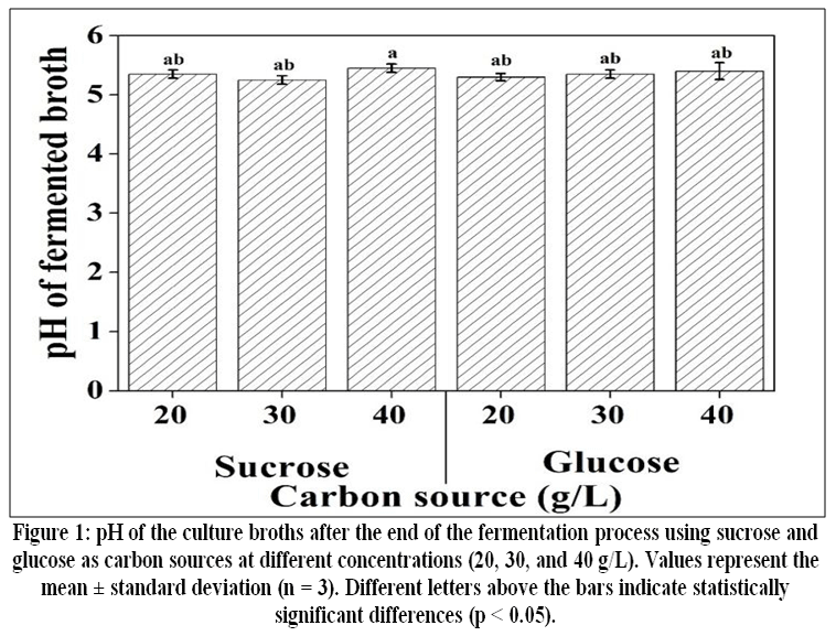 |
Figure 1: pH of the culture broths after the end of the fermentation process using sucrose and glucose as carbon sources at different concentrations (20, 30, and 40 g/L).
|
Influence of carbon source at different concentrations on mycelial biomass production
Mycelial growth varied significantly depending on the carbon source and concentration used in the fermentation medium. The highest mycelial biomass value was recorded using glucose as a carbon source at 30 g/L, reaching 8.27 ± 0.01 g/L, showing that this concentration was the most favorable for fungal development. Conversely, the lowest biomass production was observed under the condition with sucrose at 20 g/L, resulting in only 2.32 ± 0.01 g/L (Figure 2).
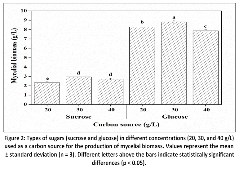 |
Figure 2: Types of sugars (sucrose and glucose) in different concentrations (20, 30, and 40 g/L) used as a carbon source for the production of mycelial biomass.
|
Yield of exopolysaccharides
The freeze-thaw method applied to the culture broth containing 40 g/L glucose resulted in the highest recovery of exopolysaccharides (EPS), reaching 4.53 ± 0.01 g/L (Figure 3a). In contrast, the exopolysaccharide precipitation technique with 95% ethanol (EPS) showed a maximum yield of 2.72 ± 0.01 g/L (Figure 3b). These results demonstrated that precipitation conditions significantly affect the yield of exopolysaccharides, and it is possible that there may be structural differences within a single polysaccharide or between different polysaccharides.
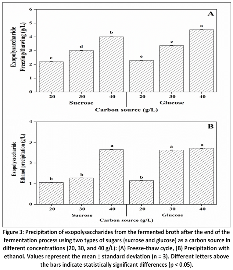 |
Figure 3: Precipitation of exopolysaccharides from the fermented broth after the end of the fermentation process using two types of sugars (sucrose and glucose) as a carbon source in different concentrations (20, 30, and 40 g/L):
|
Characterization of exopolysaccharides
SEM do EPS
In SEM analysis, the EPS structures exhibited a crystalline structure, with irregularly shaped blocks and a large, smooth surface area (Figure 4). Xu et al.37 extracted polysaccharides from G. lucidum with microstructures similar to those demonstrated in this work, with a square shape and flat surface.
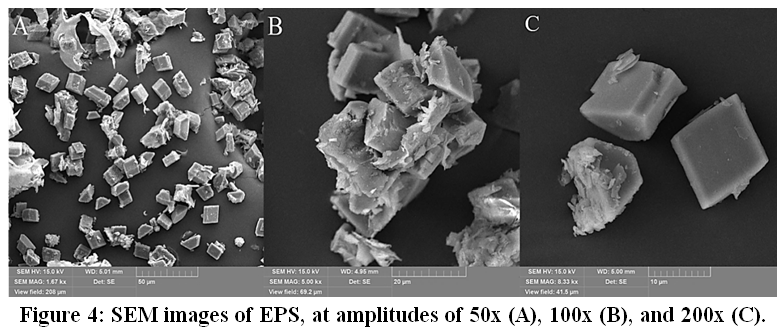 |
Figure 4: SEM images of EPS, at amplitudes of 50x (A), 100x (B), and 200x (C).
|
Fourier Transform Infrared Spectroscopy (FT-IR) of EPS
The FT-IR spectrum for EPS is shown in Figure 5. The strong signals at 3234 cm⁻¹ corresponded to the single-bond OH stretching vibration of carbohydrates.41 The next five bands in the 1404–1209 cm⁻¹ region were close to each other, and most of them were of low intensity. The signal at 1404 cm⁻¹ showed asymmetric and symmetric stretching vibrations of the carboxylate.42 The band around 1329 cm⁻¹ corresponded to the bending of the CH single bond into CH₃, while the bands at 1209 cm⁻¹ belong to the attenuated lipid.42A band at 849 cm-1 was characteristic of β-type glycosidic linkages.43,44
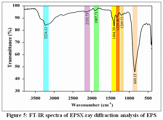 |
Figure 5: FT-IR spectra of EPSX-ray diffraction analysis of EPS
|
X-ray diffraction is a technique capable of analyzing the crystallinity of materials, being of great importance for verifying the structure of polysaccharides.45 Broad, rounded peaks typically indicate the presence of amorphous structures, while sharp diffraction peaks suggest the existence of crystalline structures.46,47 The overall diffraction peaks of EPS exhibited a broad profile with the presence of some sharp peaks (Figure 6).
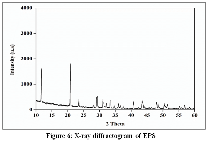 |
Figure 6: X-ray diffractogram of EPS
|
Color and UV analysis of EPS
The values for angle L*, a*, b*, chroma, and hue of EPS are shown in Table 1. The L* parameter is an approximate measure of lightness, in which each color can be considered equivalent to a member of the grayscale, between black and white.50
Table 1: CIELAB color parameters (L*, a*, and b*) of EPS.
| Color analysis | Value |
| L* = Lightness | 80.08 ± 0.59 |
| a* = redness | 0.53 ± 0.15 |
| b* = yellowness | 10.21 ± 0.58 |
| Chroma (C*) | 10.23 ± 0.55 |
| Hue angle (°) | 86.13 ± 2.17 |
The maximum value for luminosity is 100, indicating white, and L* for EPS was 80.08, which is close to luminosity. The a* parameter indicates positive values for red colors and negative values for greenish colors, while b* indicates positive values for yellow colors and negative values for bluish colors. The positive a* (0.53) and b* (10.21) values of EPS indicate reddish and yellowish colors. Chroma (C*) is considered one of the quantitative attributes of color, used to determine the degree of difference of a hue compared to a gray color with the same luminosity. The higher the chroma value, the greater the intensity of the color of the samples perceived by humans.51
The chroma values of EPS are closer to the b* values, and the hue angle was found to be 86.13. The hue angle (°) is considered the qualitative attribute of color, in which colors have traditionally been defined as red, green, etc., used to define the difference of a given color with reference to gray with the same luminosity.52 A low chroma value and a high luminosity value are desired for the product to meet consumer preferences. Thus, in this color study, EPS can be applied to food and/or pharmaceutical products as hydrocolloids due to its higher whiteness (80.08) and low chroma values (10.23). As shown in Figure 7, EPS did not absorb in the ultraviolet spectrum at 260 and 280 nm, suggesting the absence of nucleic acids and proteins.37,41
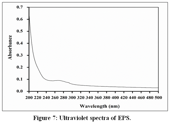 |
Figure 7: Ultraviolet spectra of EPS.
|
Analysis of monosaccharide composition
The fraction was identified as a heteropolysaccharide consisting mainly of Mannose (Man) (52%), Glucose (Glc) (24%), and Galactose (Gal) (17%) (Table 2). Many polysaccharides obtained from Ganoderma sp. are composed of similar monosaccharides.46,53 Other monosaccharides were identified as traces (Arabinose (Ara), Xylose (Xyl), and Rhamnose (Rha).54
Table 2: Neutral composition monosaccharide of polysaccharide fractions obtained from the exopolysaccharide from the submerged culture of G. sichuanense.
|
Component |
Neutral composition monosaccharide (mol%)a | ||||||
| Rha | Ara | Xyl | Man | Gal | Glc | ||
| EPS | 2.0 | 2.0 | 3.0 | 52.0 | 17.0 | 24.0 | |
NMR analysis
Comparing the results of monosaccharide analysis and NMR spectroscopy analysis, it can be inferred that the structure of the exopolysaccharide fraction obtained from the culture medium of Ganoderma sichuanense is mainly composed of mannose, galactose, and glucose units. The literature reports that polysaccharides composed of mannose and glucose, such as glucomannan, are common in fungi.57 HSQC analysis was performed, and some signals could be inferred for the units (Figure 8), which are listed in Table 3.
|
Figure 8: HSQC of EPS from Ganoderma sichuanense.
|
In the anomeric region, six signals could be identified. They were designated as residues from A to F, with the most prominent being at δ 99.3/5.12, 102.0/5.05, 99.3/4.90, 100.2/5.28, 97.7/4.98, and δ 98.1/5.09 ppm. These signals are suggested for →2,6)-α-D-Manp-(1→; →6)-α-D-Manp-(1→; →4)-α-D-Manp-(1→; →4)-α-D-Manp-(1→; →4)-α-D-Glcp-(1→; →6)-α-D-Glcp-(1→ and →6)-α-D-Galp-(1→). The units have the α configuration, which is in accordance with that reported by Cao et al.46
Table 3: 1H and 13C chemical shifts for EPS from submerged culture of G. sichuanense.
| Sugar units | Chemical shifts (ppm) | ||||||
| C1/H1 | C2/H2 | C3/H3 | C4/H4 | C5/H5 | C6/H6 | ||
| A | →2,6)-α-D-Manp-(1→ | 98.3
5.12 |
78.4
4.03 |
70.4
3.84 |
66.5
3.65 |
72.6
3.76 |
60.2
3.80 |
| B | →6)-α-D-Manp-(1→ | 102.0
5.05 |
78.3
4.11 |
76.2
4.19 |
69.8
3.98 |
72.1
3.84 |
60.7
3.75 |
| C | →4)-α-D-Manp-(1→ | 99.3
4.90 |
69.8
4.02 |
– | – | 76.2
4.21 |
69.1
3.44, 3.46 |
| D | →4)-α-D-Glcp-(1→ | 100.2
5.28 |
– | 76.2
4.11 |
81.2
3.88 |
74.0
4.06 |
62.1
3.80 |
| E | →6)-α-D-Glcp-(1→ | 97.7
4.98 |
68.9
3.48 |
– | 70.9
3.56 |
72.5
3.72 |
60.7
3.87 |
| F | →6)-α-D-Galp-(1→ | 98.1
5.09 |
69.8
3.90 |
73.8
4.03 |
70.8
3.54 |
69.2
4.21 |
66.6
3.63, 3.65 |
Discussion
The pH of the medium is a vital intrinsic factor, as it affects the ionic state of the fermentative microenvironment, as well as the structure, morphology, and physiological functions of fungal cells, and can modulate nutrient absorption and product biosynthesis.19 Studies indicate that pH stability varies according to the culture medium used in the submerged fermentation of G. sichuanense CC22. Among the media evaluated, POL showed greater stability throughout the fermentation process, with a minimal reduction in pH at the end of the cultivation (7th day) (data not shown). In contrast, the Modified Melin–Norkrans (MNM) medium underwent a more pronounced acidification, highlighting the influence of the medium composition on pH regulation during fermentation.21
The pH values observed in the Ganoderma fermented broth at the end of cultivation remained within a slightly acidic range, close to 5.3–5.5, regardless of the carbon source and concentration tested. Such pH maintenance indicates a robust homeostatic capacity of G. sichuanense CC22, effectively managing the acidic by-products of carbohydrate metabolism, even with the production of acidic metabolites resulting from carbohydrate consumption. In submerged fermentations of basidiomycetes, moderate pH reductions are common and are associated with the synthesis of organic acids, cellular respiration, and the secretion of extracellular polysaccharides, which contribute to medium acidification during mycelial growth.22
It is also observed that both glucose and sucrose resulted in statistically similar final pH values, indicating that both carbon sources were efficiently metabolized by the microorganism. This behavior has been reported in recent studies with Ganoderma, in which the assimilation of different sugars did not cause pronounced changes in the final pH, but influenced metabolic rate as well as biomass and EPS production. Maintaining the pH at moderately acidic levels is considered favorable for the activity of enzymes involved in polysaccharide biosynthesis and for mycelial growth, thereby contributing to fermentation yield.23
Thus, the results indicate that Ganoderma shows physiological robustness in response to variations in the carbon source of the medium, maintaining suitable conditions for secondary metabolism and exopolysaccharide production. This pH stability is an important factor in biotechnological processes, as it reduces the need for pH adjustments during fermentation and promotes cultivation standardization, which is essential for industrial and pharmaceutical applications involving fungal polysaccharides.24
Pessoa et al.25 evaluated the production of mycelial biomass, proteases, and intracellular protease inhibitors by G. lucidum CC22 cultivated under different submerged fermentation conditions, and observed that cultivation under agitated conditions and using glucose as a carbon source (20 g/L) showed the highest biomass production with a value of 7 g/L. Based on these studies, the literature describes that the carbon source metabolized by the fungus is directly related to the monomeric composition of the polysaccharide produced, although not always in the same proportion.26
The different extraction methods used in the precipitation of polysaccharides can impact both the yield and purity of the compounds obtained, being an essential step for the characterization and future applications of these biomolecules.27The precipitation of exopolysaccharides with ethanol resulted in lower yields than those obtained by the freeze-thaw method. One hypothesis for this difference is the removal of impurities and small lipophilic particles associated with the polysaccharides during the process.28,29 Additionally, Figure 3b shows the effect of precipitation by ethanol, since it is based on the reduction of the dielectric constant of the medium, forcing the precipitation of EPS due to the reduction of its solubility.
Submerged cultivation using high glucose concentrations results in elevated concentrations of exopolysaccharides (EPS) excreted by fungi.30-32 Studies demonstrate that the use of glucose is advantageous in EPS yield when compared to other sugars.18,32 In addition to the carbon source, the presence of Ca+2 ions in the culture medium favors the excretion of polysaccharides into the fermented medium, resulting in a significant increase in the production of exopolysaccharides (EPS) by various basidiomycetes.33,34
The influence of the initial pH value in the culture medium is another determining factor that can influence EPS production by G. sichuanense. According to Kim et al.,35 pH 6.0 favored optimal EPS production. García-Cruz et al.36 described pH 4.5 as optimal for EPS production by Lentinula edodes. These differences in pH favoring EPS may be linked to the morphology and physiology of Ganoderma sp. strains.
Due to its superior yield, the EPS fraction obtained by the freeze-thaw method with 40% glucose was prioritized for further structural characterization.
In SEM analysis, the structure of polysaccharides exhibits great diversity, making it difficult for the same conformation to occur among different compounds. However, structural alterations resulting from treatments, such as precipitation methods, can influence the morphology of polysaccharides.38-40 Previous studies have shown that ultrasound can alter the apparent structure of G. lucidum polysaccharides, significantly increasing their surface area.37
The material possibly exhibits both crystalline and amorphous structures, with the amorphous state representing a relatively larger proportion.48 This pattern reflects the presence of a variety of bioactive components, such as triterpenoids and polysaccharides, which contribute to its unique biochemical composition. This combination indicates that G. sichuanense has a more complex and heterogeneous structure with both crystalline and amorphous phases.46 Due to their large and irregular molecular arrangements, polysaccharides typically form amorphous or semicrystalline regions, findings that align with those of Gao et al.49regarding the structural characteristics of glycoproteins in shiitake mushrooms.
The influence of the culture medium composition on structural characteristics can be a factor that expresses the variation in the percentages of monosaccharides that form EPS, as well as the cultivation conditions. Nitrogen sources can affect the ability of Ganoderma sp. to produce EPS under fermentation conditions, as well as combinations of carbon and nitrogen sources that vary the molecular mass of the EPS produced.18 In the submerged fermentation of Ganoderma sp. under various cultivation conditions with glucose as the sole carbon source, the main monosaccharide components in EPS are consistently glucose, galactose, and mannose.55
In EPS overexpression experiments in Ganoderma species conducted by Zheng et al.56 and Ai-lati et al.,57 several fractions were isolated and analyzed according to their monosaccharide composition, which were constructed from glucose, mannose, and galactose, thus corroborating what was found in this study.
According to NMR analyses, a signal is observed at δ 57.4/3.63, which may be associated with the presence of O-Me substitution in galactose units. Thus, the unit described for →6)-α-D-Galp-(1→) could be identified as →6)-3-O-Me-)-α-D-Galp-(1→).58
All signals were assigned by comparing them with literature values for similar polysaccharides.43,59-62 Unlike cell wall polysaccharides, the exopolysaccharides found in fungi cultivated in liquid medium have a mannose and glucose backbone with galactose substitutions in proportions quite close to those described for the fraction under study, also implying quite similar 13C-1H HSQC signals.
Conclusion
Our findings demonstrate that both the choice of carbon source and the EPS recovery strategy are critical determinants of exopolysaccharide yield, structural characteristics, and fungal biomass production by G. sichuanense. Glucose proved to be a more effective carbon source than sucrose, promoting greater fungal growth and EPS production, while increasing glucose concentrations in the culture medium were positively correlated with enhanced EPS yields. Furthermore, the freeze–thaw recovery method significantly improved EPS recovery and may have contributed to preserving or enhancing the structural integrity of these macromolecules. Overall, these results demonstrate the potential of G. sichuanense as a promising producer of exopolysaccharides with physicochemical characteristics that support their prospective application in pharmaceutical and biotechnological fields.
Acknowledgement
The authors express their gratitude to the Edible Fungi Cultivation Laboratory of the National Institute for Amazonian Research (LCFC-INPA), the Postgraduate Program in Biotechnology (PPGBIOTEC), and the Postgraduate Program in Biodiversity and Biotechnology of the Bionorte Network (PPGBIONORTE) for the opportunity to pursue their doctoral studies.
Funding Sources
National Council for Scientific and Technical Development (CNPq) (Process number DTI-A 382922/2024-7), the Amazonas State Research Support Foundation (FAPEAM) (Process (Resolution nº 005/2022-CD/FAPEAM and nº 002/2023-CD/FAPEAM) and the Coordination for the Improvement of Higher Education Personnel (CAPES) for granting research and doctoral scholarships. To the FAPEAM Project (PROSGRAD/2022-2023/PPGBIOTEC), for funding the publication and research. Acknowledgments to the LABMAT Laboratories of UFAM, to LAMESP, and to the NMRLab of the Analytical Center of UFAM, the Nanotechnology Laboratory of IFAM, to FINEP, FAPEAM (FIXAM/AM grant number 062.00917/2015), and to CNPq.
Conflict of Interest
The authors do not have any conflict of interest.
Data Availability Statement
This statement does not apply to this article.
Ethics Statement
This research did not involve human participants, animal subjects, or any material that requires ethical approval.
Informed Consent Statement
This study did not involve human participants, and therefore, informed consent was not required.
Clinical Trial Registration
This research does not involve any clinical trials.
Permission to reproduce material from other sources
Not Applicable.
Author Contributions
- Sérgio Oliveira Júnior – Conceptualization, Data curation, Formal analysis, Investigation, Methodology, Validation, Visualization, Writing – original draft, Writing – review & editing
- Larissa Soares – Data curation, Formal analysis, Investigation, Methodology, Validation, Writing – original draft, Writing – review & editing
- Aldenora Vasconcelos – Conceptualization, Data curation, Formal analysis, Investigation, Validation, Writing – original draft, Writing – review & editing
- Lorena Aguiar – Conceptualization, Data curation, Formal analysis, Investigation, Validation, Writing – original draft, Writing – review & editing
- Luciano Campestrini – Conceptualization, Data curation, Formal analysis, Investigation, Methodology, Writing – original draft, Writing – review & editing
- Jaqueline Bezerra – Data curation, Formal analysis, Methodology, Visualization, Writing – original draft, Writing – review & editing
- Larissa Ramos Chevreuil – Conceptualization, Data curation, Formal analysis, Writing – original draft, Writing – review & editing
- Everaldo Silvino dos Santos – Data curation, Formal analysis, Visualization, Writing – original draft, Writing – review & editing
- Ceci Sales-Campos – Conceptualization, Data curation, Formal analysis, Funding acquisition, Project administration, Resources, Supervision, Visualization, Writing – original draft, Writing – review & editing
References
- Yang Z, Chen C, Zhao J, et al. Hypoglycemic mechanism of a novel proteoglycan, extracted from Ganoderma lucidum, in hepatocytes. European Journal of Pharmacology. 2018;820:77-85. doi:10.1016/j.ejphar.2017.12.020
CrossRef - Sheikha E, Farag A. Nutritional Profile and Health Benefits of Ganoderma lucidum “Lingzhi, Reishi, or Mannentake” as Functional Foods: Current Scenario and Future Perspectives. Foods. 2022;11(7):1030. doi:10.3390/foods11071030
CrossRef - Ekiz E, Oz E, Abd El-Aty AM, et al. Exploring the Potential Medicinal Benefits of Ganoderma lucidum: From Metabolic Disorders to Coronavirus Infections. Foods. 2023;12(7):1512. doi:10.3390/foods12071512
CrossRef - Li X, Sossah F.L, Tuo Y, et al. Characterization and fungicide sensitivity of Trichoderma species causing green mold of Ganoderma sichuanense in China. Front Microbiol. 2023;14. doi:10.3389/fmicb.2023.1264699
CrossRef - Apiwatanapiwat W, Janchai P, Vaithanomsat P, et al. A. Production of polysaccharide, triterpenoid, and phenolic compounds in antler-type Ganoderma sichuanense grown with fruit peels supplementation in liquid culture. Discov Food. 2025;5(1):157. doi:10.1007/s44187-025-00451-1
CrossRef - Cateni F, Gargano M.L, Procida G, Venturella G, Cirlincione F, Ferraro V. Mycochemicals in wild and cultivated mushrooms: nutrition and health. Phytochem Rev. 2022;21(2):339-383. doi:10.1007/s11101-021-09748-2
CrossRef - Shevchuk Y, Kuypers K, Janssens G.E. Fungi as a source of bioactive molecules for the development of longevity medicines. Ageing Research Reviews. 2023;87:101929. doi:10.1016/j.arr.2023.101929
CrossRef - Ionescu M, Dincă M.N, Ferdeș M, Zăbavă B. Ș, Paraschiv G, Moiceanu G. Proteins from Edible Mushrooms: Nutritional Role and Contribution to Well-Being. Foods. 2025;14(18):3201. doi:10.3390/foods14183201
CrossRef - Niego A.G, Rapior S, Thongklang N, et al. Macrofungi as a Nutraceutical Source: Promising Bioactive Compounds and Market Value. Journal of Fungi. 2021;7(5):397. doi:10.3390/jof7050397
CrossRef - Wen L, Sheng Z, Wang J, Jiang Y, Yang B. Structure of water-soluble polysaccharides in spore of Ganoderma lucidum and their anti-inflammatory activity. Food Chemistry. 2022;373:131374. doi:10.1016/j.foodchem.2021.131374
CrossRef - Zhao Y, Li Q, Wang M, et al. Structural characterization of polysaccharides after fermentation from Ganoderma lucidum and its antioxidant activity in HepG2 cells induced by H2O2. Food Chemistry: X. 2023;18:100682. doi:10.1016/j.fochx.2023.100682
CrossRef - Karunarathna S.C, Ediriweera A, Prasannath K, Mingfei Y, Hapuarachchi K.K. Exploring the health benefits of Ganoderma: bioactive compounds and mechanisms of action; immunomodulatory, and anti-tumour activities. New Zealand Journal of Botany. 2025;63(5):2325-2409. doi:10.1080/0028825X.2024.2375996
CrossRef - Petryn T, Nagalievska M, Chrzanowski G, Wasser S, Sybirna N. Therapeutic potential of the medicinal mushroom Ganoderma lucidum in metabolic syndrome: Antioxidant, hypoglycemic, and hepatoprotective activities. Biomedicine & Pharmacotherapy. 2026;196:119138. doi:10.1016/j.biopha.2026.119138
CrossRef - Wang M, Guo X, Chen J, et al. Antitumor Effects of Ganoderma lucidum polysaccharides and their Clinical Applications: An Updated Review. Natural Product Communications. 2026;21(3):1934578X261429763. doi:10.1177/1934578X261429763
CrossRef - Leng Y, Wang F, Chen C, et al. Protective Effect of Ganoderma lucidum Spore Powder on Acute Liver Injury in Mice and its Regulation of Gut Microbiota. Front Biosci (Landmark Ed). 2023;28(2). doi:10.31083/j.fbl2802023
CrossRef - Wang X, Wang X, Zhao L, Zhou F. Clinical Evaluation of Ganoderma lucidum Spore Oil for Triglyceride Reduction: A Randomized, Double-Blind, Crossover Study. Nutrients. 2025;17(5):844. doi:10.3390/nu17050844
CrossRef - Kumla J, Thangrongthong S, Kaewnunta A, Suwannarach N. Research advances in fungal polysaccharides: production, extraction, characterization, properties, and their multifaceted applications. Front Cell Infect Microbiol. 2025;15. doi:10.3389/fcimb.2025.1604184
CrossRef - Liu L, Feng J, Gao K, et al. Influence of carbon and nitrogen sources on structural features and immunomodulatory activity of exopolysaccharides from Ganoderma lucidum. Process Biochemistry. 2022;119:96-105. doi:10.1016/j.procbio.2022.05.016
CrossRef - Shen M, Xu M, Liu J, et al. Carbon starvation drives biosynthesis of high mannose-composition polysaccharides in Ganoderma lucidum. International Journal of Biological Macromolecules. 2024;282:137168. doi:10.1016/j.ijbiomac.2024.137168
CrossRef - Cavazzoni V, Adami A. Exopolysaccharides produced by mycelial edible mushrooms. Italian Journal of Food Science. 1992:9-15.
- Oliveira-Junior S.D, Silva G.L, Pessoa V.A, et al. Adding-value to Ganoderma lingzhi by producing enzymes and antioxidant compounds under submerged fermentation using different culture media. Braz J Biol. 2024;84:e283882. doi:https://doi.org/10.1590/1519-6984.283882
CrossRef - Fang Q.H, Zhong J.J. Effect of initial pH on production of ganoderic acid and polysaccharide by submerged fermentation of Ganoderma lucidum. Process Biochemistry. 2002;37(7):769-774. doi:10.1016/S0032-9592(01)00278-3
CrossRef - Supramani S, Ahmad R, Ilham Z, Annuar M.S.M, Klaus A, Wan-Mohtar W.A.A.Q.I. Optimisation of biomass, exopolysaccharide and intracellular polysaccharide production from the mycelium of an identified Ganoderma lucidum strain QRS 5120 using response surface methodology. AIMSMICRO. 2019;5(1):19-38. doi:10.3934/microbiol.2019.1.19
CrossRef - Guo J, Tang C, Liu Y, et al. Innovative submerged directed fermentation: Producing high molecular weight polysaccharides from Ganoderma lucidum. Food Chem. 2025;471:142759. doi:10.1016/j.foodchem.2025.142759
CrossRef - Pessoa V.A, Soares L.B.N, Silva G.L, et al. Production of mycelial biomass, proteases and protease inhibitors by Ganoderma lucidum under different submerged fermentation conditions. Braz J Biol. 2023;83:e270316. doi:https://doi.org/10.1590/1519-6984.270316
CrossRef - Mahapatra S, Banerjee D. Fungal Exopolysaccharide: Production, Composition and Applications. Microbiol Insights. 2013;6:1-16. doi:10.4137/MBI.S10957
CrossRef - Sun Y, He H., Wang Q, Yang X, Jiang S, Wang D. A Review of Development and Utilization for Edible Fungal Polysaccharides: Extraction, Chemical Characteristics, and Bioactivities. Polymers. 2022;14(20):4454. doi:10.3390/polym14204454
CrossRef - Donot F, Fontana A, Baccou J.C, Schorr-Galindo S. Microbial exopolysaccharides: Main examples of synthesis, excretion, genetics and extraction. Carbohydrate Polymers. 2012;87(2):951-962. doi:10.1016/j.carbpol.2011.08.083
CrossRef - Yang R.F, Zhao C, Chen X, Chan S. W, Wu J.Y. Chemical properties and bioactivities of Goji (Lycium barbarum) polysaccharides extracted by different methods. Journal of Functional Foods. 2015;17:903-909. doi:10.1016/j.jff.2015.06.045
CrossRef - Xu S, Xu J, Zeng W, Shan X, Zhou J. Efficient biosynthesis of exopolysaccharide in Candidaglabrata by a fed-batch culture. Front Bioeng Biotechnol. 2022;10. doi:10.3389/fbioe.2022.987796
CrossRef - Torun G, Yeşilada Ö, Boran F. Investigation the Exopolysaccharide Production Potential of Newly Isolated Trametes versicolor during Repeated-Batch Fermentation. ADYU J SCI. 2025;15(1):35-50. doi:10.37094/adyujsci.1619252
CrossRef - Prathumpai W., Pinruan U., Sommai S., Komwijit S., Malairuang K. Exopolysaccharide (EPS) Production by Endophytic and Basidiomycete Fungi. Fermentation. 2025;11(4):183. doi:10.3390/fermentation11040183
CrossRef - Ye L, Liu S, Xie F, Zhao L, Wu X. Enhanced production of polysaccharides and triterpenoids in Ganoderma lucidum fruit bodies on induction with signal transduction during the fruiting stage. PLOS ONE. 2018;13(4):e0196287. doi:10.1371/journal.pone.0196287
CrossRef - Adil B, Xiang Q, He M, et al. Effect of sodium and calcium on polysaccharide production and the activities of enzymes involved in the polysaccharide synthesis of Lentinus edodes. AMB Expr. 2020;10(1):47. doi:10.1186/s13568-020-00985-w
CrossRef - Kim H.M, Park M.K, Yun J.W. Culture pH affects exopolysaccharide production in submerged mycelial culture of Ganoderma lucidum. Appl Biochem Biotechnol. 2006;134(3):249-262. doi:10.1385/ABAB:134:3:249
CrossRef - García-Cruz F, Durán-Páramo E, Garín-Aguilar M.A, Valencia del Toro G., Chairez I. Parametric characterization of the initial pH effect on the polysaccharides production by Lentinula edodes in submerged culture. Food and Bioproducts Processing. 2020;119:170-178. doi:10.1016/j.fbp.2019.10.016
CrossRef - Xu Y, Zhang X, Yan X.H, et al. Characterization, hypolipidemic and antioxidant activities of degraded polysaccharides from Ganoderma lucidum. International Journal of Biological Macromolecules. 2019;135:706-716. doi:10.1016/j.ijbiomac.2019.05.166
CrossRef - Li Y, Yang X, Yang W, et al. Physicochemical characterization and immune activity of water-extract polysaccharides by stepwise ethanol precipitation from wild Cordyceps sinensis. Chem Biol Technol Agric. 2025;12(1):148. doi:10.1186/s40538-025-00866-z
CrossRef - Xu J, Yue R.Q, Liu J, et al. Structural diversity requires individual optimization of ethanol concentration in polysaccharide precipitation. International Journal of Biological Macromolecules. 2014;67:205-209. doi:10.1016/j.ijbiomac.2014.03.036
CrossRef - Xue H, Li P, Bian J, Gao Y, Sang Y, Tan J. Extraction, purification, structure, modification, and biological activity of traditional Chinese medicine polysaccharides: A review. Front Nutr. 2022;9. doi:10.3389/fnut.2022.1005181
CrossRef - Zhao L, Dong Y, Chen G, Hu Q. Extraction, purification, characterization and antitumor activity of polysaccharides from Ganoderma lucidum. Carbohydrate Polymers. 2010;80(3):783-789. doi:10.1016/j.carbpol.2009.12.029
CrossRef - Seedevi P, Moovendhan M, Vairamani S, Shanmugam A. Structural characterization and biomedical properties of sulfated polysaccharide from the gladius of Sepioteuthis lessoniana (Lesson, 1831). International Journal of Biological Macromolecules. 2016;85:117-125. doi:10.1016/j.ijbiomac.2015.12.066
CrossRef - Li J, Gu F, Cai C, et al. Purification, structural characterization, and immunomodulatory activity of the polysaccharides from Ganoderma lucidum. International Journal of Biological Macromolecules. 2020;143:806-813. doi:10.1016/j.ijbiomac.2019.09.141
CrossRef - Zheng Q, Ren D, Yang N, Yang X. Optimization for ultrasound-assisted extraction of polysaccharides with chemical composition and antioxidant activity from the Artemisia sphaerocephala Krasch seeds. International Journal of Biological Macromolecules. 2016;91:856-866. doi:10.1016/j.ijbiomac.2016.06.042
CrossRef - Jeddou K.B, Chaari F, Maktouf S, Nouri-Ellouz O, Helbert C.B, Ghorbel R.E. Structural, functional, and antioxidant properties of water-soluble polysaccharides from potatoes peels. Food Chemistry. 2016;205:97-105. doi:10.1016/j.foodchem.2016.02.108
CrossRef - Cao C, Liao Y, Yu Q, et al. Structural characterization of a galactoglucomannan with anti-neuroinflammatory activity from Ganoderma lucidum. Carbohydrate Polymers. 2024;334:122030. doi:10.1016/j.carbpol.2024.122030
CrossRef - Meftah N, Hani A, Merdas A. Extraction and Physicochemical Characterization of Highly-pure Amorphous Silica Nanoparticles from Locally Available Dunes Sand. Chemistry Africa. 2023;6(6):3039-3048. doi:10.1007/s42250-023-00688-2
CrossRef - Liang Z, Yin Z, Liu X, et al. A glucomannogalactan from Pleurotus geesteranus: Structural characterization, chain conformation and immunological effect. Carbohydrate Polymers. 2022;287:119346. doi:10.1016/j.carbpol.2022.119346
CrossRef - Gao J, Li X, Jia S, Zeng H, Zheng B. Structural characterization and antioxidant activity of a glycoprotein isolated from shiitake mushrooms. Food Bioscience. 2023;53:102608. doi:10.1016/j.fbio.2023.102608
CrossRef - Granato D, Masson M.L. Instrumental color and sensory acceptance of soy-based emulsions: a response surface approach. Food Sci Technol. 2010;30:1090-1096. doi:https://doi.org/10.1590/S0101-20612010000400039
CrossRef - Devi P.B, Kavitake D, Shetty P.H. Physico-chemical characterization of galactan exopolysaccharide produced by Weissella confusa KR780676. International Journal of Biological Macromolecules. 2016;93:822-828. doi:10.1016/j.ijbiomac.2016.09.054
CrossRef - Pathare P.B, Opara U.L, Al-Said F.A.J. Colour Measurement and Analysis in Fresh and Processed Foods: A Review. Food Bioprocess Technol. 2013;6(1):36-60. doi:10.1007/s11947-012-0867-9
CrossRef - Bleha R, Třešnáková L, Sushytskyi L, et al. Polysaccharides from Basidiocarps of the Polypore Fungus Ganoderma resinaceum: Isolation and Structure. Polymers. 2022;14(2):255. doi:10.3390/polym14020255
CrossRef - Wang Z, Zhang H, Shen Y, et al. Characterization of a novel polysaccharide from Ganoderma lucidum and its absorption mechanism in Caco-2 cells and mice model. International Journal of Biological Macromolecules. 2018;118:320-326. doi:10.1016/j.ijbiomac.2018.06.078
CrossRef - Wu Z.W, Zhao X.F, Quan C.X, et al. Structure–function insights of natural Ganoderma polysaccharides: advances in biosynthesis and functional food applications. Nat Prod Bioprospect. 2025;15(1):15. doi:10.1007/s13659-025-00496-w
CrossRef - Zheng W, Lan S, Zhang W, et al. Polysaccharide structure evaluation of Ganoderma lucidum from different regions in China based on an innovative extraction strategy. Carbohydrate Polymers. 2024;335:122079. doi:10.1016/j.carbpol.2024.122079
CrossRef - Ai-lati A, Liu S, Ji Z, Zhang H, Mao J. Structure and bioactivities of a polysaccharide isolated from Ganoderma lucidum in submerged fermentation. Bioengineered. 2017;8(5):565-571. doi:10.1080/21655979.2017.1283459
CrossRef - Silveira M.L.L, Smiderle F.R, Agostini F, et al. Exopolysaccharide produced by Pleurotus sajor-caju: Its chemical structure and anti-inflammatory activity. International Journal of Biological Macromolecules. 2015;75:90-96. doi:10.1016/j.ijbiomac.2015.01.023
CrossRef - Wang N, Jia G, Wang C, et al. Structural characterisation and immunomodulatory activity of exopolysaccharides from liquid fermentation of Monascus purpureus (Hong Qu). Food Hydrocolloids. 2020;103:105636. doi:10.1016/j.foodhyd.2019.105636
CrossRef - Lu H, Liu S, Zhang S, Chen Q. Light Irradiation Coupled with Exogenous Metal Ions to Enhance Exopolysaccharide Synthesis from Agaricus sinodeliciosus ZJU-TP-08 in Liquid Fermentation. Journal of Fungi. 2021;7(11):992. doi:10.3390/jof7110992
CrossRef - Huang J, Su Y, Wang Q, et al. A glucomannan from defatted Ganoderma lucidum spores: structural characterization and immunomodulatory activity via activating TLR4/MyD88/NF-κB signaling pathway. International Journal of Biological Macromolecules. 2025;294:139195. doi:10.1016/j.ijbiomac.2024.139195
CrossRef - Manna D.K, Maity P, Nandi A.K, et al. Structural elucidation and immunostimulating property of a novel polysaccharide extracted from an edible mushroom Lentinus fusipes. Carbohydrate Polymers. 2017;157:1657-1665. doi:10.1016/j.carbpol.2016.11.048
CrossRef
Accepted on: 06-07-2026
Second Review by: Dr. Kirti Dubli
Final Approval by: Dr. Eugene A. Silow