

Manuscript accepted on :
Published online on: --
S.H.A. Ehtisham1 and S.U. Gulavane2
1Department of Animal Husbandry Govt. of Jammu and Kashmir - 192 301, India 2Bombay Veterinary College, Associate Professor in Animal Reproduction Parel Mumbai - 400 012, India.
DOI : http://dx.doi.org/http://dx.doi.org/10.13005/bbra/1081

ABSTRACT:
The study was performed on 60 goats at Livestock farm at Unit No. 3, Arrey milk Colony Goregaon and adjoining areas of Mumbai. A total of 60 cross bred and non-descript does of which, 20 does were had history of known dates of mating at least one month back and 50 does had history of mating with unknown dates. Three scans were performed on each doe 15 to 30 days apart with Aloka SD 500 equipment having transabdominal 3.5 MHz sector transducer. The accuracy, sensitivity and specificity of the method were evaluated. An accuracy of 93.33 to 100% was detected. It was found out that the visualization of placentomes, the embryo with detection of cardiac activity and foetal movements were reliable criteria for ruling out the occurrence of early embryonic foetal death.
KEYWORDS: Goats; Pregnancy; Ultrasonographic.
| Copy the following to cite this article: Ehtisham S. H. A, Gulavane S. U. Transabdominal Ultrasonographic Diagnostics of Early Pregnancy in Goats from Mumbai and Adjoining Areas. Biosci Biotech Res Asia 2012;9(2) |
| Copy the following to cite this URL: Ehtisham S. H. A, Gulavane S. U. Transabdominal Ultrasonographic Diagnostics of Early Pregnancy in Goats from Mumbai and Adjoining Areas. Biosci Biotech Res Asia 2012;9(2). Available from: https://www.biotech-asia.org/?p=10225 |
Introduction
The early and precise detection of pregnancy in goats is especially important from an economical point of view. A reliable means of distinguishing pregnant from non-pregnant goats would enable herdsmen to remove non-pregnant animals from the flock and so save on expenses for feed, labour, vaccinations, etc. Moreover, special attention and supplementary feed can then be given to the pregnant animals. The separation of flocks into pregnant and non-pregnant permits a scheduling of the technology of breeding, reduction of reproductive and production losses in form of abortions, stillbirths and production of weak lambs and optimization of the live weight of mothers and neonates (Fowler & Wilkins, 1984; Bretzlaff et al., 1993; Haibel, 1990). Using the transabdominal method, Gearhart et al. (1988) detected without doubt pregnancy by day 25 on the basis of enlarged uterine lumen and the embryo whereas Kähn et al. (1993) reported an accuracy of 95%, sensitivity of 95% and specificity of 91% between post insemination days 22 and 90. Kaulfuss et al. (1996a) reported an accuracy of 80% by post insemination day 21 and 100% by post insemination day 60. According to most authors (Kähn et al., 1992; Kaulfuss et al., 1996b), the precise diagnosis in the early stage of the pregnancy should be based not only on the detection of an enlarged uterine lumen but on the presence of an embryo as well in order to eliminate the false positive findings. Furthermore, they reported that the approach and the frequency of used transducer were of primary significance for the accuracy, sensitivity and specificity of the method.
Real-time (B-mode) ultrasonography, available since the early 1980s, but initially considered too time consuming and expensive for use in farm animals (Lindahl, 1966, Memon and Ott, 1980 and Thwaites, 1981), has proved to be a reliable means of pregnancy detection in a number of domestic species (Kaehn, 1991 and Kaehn, 1992). Although there are many reports on transcutaneous and transrectal ultrasonography in sheep (Taverne et al., 1985, Buckrell, 1988, Garcia et al., 1993, Kaehn et al., 1993 and Schrick and Inskeep, 1993), there is a paucity of information on the suitability of this technique for use in goats. The aim of the present study was to examine the possibilities of transabdominal ultrasonography for detection of early pregnancy and to follow out the changes occurring in the enlarging uterine lumen and the embryo as parameters for confirmation of pregnancy in goats at Livestock farm at Unit No. 3, Arrey milk Colony Goregaon (Mumbai) and adjoining areas of Mumbai.
Materials and Methods
The study was performed on 60 goats at Livestock farm at Unit No. 3, Arrey milk Colony Goregaon and adjoining areas of Mumbai.
Ultrasonographic examination.
A real time, B mode, portable Ultrasonographic machine SD – 500 Aloka Co. Ltd., Japan. with 3.5MHz transabdominal sector transducer was used for scanning of the uterus. During scanning the relevant images\events were frozen. All relevant pictures were printed using electronic thermal printer attached to the machine. The selected animals needed no special preparation except does which had long hairs need shaving of area cranial to udder. Sometimes in advanced gestation clipping of hair cranial to udder was carried out to get better image. Animals were held firmly by two attendants in dorsal recumbency for proper restraint which was necessary to get better Ultrasonographic images. All the does were scanned in dorsal recumbency. Each horn was scanned separately by tilting the does to right and left side. In late gestation it was sometimes helpful to manually push the abdomen towards the transducer to get the uterus within range of transducer as reported by Habiel (1990) and Kahn (1992). An abundant ultrasound coupling gel was smeared on abdomen and on probe to increase the conductivity of ultrasound waves and to avoid clipping of hair. Clipping of an area just above the udder was done in some cases in late gestation when the uterus was pushed more cranially into the abdomen.
The bladder served as point of reference and the uterine horns were located slightly cranially. The entire reproductive tract was inspected by moving the probe gently in different directions and rotating it 180° clockwise and counters clockwise.
Pregnancy Diagnosis
The pregnancy was detected using the following parameters: Presence of an enlarged uterine lumen with amniotic fluid and presence of an embryo. The following indices were determined: Accuracy (number of confirmed diagnoses ÷ total number of diagnoses), sensitivity (number of confirmed positive diagnoses ÷ total number of positive diagnoses) and specificity (number of confirmed negative diagnoses ÷ total number of negative diagnoses).
Results
In 60 animals studied, an enlarged uterine lumen as an anechoic zone located cranially or ventrally to the urinary bladder was visible. This finding was absent in non pregnant animals. An animal was considered pregnant when the fluid filled gestational sac in the uterus, and the cotyledons and/or foetal parts were recognized. On this ground, positive diagnosis of pregnancy was assumed in 27 goats, whereas 33 were determined as non-pregnant. During the next examination 15 days apart, 24 out of 27 were found pregnant and 32 out of 33 were found non pregnant. All the goats were scanned third time to confirm diagnosis based on foetal- movements, heartbeats and placentomes detection. This was confirmed at delivery. The accuracy, sensitivity and specificity of the Ultrasonographic pregnancy diagnosis on second scan were 93.33%, 88.89% and 96.96% respectively. On third scan it was 100% on all the three parameters.
Discussion
The detection of early pregnancy in goats via ultrasonography depends primarily on the approach, the frequency of used transducer and the experience of the investigator (Fowler and Wilkins, 1984; Karen et al., 2001). The accuracy, sensitivity and specificity on second scan observed were similar to those reported by Kaulfuss et al. (1996a) and Yotov (2005) but different from those of Kähn et al. (1993) in Merino sheep who used a transducer with 5 MHz frequency. The accuracy, sensitivity and specificity on third scan were similar to Amer (2008), Medan et al. (2004), and Yotov (2005) observed were similar to those reported by Kaulfuss et
|
Ultrasound image of early pregnant uterus. Arrows showing fluid filled sacs oblong or round in shape. |
Ultrasound image of non pregnant uterus filled but filled with fluid but not oblong or round in shape (hydrometra).
|
|
Ultrasound image of late pregnant uterus. Arrows showing Large ‘C’ shaped placentome.
|
Ultrasound image of early pregnant uterus. Arrow showing hyperechoic embryo.
|
Ultrasonographic scans of pregnant uterus of goats.
(1996a) but different from those of Kähn et al. (1993) in Merino sheep that used a transducer with 5 MHz frequency. The differences, in our opinion, are transducer, species and breed dependent. The early embryonic death that, according to Michels et al. (1998) is the highest between postinsemination days 3_26 could be assumed as cause for the false positive findings In such cases, the anechoic area within the uterus is not observed during a second examination, and when an embryo or a placentome is present, they do not grow. The false negative findings could probably be attributed to the early period of the study when the embryo is commonly not visualized and the enlarged uterine lumen could be due to accumulation of
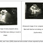 |
Figure 1
|
Table 1: Comparison of ultrasonographic findings with those observed at parturation in 60 goats.
| Findings |
1st scan |
2nd scan |
3rd scan |
Parturient goats |
| Number of positive diagnosis | 27 | 24 | 24 | 24 |
| Number of negative diagnosis | 33 | 32 | 32 | 32 |
| Accuracy (%) | – | 93.33 | 100 | – |
| Sensitivity (%) | – | 88.99 | 100 | |
| Specificity (%) | 96.96 | 100 |
oestral secretion or due to some kind of uterine pathology. The values of Ultrasonographic parameters, obtained in the present study allowed to assume that the increase detection of heart rate, foetus, foetal movements and placentome were precise criteria for ruling out the embryonic, and the foetal death. Our data could be used for elaboration of an embryonic foetal growth profile and for determination of the gestation age of the foetus in the studied goat breeds.
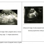 |
Figure 2
|
Conclusions
Transabdominal ultrasonography with a 3.5 MHz transducer could be used for detection of early pregnancy in the goat breeds with accuracy of 100%. It was found out that the visualization of placentomes, the embryo with detection of cardiac activity and foetal movements were reliable criteria for ruling out the occurrence of early embryonic foetal death.
References
- Amer A H, 2008. Determination of first pregnancy and foetal measurements in Egyptian Baladi goats. In Veterinaria Italia. 44(2), 429-37.
- Baronet, D. & D. Vaillancourt, 1989. Diagnostic de gestation par échotomographie chez la chèvre. Médicine Vétérinaire Québec, 19, 67_72.
- Bretzlaff, K., J. Edward, D. Forrest & L. Nuti, 1993. Ultrasonographic determination of pregnancy in small ruminants. Veterinary Medicine, 88, 12_24.
- Chalhoub, M., M. D. Lopes, C. N. Prestes & I. A. Ribeiro Filho, 2001. Prefil ultrasonografico
- do crescimento embrionario/ fetal ovino do 21 ao 41 dia de gestacao. Revista Brasiliera Saude Reprodução Animal, 2, 65_68.
- Fowler, D. G & F. J. Wilkins, 1984. Diagnosis of pregnancy and number of the fetuses in sheep by real-time ultrasonic imaging. 1. Effect of number of fetuses, stage of gestation, operator and breed of ewe on accuracy of diagnosis. Livestock Production Science, 11, 437_450.
- Gearhart, M. A., E. W. Wingfield, A. P. Knight, A. J. Smith, A. D. Daraatz, A. J. Boon & A. C. Stokes, 1988. Real-time ultrasonography for determining pregnancy status and viable fetal numbers in ewes. Theriogenology, 30, 323 – 337.
- Haibel, G. K., 1990. Use of ultrasonography in reproductive management of sheep and goat herds. Veterinary Clinics of North America _ Food Animal Practice, 3, 597_613.
- Kähn, W., B. Kähn, A. Richter, J. Schulz & M, Wolf, 1992. Sonography during the pregnancy of sheep. I. Fetometry for determination of the stage of gestation and prediction of the time of parturition. Deutsche Tierärztliche Wochenschrift, 99, 449–452.
- Kähn, W., J. Achtzehn, B. Kahn, A. Richter, J. Schulz & M. Wolf, 1993. Sonography of pregnancy in sheep. II Accuracy of transrectal and transcutaneous pregnancy diagnosis. Deutsche Tierärztliche Wochenschrift, 100, 29–31.
- Karen, A., P. Kovacs, F. J. Beckers & O. Szenci, 2001. Pregnancy diagnosis in sheep. Review of the most practical methods. Acta Veterinaria (Brno), 70, 116_126.
- Kaulfuss, K. H., N. Zipper, J. May, & R. Sub, 1996a. Ultrasonic pregnancy diagnosis (Bmode) in sheep, Comparative studies using transcutaneous and transrectal pregnancy diagnosis. Tierärztliche Praxix, 24, 559–566.
- Kaulfuss, K. H., K. Uhlich, S. Brabant, K. Blume & K. Strittmatter, 1996b. Real-time ultrasonic pregnancy diagnosis (B-mode) in sheep. 1. Frequent examination during the first month of pregnancy. Tierärztliche Praxix, 24, 443–452.
- Kaulfuss, K. H., K. Uhlich & U. Gille, 1999. Ultrasonographic examination of fetal growth of sheep between day 20 and day 50 of gestation. Deutsche Tierärztliche Wochenschrift, 106, 433–438.
- Michels, H., D. Vanmonfort, E. Dewil & E. Decuypere, 1998. Early prenatal survival in relation to the parental enviroment in sheep: A Review. Small Rumant Research, 29, 143_156.
- Mohammed, M., Watnabe, G., Absy, G., Sasaki, K., Sharawy, S., and Taya, K. (2004) Early pregnancy diagnosis by means of ultrasonography as a method of improving reproductive efficiency in goats. J. of Reprod. and Dev.,50 (4): 391-7.
- Schrick, F. N & K. E. Inskeep, 1993.Determination of early pregnancy in ewes utilizing transrectal ultrasonography. Theriogenology, 40, 295_306.
- Yotov S (2005) determination of pregnancy in Stara zagora dairy sheep breed. Bulgaaarian Journal of Veterinary Medicine. 8(1), 41-45.

This work is licensed under a Creative Commons Attribution 4.0 International License.


