

Manuscript accepted on : October 05, 2011
Published online on: 28-12-2011
S. K. Ilonga and P. M. Chimwamurombe*
Department of Biological Sciences, Faculty of Science, University of Namibia, Private Bag 13301, 340 Mandume Ndemufayo Avenue, Windhoek Namibia.
Corresponding Author E-mail: pchimwa@unam.na
ABSTRACT: Ficus cordata is an angiosperm tree which belongs to the family Moraceae and genus Ficus. Ficus cordata has a wide distribution in Namibia. Fig trees are very useful plants ecologically, agriculturally or economically. Figs play an important role in maintaining the ecological balance. They produce fruits which are edible to both human and animals, and they provide shelter to grazing animals. F. cordata can be planted as a living fence and its leaves are used as mulch. In addition, its bark is used for tanning skins and for dyeing hides and making. The objective of the study was to isolate and identify the fungal species associated with gall formation on the Ficus cordata in the Otavi mountains of Namibia. Fungal species were isolated from the galls on the infected twigs and grown into pure cultures using Sabouraud Dextrose Agar (SDA) and later on Malt Extract Broth (MEB). DNA was extracted from dried mycelia and conserved internal transcribed spacer regions of the fungal species were amplified using the ITS1 and ITS4 primers. DNA amplicons were sequenced and compared to sequences of known organisms in the GenBank using nucleotide BLAST (Basic Local Alignment Search Tool). The BLAST search program revealed the identity of fungal species isolated consistently cultured as an isolate of Dothideomycete species (100 % similarity). However, further work of testing Koch’s postulates is needed to verify whether the Dothideomycete species is directly causing the formation of galls on the Ficus cordata.
KEYWORDS: Ficus cordata; Gall formation; ITS; PCR; Fungal pathogens
Download this article as:| Copy the following to cite this article: Ilonga S. K, Chimwamurombe P. M. Molecular Identification of a Fungus Associated with Galls Formation on Ficus cordata in the Otavi Mountains of Namibia. Biosci Biotech Res Asia 2011;8(2) |
| Copy the following to cite this URL: Ilonga S. K, Chimwamurombe P. M. Molecular Identification of a Fungus Associated with Galls Formation on Ficus cordata in the Otavi Mountains of Namibia. Biosci Biotech Res Asia 2011;8(2). Available from: https://www.biotech-asia.org/?p=9268/ |
Introduction
Ficus cordata is an angiosperm tree which belongs to the family Moraceae and genus Ficus. About 2000 species of this genus are known and 70 of these species are found in southern Africa. Common names for Ficus cordata include Namaqua fig (English), Namakwavy (Afrikaans), Herzfeige (German), omunkumbwa (Herero) and Omukwiyu (Oshiwambo) (Curtis and Mannheimer, 2005; Craven and Marais, 1989). This fig tree can grow up to a height of 8m tall and have pale grey bark which appears white from a distance. The leaves are heart shaped, 4-10 cm in diameter and are dark green on both sides with prominent veins. The petioles are slender and can be up to 3 cm long. The figs are slightly downy to hairless and are arranged in small axillary groups near the end of the branch. The figs are yellow-green in color when ripen and is less than 1cm in diameter (Curtis and Mannheimer, 2005; van Wyk B and van Wyk P, 1997; van Wyk et al, 2000). Ficus cordata grow mostly on hilly slopes, rocks and along dry rivers. Apart from being found in Namibia in areas like the Namaqualand, Waterberg, Kaokoveld, in the Great Fish River Canyon, along the Namib Desert and at the Augrabies Falls, it is sparsely distributed in other areas of southern Africa like in Botswana and in Northern Cape (Craven and Marais, 1989; Keith, 1991).
Figs are very useful plants be it ecologically, agriculturally or economically. Figs play an important role in maintaining the ecological balance. They produce fruits which are edible to both human and animals, and they provide shelter to grazing animals. The sap (latex) and bulk extracts of F. sycomorus can be used for chest and glandular problems, sore throat and diarrhea treatment. The leaves of F. thorningii are poisonous and are used to poison rats (van Koenen, 1996). F. cordata can be planted as a living fence and its leaves are used as mulch. In addition, its bark is used for tanning skins in Kaokoveld of Namibia and for dyeing hides and also good chairs can be made from them (Craven and Marais, 1989; van Wyk et al., 2000).
Fungi infect plant either as spores or through different infection structures. Penetration into plant tissues can either be by physical means (Agrios, 1997, Moore-Landecker, 1996; Moore-Landecker, 1972), via chemical mechanisms or via enzymatic penetration of cell walls (Isaac, 1992). Infection through physical means can be through wounds, natural openings such as stoma and hyphophodia, or by direct penetration. Plant wounds may result from a number of factors. Surface breaking, environmental influences such as wind, animal and insect activities and natural processes such as leaf scar may result in wounding of plants. These wounds may serve as the penetration site for fungal hyphae. Natural openings especially the stomata which open during the day and close at night, are the highest potential for plant fungal attacks either by fungal hyphae or germ tubes, which are sometimes chemically attracted (Epstein and Nicholson, 1997). Identification of fungal pathogens is a important obligatory step in the treating and prevention of infections since it is critical to understand the biology of the causal organisms in order to design effecting control means.
On the Otavi mountains of Namibia, bizarre galls were observed on Ficus cordata. Galls are abnormal growths resulting from works of insects, fungi and other organisms. Isaac (1992) defined galls as localized swellings which occur on the host and have altered morphology from the normal host tissues at that site. Many are formed as a result of a variety of causes e.g. insect actions but some are produced by fungi. In most cases, fungi acts optimistically in the sense that they only invade the plant or plant part when it is already attacked by other organisms. Figure 1 below show some galls on a Ficus cordata twig.
The ITS region is a conserved region of the ribosomal DNA (rDNA) which is present both in the nucleus and the mitochondria of all organisms in a considerable amount. In eukaryotes, the nuclear rDNA is consisted of three genes; the large subunit gene (28S), the small subunit gene (18S) and the 5.8 S gene. These three genes are interspersed by internal transcribed spacer (ITS) regions, which are arranged in tandemly repeated arrays. This region is important for it codes for the ribosomes. Unlike the rRNA region which is relatively conserved in evolution, the ITS region evolves at a slower pace and it may have some notable variances across different species. For this reason, the ITS region is considered as a powerful diagnostic tool and it is widely used in the identification of different and even closely related species. Comparing the ITS region of the organism in question to those of the known organisms reveals the identity of the organism (if this specific organism was previously studied). The constant areas of this region are used in the construction of primers and for this reason primers can be used over a range of related organisms (Navajas et al, 1999; Ward, 1994).
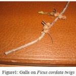 |
Figure 1: Galls on Ficus cordata twigs.
|
Fig trees are a vital component of the ecology of the Otavi Mountains, it then imperative that the causal agents of these galls be investigated. The galls can have negative effects on the growth of these figs and in a long run lead to death of the population of trees for this specific species. For this reason, diagnostic methods are required to reveal the identity of the causal agent. So, the main objective of the research was to identify the causal agents associated with gall formation in Ficus cordata using the ITS-PCR technique as this may help in the implementation of effective control methods.
Materials and Methods
Culturing and Isolation of Fungi
Sabouraud Dextrose Agar 4% (SDA) medium was prepared by dissolving 60g of SDA powder in 1l of distilled water. The medium was then sterilized in an autoclave at 121oC for 15 minutes. It was then cooled to a hand touch then the content of each flask was poured into a maximum of 20 plates which were allowed to cool and solidify. The plates were inoculated the next day. Galls from the twig of an infected plant were cut into 3 pieces which were first immersed in 70% alcohol solution for 2 minutes and then washed with distilled water were used to inoculate the plate in which they were placed with the cut edges faced the medium. The fungi were left for 7 days before being subcultured and subculturing was done until pure cultures were obtained from single spores of the fungus. Malt extract broth was used. This was prepared by dissolving 17g of Malt Extract powder and 3g of Peptone in 1l of distilled water. This solution was distributed to a maximum of seven 1l flasks which were then covered with a foil and autoclaved at 115 oC for 10 minutes. The broth was allowed to cool and was inoculated the next day with samples of pure culture. The fungi were allowed to grow for a minimum of 7 days before they were filtered using sterilized funnels and filter papers. The mycelium was air-dried for 3 days.
DNA Extraction
The dried mycelium was powderised under liquid nitrogen using sterilized mortar and pestle. To the powdered mycelium, 3000µl and then 2000 µl of CTAB isolation buffer which was preheated to 60 oC was added followed by thorough mixing. Using a micropipette, 500µl of this solution was put into an eppendoff tube and it was incubated at 60 oC for an hour. After incubation, 500µl of chloroform was added, and the mixture was extracted for 5 minutes and it was centrifuged for 20 minutes at 8000rpm. The top layer was transferred to a new tube and 1.5 times the volume of the 96% ethanol was added prior to centrifuging for 15 minutes at 13 000rpm. The ethanol was discarded, carefully not to pour the DNA out, and the DNA was washed with 100 µl of 70% ethanol. The mixture was centrifuged for 2 minutes at 10 000rpm and then the ethanol was again discarded and the DNA was dissolved in 75 µl of T.E buffer over night. The extracted DNA samples were kept at -20oC. The fungal genomic DNA was visualized under UV light in a UV transilluminator.
Polymerase Chain Reaction (PCR) and Automated Sequencing
In order to amplify the ITS regions from the genomic DNA, ITS1 and ITS4 primers were used. The sequences for the primers used were ITS1 (5ˈ-CGTAGGTGAACCTGCG-3ˈ) and ITS4 (5ˈ-TCCTCCGCTTATTGATAT-3ˈ). The PCR was run using a Master Mix solution prepared by mixing 12.5µl Go green buffer, 2µl ITS1 and ITS 4, 3.5µl double distilled water and 2µl DNA in a total volume of 20 µl. A control was prepared using only the Go green buffer and water, no DNA was added. The PCR machine was set under the amplification profile: an initial of 94 oC for 4 minutes followed by 32 cycles of 94 oC for 30 seconds; 30 seconds at 62 oC and 1 minute at 72 oC and then it was held at 4 oC. The PCR amplifications were attempted several times until a clear distinct amplicons were obtained. The PCR products were analyzed using a 1% gel.
The PCR products were cleaned and sent to Inqaba Biotec Industries in South Africa for sequencing using an automatic sequencer. DNA samples amplified using primers ITS1 and 4 were sequenced in the forward and reverse direction using primers ITS1 and ITS4.
Results and Discussion
The objective of this study was to isolate and identify the causal agent associated with gall formation on Ficus cordata in the Otavi Mountains using ITS-PCR technique. A single pure cure was consistently obtained in all the isolate of the Ficus cordata fungal cultures. Sequencing and analysis of the DNA samples revealed the identity of the fungus as a crop pathogenic fungi belonging to the Dothideomycetes species. Pair-wise sequence alignment of the obtained sequence with the sequences already existing in the Genbank through nucleotide BLAST searches revealed that the successfully sequenced fungus was a Dothideomycete species (Figure 2) with similarity at 100%.
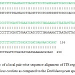 |
Figure 2: A partial display of a local pair-wise sequence alignment of ITS regions of the isolated fungus associated with galls of Ficus cordata as compared to the Dothideomycete species. |
The vertical line connecting the query sequence to the Dothideomycete species (Dothid. spp) sequence indicates the similarity in the bases.
Gall formation can be as a result of many different factors. Attacks from gall midges, gall wasps, plant lices, plant mites and fungi may all result in the formation of galls. Felt (1940) classified the first four as the principal gall producers, including fungi. Dothideomycete species isolated are for the first time being associated with formation of galls on Ficus cordata. According to Hane et al (2007) Dothideomycetes forms a large fungal taxon that includes many important plant pathogens affecting all major crop plant families. Infection by these fungi results in major economic losses. Plant diseases caused by members of the Dothideomycetes species include black leg of wheat caused by Leptosphaeria maculans and Stagonospora nodorum, southern maize leaf blight caused by Cochilobolus heterostrophus, apple scab caused by Venturia inaequalis, infection of barley by Hordeum vulgare and black sigatorka of banana caused by Mycosphaeria fijiensis (Hane et al., 2007). However, though members of this taxon may be directly associated with gall formation on Ficus cordata, no case of gall formation associated with members of this taxon (Dothideomycetes) was reported in earlier literature. To verify whether members of this taxon are indeed associated with gall formation on Ficus cordata Koch’s postulates need to be tested as a follow up step.
Acknowledgments
This study was supported by BIOTA under Biolog III project.
References
- Agrios, GN. 1997. Plant Pathology. Academic Press. USA
- Curtis, BA and Mannheimer, CA. 2005. Trees Atlas of Namibia. Windhoek: Natural Botanical Research Institute.688pp.
- Craven, P and Marais, C. 1989. Waterberg Flora; Footpaths in and around the camp. Gamsberg, Namibia.
- Epstein, L and Nicholson RL. 1997. Adhesion of spores and hyphae to plant surfaces. The Mycota: Plant relationships, V (1): 11-15.
- Felt, EP. 1940. Plant galls and Gall makers. Comstock Publishing Company, Inc. New York.
- Hane, JK; Lowe, RGT; Solomon, PS; Tan, K-C; Schoch, CL; Spatafora, JW;. Crous, PW; Kodira, C; Birren, BW; Galagan, JE; Torriani, SFF; McDonald, BA and Olivera, JK. 2007. Dothideomycete–Plant Interactions Illuminated by Genome Sequencing and EST Analysis of the Wheat Pathogen Stagonospora nodorum. The Plant Cell. 19(11): 3347–3368.
- Isaac, S. 1992. Fungal-plant interactions. Chapman and Hall. USA.
- Keith, CP. 1991. Trees of Southern Africa. Struik Publishers. Cape Town.
- Moore-Landecker, E. 1996. Fundamentals of the Fungi. Prentice Hall, Inc. USA.
- Moore-Landecker, E. 1972. Fundamentals of the Fungi. Prentice Hall, Inc. USA.
- Navajas, M; Lagnel, J; Fauvel, G and de Moares, G. 1999. Sequence Variation of Ribosomal Internal Transcribed Spacer (ITS) in Commercially Important Phytoseiidae mites. Experimental and Applied Acarology, 23: 851-859
- Van Koenen, E. 1996. Medicinal, Poisonous and Edible Plants in Namibia. Klaus Hess Verlag. Windhoek.
- Van Wyk, B and van Wyk, P. 1997. Field guide to Trees of Southern Africa. Strunk Publisher.SA.
- Van Wyk, B, van Wyk P and van Wyk, BE. 2000. Photographic guide to Trees of Southern Africa. Briza Publications. SA.
- Ward, E. 1994. Use of the Polymerase Chain Reaction for Identifying Plant Pathogens. Ecology of Plant Pathology, 143-159.

This work is licensed under a Creative Commons Attribution 4.0 International License.


