

Manuscript accepted on : 12 August 2016)
Published online on: --
Plagiarism Check: Yes
Anatomical Structure Study of Aerial Organs of Crataegus Ambigua C. A. Mey
Ainur T. Tuyakova, Akzhunis A. Imanbayeva, Nurzhaugan I. Duysenova and Margarita Yu. Ishmuratova
Mangyshlak Experimental Botanical garden 10th Micro-region, Aktau 130000, Kazakhstan.
DOI : http://dx.doi.org/10.13005/bbra/2270

ABSTRACT: The purpose of the present research was to study the peculiarities of the anatomical structure of some vegetative and genesic organs of Crataegus ambigua C.A.Mey. growing in the territory of Mangistau region. The findings of investigation allowed determining that the annual sprouts and pedicel of this species have a primary anatomical structure with the transition to secondary growth. Leaf of the Crataegus ambigua is of a dorsoventral type of light mesophytic structure, hypostomatic in terms of stomata arrangement. The fetal epidermis is presented by polygonal cells of the epidermis without additional elements. The pulp consists of large and loosely-arranged parenchymal cells. The epidermis of the corolla is represented by oval thick-walled cells. The pericarp of the seed consists of three well-defined layers (exocarp, mesocarp and endocarp). The corcule occupies the entire internal cavity. In terms of the cover and pulp structure, Crataegus ambigua has a structure similar to other species. Characteristic features for this species are the shape and the arrangement of the main cells of the epidermis, stomata and simple unicellular trichomes.
KEYWORDS: Crataegus ambigua; anatomical structure; aerial parts; rare species; Mangyshlak
Download this article as:| Copy the following to cite this article: Tuyakova A. T, Imanbayeva A. A, Duysenova N. I, Ishmuratova M. Y. Anatomical Structure Study of Aerial Organs of Crataegus Ambigua C.A.Mey. Biotech Res Asia 2016;13(3). |
| Copy the following to cite this URL: Tuyakova A. T, Imanbayeva A. A, Duysenova N. I, Ishmuratova M. Y. Anatomical Structure Study of Aerial Organs of Crataegus Ambigua C.A.Mey. Biotech Res Asia 2016;13(3). Available from: https://www.biotech-asia.org/?p=15812 |
Introduction
Plants of the hawthorn genus (Crataegus L., Rosaceae) are medicinal, melliferous, decorative and phyto-reclamation plants [1-5]. The fruits and flowers of 12 species (Crataegus sanguinea, C. laevigata, C. korolkovii, C. chlorocarpa, C. dahurica, C. monogina, S. alemanniensis, C. pentagyna, orientobaltica, S. curvisepala, S. curonica, C. dunensis) are used to prevent the weakening of the heart muscle and sclerotic changes in the coronary vessels. They enhance immunity, normalize blood pressure, prevent infectious lesions and the intestinal disorders [6-10], and are part of the vitamin containing combinations of ataractic and herbal medicinal products.
Anatomical features of Crataegus genus plants are underinvestigated. Thus, there are just single works on wood structure and stamens of the flowers [11, 12]. The fruit structure is a more studied issue as a pharmacognostic character of medicinal raw material [13, 14]. However, the anatomical structure of the leaves and young sprouts are completely absent.
Crataegus ambigua C.A.Mey. is a rare and endemic plant of the flora of Mangystau region. The habitat of this species is confined to the grounds and gentle slopes of the gorges of the Mangyshlak Peninsula and Tubkaragan [15]. Anatomic features of this plant are to date unexplored.
The purpose of the present research was to study the peculiarities of the anatomical structure of some vegetative and genesic organs of Crataegus ambigua growing in the territory of Mangistau region.
Methodology
The investigation objects were aerial vegetative (leaf and annual sprouts) and genesic (flowers and fruit) organs of middle-age Crataegus ambigua plants growing in Mangystau (Akmysh, Samal and Kezym mountain gorges).
Sampling was carried out during the vegetation periods of the years 2015-2016 in the phase of large flowering and fruiting. Freshly harvested organs were preserved in a mixture of alcohol (70%), glycerin and distilled water in 1:1:1 ratio (Strauss-Fleming solution). When determining the anatomical features of the leaf blade of the studied species, we selected the most developed intact leaves in the middle part of the sprouts, as well as analyzed the fragments (surface preparations and transverse sections) in the middle part between the main nervure and the edge. When studying flowers, we investigated the surface preparation of the corolla and cross-section of the pedicle. Transverse sections of annual sprouts were made throughout their length every 2-3 cm. For fruit, we investigated the peel surface preparation and transverse sections of pulp and seeds. Not less than 10-15 micro specimens were prepared for each organ.
Anatomical study of the plant was conducted according to the recommended practices described by M.N. Prozina [16], A.A. Dolgova and E.Ya. Ladygina [17], V.N. Vachov and L.I. Lotov [18]. Fabrication of temporary preparations (surface and pressure preparations, transverse sections) were performed according to standard techniques [19-21] using “MZP-01 Tehnokom” freezing microtome. Bleaching of the preparations was performed using the glycerol. To obtain surface preparations, leaves were boiled in 10% potassium hydroxide solution.
The obtained preparations were studied by means of Melji-Techno “MT 4310 L” scanning microscope and BisionCamV 500B camera. Digital photographs were obtained at the magnification of eyeglass and lens by 10х4, 10×10, 10×40, employing the VisualBio program. When studying leaf epidermis, drawings were performed from photos manually.
When describing the anatomical structure, we used the terminology proposed by K. Esau [22, 23], N.A. Anely [24], and L.I. Lotova [25].
Results and discussion
Crataegus ambigua is a shrub or small tree [26]. Sprouts are glabrous, inerm, rarely with spines, branches are brown with gray patches of preserved peel, leaves are bright green, the lowermost leaves are brevipetiolate, cuneate obovate, trilobate or incisodentate at the apex, upper leaves are petiolate, ovate, with broadly cuneate or rounded base, 5-7-lobed. Inflorescences are dense with up to 20 flowers. Fruits are globose or broadly elliptical, purple-black with light dots, fleshy; bones in outline are broadly elliptical.
The anatomical structure of Crataegus ambigua annual sprouts
Cross-sections of young sprouts of Crataegus ambigua have a primary anatomical structure with the transition to secondary growth (Fig.1). Outside, the stem is covered with one-layered epidermis 19-23 µm thick consisting of the cells with rounded-rectangular structure, which are covered on top with thick cuticle. Under the epidermis there is a three-layer lamellar collenchyme 83-98 µm thick stained in a dark color.
The area between the endoderm and the cortex is filled with 5-6 rows of loose parenchymal cell. The thickness of this zone ranged from 148 to 240 µm. Endoderm is arranged in a single row and its cells are tangentially thickened.
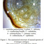 |
Figure 1: The anatomical structure of annual sprouts of Crataegus ambigua. The cross-section, magnification 10×10.
|
There are numerous conducting bundles with the structure of transitional type between the primary and secondary anatomical structures. Bundles are of collateral open type with the area of 360-530 10-3 mm2. The number of conducting bundles is 16-18. The primary xylem is arranged in smooth columns, while the secondary xylem is arranged in the form of uneven areas with smaller lumens. There are small phloem cells outside the xylem, which are covered by sclerenchyma in the form of “caps”. The central part is filled with loosely arranged rounded cells of medullar parenchyma with a diameter of 56-61 µm.
The anatomical structure of Crataegus ambigua leaf
Crataegus ambigua leaf is flat with a dorsoventral light type structure. The cells of the upper and lower epidermis are well seen in a cross-section (Fig. 2). They are thin-walled, covered from outside with a thin layer of cutin. The thickness of the upper epidermis was 23-25 µm, while that of the lower one was 18-20 µm.
The mesophyll is differentiated into palisade and spongy tissues. Cells of palisade mesophyll are arranged in 1-2 rows 45-51 µm thick, the intercellular ducts are not expressed. Palisade mesophyll over midrib is interrupted by angular collenchyma. The transition to the spongy mesophyll is clear, intercellular ducts are of aerenchyme type 78-90 µm thick. Collateral bundles of closed type (cambium is absent) are formed only along the nervures and consist of 10-15 rows of xylem and 5-8 rows of phloem. From the phloem side, conducting bundle is lined with 2-3 rows of sclerenchyma as a cap. The bundles area made up 405-453 10-3 mm2. The hawthorns are characterized by variability in shape of the lower part of the main nervure. Some leaves are rounded in cross section, while others have projections in the shape of the “ears”.
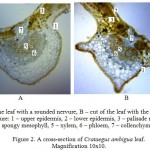 |
Figure 2: A cross-section of Crataegus ambigua leaf. Magnification 10×10
|
The cells of the upper epidermis have slightly sinuous membranes (Fig. 3) with expressed thickened walls. Cells length is 0.46-0.70 µm, width – 0.30-0.35 µm. Some rare simple single-celled trichomes about 4 µm in length with the base thickness of 0.38-0.42 µm are effused over the surface.
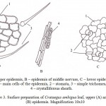 |
Figure 3: Surface preparation of Crataegus ambigua leaf, upper (A) and lower (B) epidermis. Magnification 10×10
|
Cells of the lower epidermis are more elongated with strongly sinuous membranes 0.47-0.59 µm in length and 0.19-0.30 µm in width. Walls are thin and surface is covered with a cuticle layer. On the underside of the leaf there are anomocytic stomata, 0.40-0.47 µm in length and 0.16-0.22 µm in width. The number of stomata is 9-10 pieces/mm2. Trichomes are absent. Thus, Doubtful Hawthorn is characterized by hypostomatic type of leaf.
Above the nervure, the cells of epidermis have prosenchymatous shape. Nervures on both sides of the leaf are surrounded by the rows of sheath, whose cells contain crystals of calcium oxalate.
Anatomical structure of Crataegus ambigua fruits
Drupe of the hawthorn contains 2 to 3 seeds, 5-6 mm long and 3-4 mm wide. The weight of 1,000 seeds ranges from 12 to 16.5 g. The seed is oblong with the slightly recurved tip, bluntly cut at upper end. It has 1-2 vague ribs with color ranged from yellow to light brown (Fig. 4).
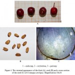 |
Figure 4: The external appearance of the fruit (A), seed (B) and a cross-section of the seed (C) of Crateagus ambigua. Magnification 10×10.
|
In the structure of the seed one can clearly distinguish the pericarp, endosperm and cotyledons. Pericarp of the seeds consists of 3 layers (exocarp, mesocarp and endocarp) 10-16 µm thick.
Exocarp is thin, covered with cuticle. It consists a single layer of dead cells. Cells of mesocarp are thin-walled, densely arranged, parenhimy. Endocarp is very weak, comprises of thin-walled living cells arranged in a single layer. The cells of the endosperm 109-116 µm thick have the same rounded shape arranged in 8-10 rows.
The epidermal cells of the cotyledon are elongated, large, arranged in a single layer 570-575 µm thick. Embryo is 2-cotyledonous, elongated, disk-shaped, the cut completely captures the internal cavity.
The rind of the hawthorn fruit (Fig. 5) consists of polygonal cells with thickened walls and yellow-brown contents, tightly adjacent to each other.
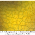 |
Figure 5: Surface preparation of the epidermis of Crataegus ambigua fruit. Magnification 10×10.
|
The cells length was 0.20-0.37 µm, width – 0.19-0.26 µm. The surface of epidermis is covered with a layer of cuticle.
The pulp of the fruit (Fig. 6) consists of round or oval cells, 0.96-1.15 µm in length and 0.61-0.90 µm in width, containing inclusions of orange-red or brownish-yellow color (carotenoids), small druses and prismatic crystals of calcium oxalate.
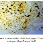 |
Figure 6: A cross-section of the fruit pulp of Crataegus ambigua. Magnification 10×10.
|
The inner part of the fruit pulp is shot through with collateral bundles and solitary sclereids. Layers of stone cells are arranged near the large bundles; calcium oxalate crystals form in patches crystalliferous sheath.
The anatomical structure of Doubtful Hawthorn flower
The corolla of the flower on the surface preparation consists of oval cells of 0.28-0.36 µm in length and 0.15-0.22 µm in width (Fig. 7). Walls are thick and surface is covered with a layer of cuticle. Stomata and trichomes were not identified.
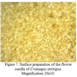 |
Figure 7: Surface preparation of the flower corolla of Crataegus ambigua. Magnification 10×10.
|
At cross-section, pedicel of hawthorn is rounded, 9-10 µm in diameter (Fig. 8). Cells of the epidermis, rounded at cross section, 0.16-0.18 µm in diameter are arranged peripherally. Beneath are 3-4 layers of collenchyma laid after 3-5 layers of primary cortex 1.22-1.49 µm thick. Cells are of parenchymal type with large lumens and loose arrangement.
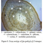 |
Figure 8: Cross section of the pedicel of Crataegus ambigua. Magnification 10×10
|
Primary cortex is isolated from the central cylinder by 0.20-0.41 µm thick endodermis layer, whose cells are tangentially thickened. Conducting system is of a bundle type. The bundles are collateral, closed, with area of 0.63-0.88 10-3 mm2. Each beam is covered by the sclerenchyma “cap” 0.26-0.32 µm thick. The central part is filled with medullar parenchyma with the cells diameter of 0.28-0.32 µm.
Conclusion
Thus, on the basis of conducted research we can draw the following conclusions:
The stem and pedicel of Crataegus ambigua are of primary anatomical structure, while transition to secondary growth was observed for stem.
The leaf of Crataegus ambigua is of a dorsoventral type of light mesophytic structure, hypostomatic in terms of stomata arrangement. Its characteristic features are the shape and arrangement of the main epidermal cells, stomata and single-cellular trichomes.
The fetal epidermis is represented by polygonal cells of the epidermis without additional elements. The pulp consists of large and loosely-arranged parenchymal cells.
Pericarp of the seed consists of 3 well-defined layers (exocarp, mesocarp and endocarp); the corcule occupies the entire internal cavity.
Epidermis of the corolla is represented by oval thick-walled cells without additional structures.
Acknowledgment
The research was carried out within the grant project of the Science Committee of the Ministry of Education and Science of the Republic of Kazakhstan “Evaluation of the gene pool of natural populations of Crataegus ambigua, rare endemic species to Kazakhstan” (2015-2017).
References
- Abaimov, V.F., 2009, Dendrologiya [Dendrology] [Text]. Moscow.
- Rastitel’nye resursy Rossii. Polevye cvety, ih sostav i biologicheskaya aktivnost’ [Plant resources of Russia. Wild flower plants, their compositions and biological activity. Fabaceae – Apiaceae family] [Text], 2010, 3, Saint-Petersburg – Moscow.
- Ljubuncic, P., Portnaya, I., and Bomson, A., 2005, Antioxidant activity of Crataegus aronia aqueous extract used in traditional Arab medicine in Israel. Journal of Ethnopharmacology, 101(1), pp. 153-161.
CrossRef - Wang, I., Xiogjiang, X., and Feng, B., 2013, Effect of Crataegus usage in cardiovascular disease prevention: an evidence-based approach. Evidence-Based Complementary and Alternative Medicine, pp. 23-39.
- Grunzinskaya, L.M., Gemedjieva, N.G., Nelina, N.V., and Karzhaubekova, Zh.Zh., 2014, Spisok poleznyh trav Kazahstana [Noted list of herbs of Kazakhstan] [Text], Almaty.
- Méndez-Iturbide, D., Banderas-Tarabay, J.A., Nieto-Camacho, A., Rojas-Chávez, A., and García-Meza, M., 2013, Antioxidant capacity of extracts from hawthorn (Crataegus mexicana) skin. Journal of Food Science, 7(6), pp. 150-158.
- Hernández-Pérez, А., Moustapha Bah, Ibarra-Alvarado, С., Rivero-Cruz, G.F., Rojas-Molina, A., Rojas-Molina, J.I., and Cabrera-Luna, J.A., 2014, Aortic relaxant activity of Crataegus gracilior Phipps and identification of some of its chemical constituents, Molecules, 19, pp. 20962-20974.
- Swaminathan, J.K., Khan, M., Mohan, I.A., Selvendiran, K., Devara,j S.N., Rivera1, B.K., and Kuppusamy P., 2015, Cardioprotective properties of Crataegus oxycantha extract against ischemia-reperfusion injury, Phytomedicine, 17(10), pp. 744-752.
CrossRef - Nichita, C., Neagu, G., Vulturescu, R., Pirvu, L., Badea N.M., Albulescu, R., Giurginca, M., 2009, Evaluation of scavenger properties of some flavonoidic vegetal extracts obtained from Crataegus monogyna Jacq. Key Engineering Materials, 45, pp. 41-44.
CrossRef - Sokolov, Ya., 2000, Fitoterapiya i fitofarmakologiya [Phytotherapy and phytopharmacology] [Text], Moscow.
- Saribas, M., and Yaman, B., 2005, Wood anatomical of Crataegus tanacetifolia (Lam.) Pers. (Rosaceae), endemic to Turkey, International Journal of Botany, 1(2), pp. 158-162.
CrossRef - Weryszko-Chmielewska, E., and Konarska, A., 2004, Anatomical of the floral nectarines of nine species from Sunf. Pomoidea (Rosaceae), Acta Agrobotanica, 49(1-2), pp. 95-105.
CrossRef - Gosudarstvennaya Farmakopeya Respubliki Kazahstan [The state pharmacopeia of Republic of Kazakhstan] [Text], 2009, 2, Astana.
- Muravjev, D.A., Samylina, I.A., and Yakovleva, G.P., 2002, Farmakognoziya [Pharmacognosy] [Text], Moscow.
- Katalog redkih i ischeznuvshih rastenij Mangistauskoj oblasti (Krasnaya kniga) [Catalog of rare and disappeared plants of Mangystau region (The Red Book)] [Text], 2006 Akteu.
- Prosina, M.N., 1960, Botanicheskie mikrometody [Botanical microtechniques] [Text], Moscow.
- Dolgova, А.А., and Ladygina, E.Ya., 2005, Rekomendacii k prakticheskim rabotam po farmakognozii [Recommendation to practical works on pharmacognosy] [Text], Moscow.
- Vekhov, V.N., Lotova, L.I., and Filin, V.R., 1980, Praktikum po anatomii i morfologii rastenij [Practicum on anatomical and morphology of vascular plants], Moscow.
- Barykina, R.P., Veselova, T.D., Devyatov, A.G. et al., 2004, Spravochnik po botanicheskim mikrometodam (bazovyj i metodov) [Reference book on botanical microtechniques (base and methods)] [Text], Moscow.
- Permyakov, A.I., Mikrometody [Microtechniques] [Text], Moscow.
- Lakyn, G.F., 1990, Biometriya [Biometry] [Text], Moscow.
- Esau, K., 1977, Anatomy of Seed Plants, 1, 2nd edn., Wiley, New York.
- Esau, K., 1977, Anatomy of Seed Plants, 2, 2nd edn., Wiley, New York.
- Anely, N.A., 1975, Atlas ehpidermisa lista [Atlas of leaf epidermis] [Text], Tbilisi.
- Lotova, L.I., 2007, Botanika: Morfologiya i anatomiya rastenij [Botany: Morphology and anatomy of vascular plants] [Text], Moscow.
- Flora Kazahstana [Flora of Kazakhstan] [Text], 1961, 4, Alma-Ata.

This work is licensed under a Creative Commons Attribution 4.0 International License.


