

Manuscript accepted on : November 14, 2010
Published online on: 28-12-2010
Relation of Maxillary Teeth to the Maxillary Sinus in Normal Saudi Individuals Living In Riyadh
Essam Mattar1, Lina Hammad1, Asmaa Faden2 and Hesham Khalil2
1Department of Radiological Sciences, College of Applied Medical Sciences, King Saud University, Riyadh Saudi Arabia 2Department of Maxillofacial Surgery and Diagnostic Sciences, College of Dentistry, King Saud University, Riyadh Saudi Arabia Corresponding Author E-mail: hkhalil@ksu.edu.sa
ABSTRACT: This study investigated the relationship of the maxillary sinus floor to the roots of posterior teeth imaged by panoramic radiography in Saudi patients living in Riyadh, Saudi Arabia. Panoramic radiographs of 60 subjects were analyzed. A total of 266 teeth in the left side and 277 teeth in right side were examined in this study. The closest root to the sinus was considered and classified according to a scale from 0 to 4. The results showed that the first and second molar teeth are in a very close relation to the maxillary sinus in these subjects. Although some reports showed that the reliability of panoramic radiographs is limited in showing the relationship of the teeth roots to the sinus, these radiographs are of a great help and could be used as a primary indicator for the relation of posterior teeth for general dentists who have no access to cone beam CT scan.
KEYWORDS: Maxillary sinus; molar teeth; panoramic
Download this article as:| Copy the following to cite this article: Mattar E, Hammad L, Faden A, Khalil H. Evaluation of the Antibacterial Activity of Honeys (Including Manuka And Sedr Honeys) Against Bacteria Causing Opportunist Infections. Biosci Biotech Res Asia 2010;7(2) |
| Copy the following to cite this URL: Mattar E, Hammad L, Faden A, Khalil H. Evaluation of the Antibacterial Activity of Honeys (Including Manuka And Sedr Honeys) Against Bacteria Causing Opportunist Infections. Biosci Biotech Res Asia 2010;7(2). Available from: https://www.biotech-asia.org/?p=9046. |
Introduction
The adult maxillary sinus size and extension varies between patients. The floor of the sinus extends between adjacent teeth or between individual roots in fifty percent of the population (Hauman et al., 2002). This extension creates elevations in the antral surface which are commonly referred to as “hillocks” or protrusions of root apices into the sinus (Waite, 1971). In these cases, the thickness of the sinus floor is markedly reduced. Tooth roots that protrude into the maxillary sinus can have various implications, including oroantral fistulae or root displacement into the sinus cavity which are a frequent complication after extractions of upper molars (HARRISON, 1961). Sinus expansion after extractions can greatly decrease the bone height available for implant placement and it can affect its success rate (Wehrbein and Diedrich, 1992b). Endo-antral syndrome which is characterized by spread of pulpal disease beyond the area of the dental supporting tissues or into the maxillary sinus causing sinusitis, may affect the relation of the teeth to the sinus (Hauman et al., 2002;Wehrbein and Diedrich, 1992a). Orthodontic treatment cal also affect the relation of the upper posterior teeth to the sinus. Intrusion or bodily movement of teeth across the sinus floor by orthodontic treatment have been shown to cause moderate apical root resorption (Daimaruya et al., 2003;Wehrbein et al., 1990). Panoramic radiography images can show the body of both the mandible and the maxilla, and the maxillary sinus (Kilic et al., 2010). It is probably the most commonly utilized radiographic technique in dentistry. Studies have shown that 64% to 95% of dentists prescribe only panoramic radiography for dental implant assessment (Sakakura et al., 2003).
The percentage of teeth penetrating into the sinus or even approaching it varies widely according to the tested sample and the communities where these studies were done. Because of the clinical observation and findings that we met during practice we have the impression that Saudi people may have more pneumatized maxillary sinus than other nations, figure 1. Furthermore there is no published data about the actual size or the relation of the maxillary teeth to the floor of maxillary sinus.
Materials and Methods
Sixty patients were included in his study including 39 male and 21 female with age range from 20 to 40 years. Criteria of patient’s selection included the followings:
– Patients attending OMFS clinic, College of Dentistry, King Saud University.
– Only Saudi, Riyadh residents, patients were included.
– Free of systemic diseases e.g. diabetes and hypertension.
– No previous maxillary sinus surgery.
– No clinical or radiographically evident paranasal sinus diseases (infection, cyst or tumors) that may affect the size of the sinus.
– Normal clinical facial proportions.
Panoramic radiographs were taken for each patient. The panoramic radiograph is routinely taken for all OMFS patients undergoing third molar surgery. The relation of the roots of upper posterior tooth to floor of the maxilla sinus was determined using the panoramic radiograph according to a modified scheme of that published by Sharan and Madjar (Sharan and Madjar, 2006), figure 2. The scale used is as follow:
0 = There is no contact between any root and the cortical borders of the sinus
1 = One or more root is in contact with the cortical borders of the sinus
2 = One or more root is projecting laterally on the sinus cavity but its apex is outside the sinus boundaries
3 = One or more root apex is projecting on the sinus cavity for a distance less than 2 mm
4 = One or more root apex is projecting on the sinus cavity for a distance more than 2 mm
Data were collected and compared with other studies performed in other countries.
Results
A total of 266 teeth in the left side and 277 teeth in right side were examined in this study to determine their relations with the floor of the maxillary sinus. Table 1 and 2 show the relation of the upper posterior teeth roots to the floor of the maxillary sinus according to the modified score used in this study. Most of the first and second premolars (77-98 %) scored 0 and 1 which indicate that most of the upper premolars have no close relation to the sinus floor. Molar teeth showed closer relation to the sinus floor. There was 37% of the first molar, 55 of the second molars and 31 % of the third molar teeth scored 3 and 4, table 3.
Table 1: Teeth root apex relation to the maxillary sinus floor in the left side
| Left side
|
|||||
| number examined | Score 0 | score 1 | Score 2 | Score 3 | Score 4 |
| 55 | 50 | 4 | 1 | 0 | 0 |
| 58 | 32 | 13 | 5 | 8 | 0 |
| 55 | 9 | 11 | 1 | 26 | 8 |
| 58 | 5 | 13 | 6 | 27 | 7 |
| 40 | 4 | 13 | 6 | 15 | 2 |
| 266 | 100 | 54 | 19 | 76 | 17 |
| 37.6 | 20.3 | 7.1 | 28.6 | 6.4 | |
Table 2: Teeth root apex relation to the maxillary sinus floor in the right side
| Right side
|
|||||
| number examined | Score 0 | score 1 | Score 2 | Score 3 | Score 4 |
| 56 | 52 | 6 | 0 | 0 | 0 |
| 57 | 37 | 14 | 2 | 4 | 0 |
| 56 | 10 | 27 | 1 | 12 | 6 |
| 59 | 6 | 21 | 3 | 24 | 5 |
| 49 | 10 | 14 | 9 | 10 | 1 |
| 277 | 115 | 82 | 15 | 50 | 12 |
| 41.5 | 29.6 | 5.4 | 18.1 | 4.4 | |
Table 3: Percentage of molar teeth relations with sinus for scores 3 and 4 in both right and left maxilla
| Tooth | Total number | Score 3 | Score 4 | Total percentage |
| 1st molar | 138 | 38 | 14 | 37% |
| 2nd molar | 117 | 53 | 12 | 55% |
| 3rd molar | 89 | 25 | 3 | 31% |
Discussion
The relationship between the roots of the maxillary posterior teeth and the maxillary sinus can be investigated by various radiographic techniques, including two dimensional techniques such panoramic radiographs. This study compared the relation of maxillary posterior teeth to the floor of the maxillary sinus using panoramic radiographs. Theses radiographs are taken routinely for patients who are undergoing third molar extraction in our department. From our experience we observed a high prevalence of oroantral communication of patients living in Riyadh and attending for molar extraction in primary care clinics as these cases are referred to us for further management. The sinus of these patients looked lager than normal which could be the reason behind such high prevalence of oroantral communications. We decided to study the situation gradually starting with analyzing the relation of maxillary posterior teeth to the floor of the sinus using the panoramic radiographs then measuring the sinus volume in further studies. Analysis of the results showed a close relationship of the first and second molar teeth to the sinus floor with other teeth also having high score relation to the sinus in some patients. Sharan et al have shown that panoramic radiographs can be as 96% accurate in indentifying the relation of the roots to the maxillary sinus in case of zero and one scores when compared to the CT- scan images (Sharan and Madjar, 2006) but this percentage decreases with higher scores relations. Although Hassan showed that periapical radiographs and panoramic are not reliable in determination of exact relationship between the apex of the toothy root and the maxillary sinus compared to cone beam computed tomography, still these radiographs can give a general idea of the relation for further precautions during extraction of upper posterior teeth. For the premolars in this study most teeth scores were between zero and two which indicate that few numbers of premolars showed root apex projection in to the sinus. The same observation was seen in previous studies (Kwak et al., 2004;Sharan and Madjar, 2006).
Relations of the posterior teeth to the floor of the maxillary sinus were also analyzed according to the age (20-39 years and 40-60 years). Analysis of the difference in relation in these two groups showed that there is a significant difference (P value < 0.05) in the relation of the second premolar, first molar and second molar teeth in the 40-60 years patients group. Such difference could be related to the change in the sinus size with age and the environmental factors. There was also a difference between the right and left teeth relation to the sinus which is mainly due to the difference in sinus size. Despite of many studies concerning asymmetry in the size of the human maxillary sinus which differ mainly in whether the left or right maxillary sinus is larger, it has been suggested that although asymmetry in the human maxillary sinus is present it is usually very small (Ariji et al., 1994;Koppe et al., 1994). We also reviewed the extraction cases that were referred to us with oroantral communications and found five of the 12 cases (41 %) had scores of 3 or 4 relations with the sinus as seen in the pre extraction panoramic radiographs. This percentage is very helpful in anticipating the occurrence of ororantral communication in such cases and referring them for specialist care prior to treatment. It is very important for the general dental practitioner to avoid extracting upper molar teeth when they have score 3 or more on the panoramic radiograph. They should consult an oral surgeon in these cases to avoid further complications that can arise from such extractions.
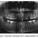 |
Figure 1: Panoramic radiograph shows a large maxillary sinus.
|
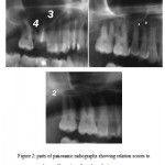 |
Figure 2: parts of panoramic radiographs showing relation scores to the maxillary sinus floor from 0 -4.
|
Conclusion
From these results it is obvious that from the panoramic radiograph there is a close relation of the first and second molar teeth to the maxillary sinus. Another observation that the sinus size looks larger than normal in these patients who are living in Riyadh compared to our experience with such radiographs in other cities in Saudi Arabia. This could be due to the weather of the Riyadh through the year. Dentists conducting extraction procedures in the posterior maxilla should take into consideration the amount of protrusion of teeth roots into the sinus in panoramic radiographs.
We are conducting another study in which we are measuring the size of the maxillary sinus in people living in different cities in Saudi Arabia and the incidence of oroantral communication after teeth extraction in these cities.
Acknowledgment
The authors would like to thank the staff of radiology department at the college of Dentistry, King Saud University for their great support.
Reference
- Ariji,Y, Kuroki,T, Moriguchi,S, Ariji,E, and Kanda,S (1994). Age changes in the volume of the human maxillary sinus: a study using computed tomography. Dentomaxillofac Radiol 23(3):163-168.
- Daimaruya,T, Takahashi,I, Nagasaka,H, Umemori,M, Sugawara,J, and Mitani,H (2003). Effects of maxillary molar intrusion on the nasal floor and tooth root using the skeletal anchorage system in dogs. Angle Orthod 73(2):158-166.
- HARRISON,DF (1961). Oro-antral fistula. Br J Clin Pract 15:169-174.
- Hauman,CH, Chandler,NP, and Tong,DC (2002). Endodontic implications of the maxillary sinus: a review. Int Endod J 35(2):127-141.
- Kilic,C, Kamburoglu,K, Yuksel,SP, and Ozen,T (2010). An Assessment of the Relationship between the Maxillary Sinus Floor and the Maxillary Posterior Teeth Root Tips Using Dental Cone-beam Computerized Tomography. Eur J Dent 4(4):462-467.
- Koppe,T, Yamamoto,T, Tanaka,O, and Nagai,H (1994). Investigations on the growth pattern of the maxillary sinus in Japanese human fetuses. Okajimas Folia Anat Jpn 71(5):311-318.
- Kwak,HH, Park,HD, Yoon,HR, Kang,MK, Koh,KS, and Kim,HJ (2004). Topographic anatomy of the inferior wall of the maxillary sinus in Koreans. Int J Oral Maxillofac Surg 33(4):382-388.
- Sakakura,CE, Morais,JA, Loffredo,LC, and Scaf,G (2003). A survey of radiographic prescription in dental implant assessment. Dentomaxillofac Radiol 32(6):397-400.
- Sharan,A and Madjar,D (2006). Correlation between maxillary sinus floor topography and related root position of posterior teeth using panoramic and cross-sectional computed tomography imaging. Oral Surg Oral Med Oral Pathol Oral Radiol Endod 102(3):375-381.
- Waite,DE (1971). Maxillary sinus. Dent Clin North Am 15(2):349-368.
- Wehrbein,H, Bauer,W, Wessing,G, and Diedrich,P (1990). [The effect of the maxillary sinus floor on orthodontic tooth movement]. Fortschr Kieferorthop 51(6):345-351.
- Wehrbein,H and Diedrich,P (1992a). [Progressive pneumatization of the basal maxillary sinus after extraction and space closure]. Fortschr Kieferorthop 53(2):77-83.
- Wehrbein,H and Diedrich,P (1992b). [The initial morphological state in the basally pneumatized maxillary sinus–a radiological-histological study in man]. Fortschr Kieferorthop 53(5):254-262.

This work is licensed under a Creative Commons Attribution 4.0 International License.


