

Manuscript accepted on : December 11, 2008
Published online on: --
Effect of Spray Application of Glyphosate on Morphological Characters of Hibiscus Cannabinus Linn
Sanjay I. Kamble
Department of Botany and Department of Seed Technology, Phulsing Naik Mahavidyalaya, Pusad, District Yavatmal (India).
ABSTRACT: In present study, the herbicidal activities of glyphosate on Hibiscus cannabinus Linn. have been studied. The Morphological responses might produce some light on the manner by which this compound affected on plants. The plants were sprayed with aqueous solution of different concentrations of herbicide from 100 to 5000 ppm. Glyphosate was efficient in killing the weed. The lethal dose of glyphosate was 1200 ppm.
KEYWORDS: Herbicide, Glyphosate, Spray application, Morphological characters and Hibiscus cannabinus Linn.
Download this article as:| Copy the following to cite this article: Sanjay I. Kamble. Effect of Spray Application of Glyphosate on Morphological Characters of Hibiscus Cannabinus Linn.Biosci Biotechnol Res Asia 2008;5(2) |
| Copy the following to cite this URL: Sanjay I. Kamble. Effect of Spray Application of Glyphosate on Morphological Characters of Hibiscus Cannabinus Linn.Biosci Biotechnol Res Asia 2008;5(2). Available from: https://www.biotech-asia.org/?p=20092 |
Materials and Methods
Plants of Hibiscus cannabinus Linn. were raised from seeds collected from naturally growing plants of different places in Nagpur and its environs. They were allowed to grow till they attained the flowering and at this stage plants were plants were sprayed with different concentrations of herbicide.
The aqueous solution of herbicide ranging from 100 to 5000 ppm was prepared. Ten pots for each concentrations (100 to 2000 ppm) containing 2 to 3 plants were sprayed. If 2000 ppm was found higher; the lower concentrations were tried to determined lethal dose. Asppe- poly sprayer of one litter capacity did spraying. A small quantity of sodium lauryl sulphate as a surfactant added in the herbicide solution. The Spraying was started in month of October 1996 and same experiments were repeated next year also. Spraying was done twice in an hour to make it more effective in the evening hours, when the wind was slow and temperature comparatively lower than that of the day. This help in less evaporation and more absorption of herbicide solution by the leaves. To avoid contamination of different concentrations of herbicide, cardboard was used at the time of spraying application. Six pots were sprayed with water used as control. Field trials were conducted on naturally growing plants in randomly designed plots of size approximately 3 O3 feet’s.
The fresh and dry weights of shoots and roots of control as well as treated plants were taken to determined desiccation of plants. Morphological changes were observed daily till the death of plants.
To study the anatomical changes induced by herbicide, plant parts like root, stem, petiole and leaf of the treated as well as control plants were fixed in F: A: A (Formalin: Acetic acid: Alcohol) solution for 24 hours and stored at 70% alcohol. The plant material was embedded in paraffin wax following customary method. Sections were cut at 6 to 9 microns. They were then stained according to crystal violet – erythrosine schedule and mounted in D.P.X. Microphotograph of various sections of both control and treated plants were taken.
Results
Control
The control stem showed defined single layered epidermis followed by ground tissue. It was divided in to three zones; the outer zone was chlorenchymatous which was made up of 2 to 3 layers of parenchymatous cells containing chloroplast. This zone was followed by 2 to 3 layers collenchymas were immediately followed by 3 to 4 layers of parenchymatous cells. Endodermis and pericycle were distinct. Vascular tissues were arranged in ring and enclose large parenchymatous pith (Fig. 1).
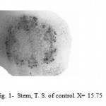 |
Figure 1: Stem, T. S. of control. X= 15.75.
|
The root showed secondary growth and consisted of an outer epidermis, parenchymatous cortex followed by ring of secondary phloem and xylem. The distinct metaxylam was observed with large vessels (Fig. 2).
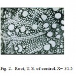 |
Figure 2: Root, T. S. of control. X= 31.5
|
The outer layer of petiole was made up of epidermis. Hypodermis was below the epidermis. Vascular bundles consisted of xylem and phloem (Fig. 3).
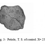 |
Figure 3: Petiole, T. S. of control. X= 25.2.
|
The leaf was composed of three types of tissue system; the epidermal, mesophyll and vascular. The lamina has upper and lower epidermis. The mesophyll comprises upper palisade parenchyma and lower spongy parenchyma. The midrib comprises of parenchymatous tissue embedding vascular elements (Fig. 4).
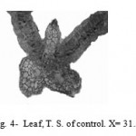 |
Figure 4: Leaf, T. S. of control. X= 31.5.
|
Glyphosate
This herbicide induced some anatomical changes in stem. It showed ruptured epidermis and disorganization of cortical cells at 400 and 600 ppm concentrations (Fig. 5 ). Later on, the cortex ruptured and lost their identity and disorganization of cambium and epidermis was observed at 800 and 1000 ppm. The pith cells destroyed at all concentrations of herbicide (Fig. 6).
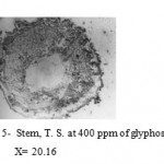 |
Figure 5: Stem, T. S. at 400 ppm of glyphosate.
|
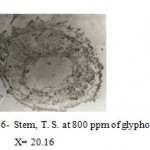 |
Figure 6: Stem, T. S. at 800 ppm of glyphosate.
|
The root showed ruptured epidermis and disorganization of cortex forming lacunae. The phloem and cambium were lost their identity at 400 and 500 ppm concentrations (Fig. 7). The pith cells were severely injured forming lacunae at some places. The cortex and epidermis sloughed off at higher concentrations i.e. 600 and 1000 ppm (Fig. 8).
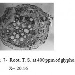 |
Figure 7: Root, T. S. at 400 ppm of glyphosate.
|
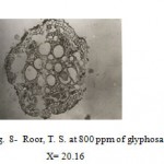 |
Figure 8: Roor, T. S. at 800 ppm of glyphosate.
|
The epidermis of petiole ruptured at some places. Cortical cells were plasmolysed at 400 and 500 ppm (Fig. 9). The cells of cortex were severely disorganized and lost their identity. Vascular tissues were not affected at all concentrations but in few places it showed disorganization at 800 and 1000 ppm concentrations (Fig.10).
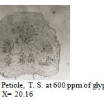 |
Figure 9: Petiole, T. S. at 600 ppm of glyphosate.
|
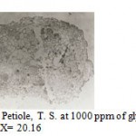 |
Figure 10: Petiole, T. S. at 1000 ppm of glyphosate.
|
Leaf showed cellular disorganization of epidermis and mesophyll tissues. The depletion of chloroplast in the palisade and spongy were observed (Fig.11). In midrib phloem and surrounding parenchymatous cells were crushed. The xylem was also disorganized at all concentrations of herbicide (Fig. 12).
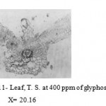 |
Figure 11: Leaf, T. S. at 400 ppm of glyphosate.
|
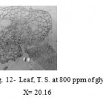 |
Figure 12: Leaf, T. S. at 800 ppm of glyphosate.
|
Discussion
In the stem, destruction of cortical cells, proliferation of cambium and phloem were observed. The proliferation of cambium and phloem resulted in the formation of meristematic masses. These masses penetrate in the cortex and ruptured the cortical and epidermal cells. Similar results were reported by Cole et. al. (1980) on some dicotyledonous weeds, Smid and Hiller (1981) on Solanum tuberosum, Canal et. al. (1990) on Cyperus esculentus, Bobde (1993) on Crotalaria juncea, and Jain (1993) on Chenopodium album.
The effects of glyphosate on leaf showed disorganization of mesophyll cells and vascular bundles. Similar results were reported by Cole et.al. (1980) on some dicotyledonous weeds, Smid and Hiller (1981) on Solanum tuberosum, Canal et. al. (1990) on Cyperus esculentus, Bobde (1993) on Crotalaria juncea, and Jain (1993) on Chenopodium album.
The root showed some anatomical changes such as desiccation and destruction of cortical cells and proliferation of cambium and phloem. The proliferation of cambium and phloem cells was resulted in the formation of meristematic masses. Canal et.al. (1990) on Cyperus esculentus, Bobde (1993) on Crotalaria juncea, and Jain (1993) on Chenopodium album reported desiccation destruction of cortex.
Petiole showed destruction of cambium and phloem cells also showed proliferation of inters cellular cambium. Bobde (1993) on Crotalaria juncea and Jain (1993) on Chenopodium album reported destruction of phloem cells.
The division and expansion of cells required the synthesis of nucleic acids and protein but after the application of glyphosate, all these process were affected due to toxicity of herbicide causing death of plants.
References
- Bobde, S. N. 1993. Comparative effects of herbicides on Crotalaria juncea Linn. Ph.D. Thesis, Nagpur Univ., Nagpur.
- Canal, M. J., Albuerne, M., Pamus, R.S. and Fernanbaz, B. 1990. Glyphosate injury on Cyperus esculentus leaves and basal bulb: Histological study. Weed Research. 30: 117-122.
- Cole, D. J., Dodge, A. D. and Caseley, J. C. 1980. Some biochemical effects of glyphosate on plant meristem. Exp. Bot. 31: 1665-1674.
- Jain, S. B. 1993. Cytomorphological effects of weedicides on weed Chenopodium album. D. Thesis, Nagpur Univ., Nagpur.
- Smid, D. and Hiller, L. K. 1981. Phytotoxicity and translocation of glyphosate in the Solanum tuberosum prior to tuber inhibition. Weed Science. 29: 218-223.

This work is licensed under a Creative Commons Attribution 4.0 International License.


