

Manuscript accepted on : May 30, 2008
Published online on: 04-03-2016
Sanjay I. Kamble
Department of Botany and Coordinator Department of Seed Technology, Phulsing Naik Mahavidyalaya, Pusad Dist. Yavatmal India.
ABSTRACT: In the present investigation, effects of glyphosate on linear growth of seedlings have been studied. The seedlings have been reported to be most sensitive to the herbicidal action. Uniform seedlings were treated with different concentrations of herbicide for 24 hours. Then they were thoroughly washed with distilled water and kept for germination in petri dishes containing double layered of moistened filter paper at room temperature for 72 hours. The effect of herbicide on linear growth of hypocotyl and radicles was noted. Glyphosate was effective in checking the linear growth of seedlings. It inhibited the linear growth of seedlings and caused swellings. The radicle was found to be more susceptible than hypocotyl to this herbicide. The lethal dose was found to be 600 ppm for hypocotyl and 500 ppm for radicle. The anatomical changes like ruptured epidermis, disorganization of cortical cells in hypocotyl and radicle and disintegration of mesophyll cells were observed in cotyledon.
KEYWORDS: Herbicide; Glyphosate; Linear growth; Anatomical characters; Hibiscus cannabinus Linn
Download this article as:| Copy the following to cite this article: Kamble S. I. Effect of glyphosate on linear growth of seedling and thier anatomical characters of Hibiscus cannabinus Lin. Biosci Biotechnol Res Asia 2008;5(1) |
| Copy the following to cite this URL: Kamble S. I. Effect of glyphosate on linear growth of seedling and thier anatomical characters of Hibiscus cannabinus Lin. Biosci Biotechnol Res Asia 2008;5(1). Available from: https://www.biotech-asia.org/?p=6943 |
Material and Methods
A large number of seeds of Hibiscus cannabinus Linn. were allowed to germinate in petridishes containing double layers of moistened filter papers at room temperature. When the seedlings attained the length of 7 to 10 mm, the seedlings of uniform length were selected for herbicidal treatment.
Each set of 10 seedlings were treated with different concentrations of glyphosate ranging from 100 to 2000 ppm for 24 hours. Lower concentrations were used, where it was found to be a higher dose. After treatment the seedlings were thoroughly washed with distilled water and allowed to grow for 72 hours in petridishes containing moistened filter papers. After 72 hours, the length of hypocotyl and radicle was measured separately in each seedling. The seedlings soaked in distilled water for 24 hours were used as control. Three replicates of each treatment were carried out.
To study the anatomical abnormalities induced by the herbicide, the seedlings of control and treated ones were fixed in F.A.A. (Formline-Acetic-acid-Alcohol) (5:5:90) solution. After 24 hours they were washed thoroughly with distilled water and preserved in 70% alcohol. For anatomical studies, the materials were embedded in paraffin wax following customary method (Sass, 1958). Sections were cut at 6 to 9 microns and stained with crystal violet and erythrosin and mounted in D.P.X.
The series of various sections of different concentrations of glyphosate were observed and recorded the anatomical abnormalities and then microphotographs were taken.
Results
Control
In control, hypocotyl of Hibiscus cannabinus Linn. showed single layered epidermis, 5 to 6 cells thick parenchymatous cortex, which was limited on the inside by single layer of endodermis and pericycle with centrally situated diarch xylem. In centre there was 4-celled thick parenchymatous pith (fig. 1).
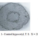 |
Figure 1: Control hypocotyl, T. S. X = 20.16.
|
The radicle showed 4 to 5 layers of parenchymatous cortex surrounded by uniseriate epiblema and immediately followed by endodermis and pericycle with centrally xylem (fig. 2).
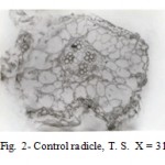 |
Figure 2: Control radicle, T. S. X = 31.5.
|
The cotyledons comprises of mesophyll cells with three layered palisade and about 4 to 5 layered spongy cells with intercellular spaces. Vascular elements were bounded by upper and lower epidermis and separated from each other by parenchymatous cells (fig. 3).
 |
Figure 3: Control cotyledon, T. S. X = 20.16.
|
Glyphosate
The herbicide showed some anatomical abnormalities in hypocotyl. The parenchymatous cells increased in size as compared to control. At and above 100 ppm, inner cortical cells disintegrated at several places. The ruptured epidermis and their wavy nature were observed. The proliferation of phloem forming was observed at 300 ppm. At higher concentrations i.e. 400 and 500 ppm, pith cells were disorganized and then cortical and pith cells ruptured (Fig. 4).
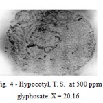 |
Figure 4: Hypocotyl, T. S. at 500 ppm of glyphosate. X = 20.16.
|
This herbicide also showed some remarkable anatomical changes in radicle. The radicle at 200 and 300 ppm, the cortical cells enlarged and then ruptured. The pith cells were disorganized and epidermal cells were ruptured at various places (Fig. 5).
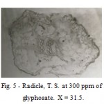 |
Figure 5: Radicle, T. S. at 300 ppm of glyphosate. X = 31.5.
|
The cotyledon dose not showed any remarkable change at lower concentrations of herbicide. At and above 300 ppm, there was dehydration of mesophyll cells occurred and lacunae were developed. As the concentrations increased, the mesophyll cells lost their identity and palisade cells were disorganized. At 500 ppm, epidermal cells lost their outline and lacunae were developed in palisade cells (Fig. 6).
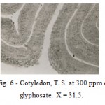 |
Figure 6: Cotyledon, T. S. at 300 ppm of glyphosate. X = 31.5.
|
Discussion
This herbicide induced some anatomical changes in hypocotyl, radicle and cotyledon. The hypocotyl showed enlargements of cortical cells and meristematic zone developed around the vascular bundles. The destruction of cortical cells, ruptured epidermis and disorganization of pith cells also observed in present study. Jain (1993) in Chenopodium album reported more strands of xylem and phloem in the seedlings due to applications of glyphosate. Bobde (1993) in Crotalaria juncea reported ruptured epidermis in hypocotyl due to enlargement of cortical cells in glyphosate treated seedlings.
The radicle showed enlargement of cortical cells, crushed pith cells and ruptured epidermis. Bobde (1993) in Crotalaria juncea reported that the growth of lateral roots primordia in radicle was inhibited due to application of glyphosate. Similar results were reported by Canal et. al. (1985) in Cyprus rotundus and recently, by Jain (1993) in Chenopodium album.
Cotyledon treated with glyphosate showed disorganization of mesophyll cells and ultimately lacunae was developed. This was probably due to dehydration of mesophyll cells. The lower and upper epidermis was also ruptured at several places. Jain (1993) in Chenopodium album and Bobde (1993) in Crotalaria juncea reported disorganization and disintegration of mesophyll cells which was probably due to dehydration and plasmolysis of mesophyll cells.
From the above discussion, it may be concluded that glyphosate inhibits the cellular growth of radicle, hypocotyl and cotyledons results in death of seedlings.
References
- Bobde, S.N. 1993. Comparative effects of herbicides on Crotalaria juncea Linn. Ph. D. Thesis, Nagpur University, Nagpur.
- Canal, M.J., Albuerne, M., Pamus, R.S. and Fernanbaz, B. 1985. Glyphosate injury on Cyperus esculentus leaves and vassal bulbs: Histological study weed. Res. 30: 117-122.
- Jain, S. B. 1993. Cytomorphological effects of weedicides on weed Chenopodium album. Ph.D. Thesis, Nagpur University, Nagpur.

This work is licensed under a Creative Commons Attribution 4.0 International License.


