

Manuscript accepted on : June 02, 2008
Published online on: 04-03-2016
Pradeep Kumar1*, Vishal Srivastava1 and Lalit Kumar2
1Department of Pharmaceutical Analysis, PES College of Pharmacy, Bangalore, Karnataka India.
2Department of Pharmaceutical Chemistry, Manipal College of Pharmaceutical Sciences, Karnataka India.
Corresponding Author E-mail: pradeep_alpine@yahoo.co.in
ABSTRACT: Mangiferin is the active constituent obtained from the dried parts such as leaves and barks of the Mango tree (Mangifera indica L.) which belongs to the family Anacardiaceae. Analytical method is developed and validated for the determination of Mangiferin in Mangifera indica by reversed-phase HPLC method. The HPLC method is described for determination of Mangiferin from leaves and barks Extract. The separation of the Mangiferin was achieved on a Lichrospher ® 100; RP- 18e (5 μm) column using mobile phase Acetonitrile: Buffer (Potassium dihydrogen orthophosphate (1.36 gm) in 900 ml HPLC grade water, adjust the PH to 2.5-2.8 with orthophosphoric acid and detection was carried at 254 nm (Isocratic Elution). The Lichrospher ® 100; RP-18e (5 μm) shows most favorable chromatographic parameters for analysis of Mangiferin in Mangifera indica. The linear range for Mangiferin was found to be 15.0-1000 μg mL-1. The Method has been successfully validated for linearity, Specificity, Precision, Accuracy, range and with good recoveries. Hence the present RP-HPLC method is suitable for assay of the Mangiferin in raw materials, extracts in quality control laboratories.
KEYWORDS: Mangiferin; leaves; bark; RP-HPLC; isocratic elution; alcoholic extracts
Download this article as:| Copy the following to cite this article: Kumar P, Srivastava V, Kumar L. Development of new reversed-phase HPLC method for the determination of Mangiferin in Mangifera indica Linn. Biosci Biotechnol Res Asia 2008;5(1) |
| Copy the following to cite this URL: Kumar P, Srivastava V, Kumar L. Development of new reversed-phase HPLC method for the determination of Mangiferin in Mangifera indica Linn. Biosci Biotechnol Res Asia 2008;5(1). Available from: https://www.biotech-asia.org/?p=6915 |
Introduction
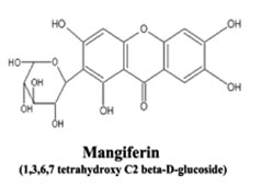
Mangiferin obtained from the dried stem bark and leaves of Mangifera indica Linn. and having a molecular formula C 19 H18 O11 and molecular weight 422 . The major bioactive constituent of Mangifera indica is a xanthone c-glucoside mangiferin. The other constituents are triterpenen viz. mangiferolic acid, isomangiferolic acid and several cycloartenod derivatives a flavonoide amantoflavone.
Dried mango flowers, containing 15% tannin, serve as astringents in cases of diarrhea, chronic dysentery, catarrh of the bladder and chronic urethritis resulting from gonorrhea. The bark contains Mangiferin and is astringent and employed against rheumatism and diphtheria in India. The resinous gum from the trunk is applied on cracks in the skin of the feet and on scabies, and is believed helpful in cases of syphilis.1-10
Experimental
Chromatographic condition
The following HPLC condition was maintained through out the experiment.
Mobile Phase
( Buffer 85% Acetonitrile 15%)
Buffer
I weigh accurately 1.36 gm. of Potassium dihydrogen orthophosphate in 900 ml of HPLC grade water; adjust the PH to 2.5 to 2.8 with dilute orthophosphoric acid than make up the volume to 1000ml. (Solvent A)
II) Acetonitrile (solvent B)
Detector: UV Detector.
Wavelength: 254 nm.
Flow rate: 1.0ml/min
Injection volume: 20µl
Run Time: For Standard: – 15 min; For Sample: – 15min
Data acquisition: Using suitable software CFR-21 Part 11 Compliant
Record the Chromatogram: Record the chromatogram with the following
Details: Retention time, Peak area, Peak height, % Area, Assigning the name to the Peaks.
Integration parameters: Integrate peaks base to base.
For calculating concentration, Mean, Standard Deviation, Relative Standard Deviation, Relative retention time, Response factor, Theoretical plate, Resolution, Asymmetry, Tailing factor by EXCELL or any other suitable Software can be used.
Plotting the Graph: EXCELL and PRISUM software.
Standard solution
Weigh accurately 10.089 mg of Mangiferin in the 10ml volumetric flask; add 1ml of D.M.F and make up the volume 10 ml by using methanol i.e. dilution I, Dilution II, III, IV, V, VI, VII were made consecutively by dissolving 5ml to 10ml in sufficient quantity of methanol in a standard flask. The Peak area & Retention time for individual standard are tabulated in the table no. 02 and 03 respectively.
The concentration of analyte in 10 ml is; Mangiferin = = 9.7358 mg.
Sample solution 🙁 for raw material) The sample solution was prepared by weighing the required quantity of sample (coarse powder) and transferred to a 100 ml beaker. Extract with 10 ml of DMF (Dimethyl form amide) and 80 ml of HPLC grade methanol sonicate for 5 minutes, by warming on water bath for about 20 min. discard the supernatant liquid (extract) to a 250 ml beaker. Repeat the procedure 4-5 times till the raw material was completely extracted or till the extract is colorless. Collected all the extract in a 250ml beaker and concentrated it to below 100ml volumetric flask and made up the volume to 100ml with methanol, mixed well and filtered through 0.45µl membrane.
Procedure
Take required quantity of Mangifera indica extract or raw material such as leaves or bark of Mangifera indica,(After extraction) weighed and transfer into 100ml volumetric flask, dissolved in 10ml dimethyl form amide and 25ml of methanol of HPLC grade, sonicated for 5 minutes. Warmed on steam water bath for 5 minutes, cool and make up the volume to 100ml with Methanol. Inject 20µLof sample and record the chromatogram. Inject 3 times and calculated the results.
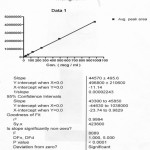 |
Graph 1: Linear Graph of Mangiferin.
|
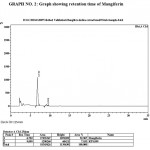 |
Graph 2: Graph showing retention time of Mangiferin.
|
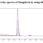 |
Graph 3: Overlay spectra of Mangiferin by using different concentration.
|
Table 1:
| SL NO | Dilution | Analyte | Concentration (mcg/ml) |
| 01 | 1 | Mangiferin | 15.212 |
| 02 | 2 | Mangiferin | 30.425 |
| 03 | 3 | Mangiferin | 60.849 |
| 04 | 4 | Mangiferin | 121.699 |
| 05 | 5 | Mangiferin | 243.397 |
| 06 | 6 | Mangiferin | 486.794 |
| 07 | 7 | Mangiferin | 973.589 |
Correlation coefficient (r2) for Mangiferin was found to be 0.9994 indicating that the method is linear between the concentration 15.212 to 973.589 µg mL-1.
Table 2: For Peak Area of Mangiferin.
| Replicate | Dilution
I |
Dilution
II |
Dilution
III |
Dilution
IV |
Dilution
V |
Dilution
VI |
Dilution
VII |
| 1 | 720827 | 1635258 | 3017575 | 6417442 | 1164290 6 | 22801006 | 43270923 |
| 2 | 720289 | 1640056 | 3070415 | 6412587 | 11553605 | 22456240 | 43943343 |
| 3 | 723671 | 1635065 | 3075631 | 6417073 | 11322855 | 22812351 | 43488394 |
| Average | 721596 | 1636793 | 3054540 | 6415701 | 11506455 | 22689866 | 43567553 |
| SD | 1817.310 | 2827.488 | 32118.975 | 2702.819 | 165152.89 | 202405.26 | 343127.98 |
| R. Factor | 47434.95 | 53798.269 | 50198.462 | 52717.966 | 47274.409 | 46610.792 | 44749.454 |
| RSD% | 0.252 | 0.173 | 1.052 | 0.042 | 1.435 | 0.892 | 0.788 |
Table 3: For Retention Time of Mangiferin.
| Replicate | Dilution
I |
Dilution
II |
Dilution
III III |
Dilution
IV |
Dilution
V |
Dilution
VI |
Dilution
VII |
| 1 | 6.801 | 6.737 | 6.789 | 6.769 | 6.684 | 6.657 | 6.614 |
| 2 | 6.787 | 6.798 | 6.780 | 6.733 | 6.676 | 6.658 | 6.611 |
| 3 | 6.789 | 6.800 | 6.786 | 6.707 | 6.679 | 6.654 | 6.611 |
| Average | 6.792 | 6.778 | 6.785 | 6.736 | 6.680 | 6.656 | 6.612 |
| SD | 0.008 | 0.036 | 0.005 | 0.031 | 0.004 | 0.002 | 0.002 |
| RSD% | 0.111 | 0.528 | 0.068 | 0.462 | 0.061 | 0.031 | 0.026 |
Average of retention time = 6.7204 minutes
Results and Discussion
The main constituents present in Mangifera indica is mangiferin. Quantitative determination of mangiferin from Raw material and extracts of Mangifera indica was done.
The Present method was done on HPLC, using Isocratic pump, C18 column with UV detector. As mangiferin is a polar molecule, its analysis was carried out using reverse phase HPLC method. The column used for separation of mangiferin was Lichrospher® C18 with a particle size 5 µl. The mobile phase used was (Buffer 85% Acetonitrile 15%). The mangiferin have Absorption maxima at 254 nm. Therefore, the wavelength was chosen as 254 nm. From the typical chromatograms the Retention time for mangiferin was found to be 6.7204 min. The method is validated in terms of Linearity, precision, repeatability, accuracy, range, Quantification and specificity. A good linear relationship was observed for mangiferin with correlation coefficient (r2) of more than 0.9994 the chromatograms indicating that the method is free from interference. The repeatability and reproducibility indicated that the proposed method is precise and reproducible. The percentage recovery of different batches of extract and raw material were found to be in between 95-110%. This demonstrates that the developed HPLC method is simple, precise and accurate
Acknowledgements
The authors are grateful to Natural Remedies Private Limited, Bangalore for providing gift samples of Extract and Raw materials and for providing laboratory facility and constant encouragement.
References
- The Ayurvedic Pharmacopoeia of India, by government of india Ministry of health and family welfare department of ISM and H, 1st Edition, Part -1, vol. –III, 9.
- Molecular formula and molecular weight of mangiferin. (Source: http://www.made-in-china.com/showroom/byro 2000/product-list/Sap-Extract-1.html).
- Clasificación of Mangifera indica. (Source :http://en.wikipedia.org/wiki/Mango)
- Parts used for the estimation of mangiferin in Mangifera indica. (Source: http://www.rain-tree.com/mango.htm)
- Ivan Ross A, Medicinal plants of the world, Published by Humana Press Totowa, New Jersey, 197.
- Rastogi RP, Mehrota BN, Compendium of Indian Medicinal Plants, Central Drug Research Institute Lucknow; India, 1991; 265-6.
- Rastogi RP, Mehrota BN, Compendium of indian Medicinal Plants, Central Drug Research Institute Lucknow; India, 1993; 407-9.
- Rastogi RP, Mehrota BN, Compendium of indian Medicinal Plants, Central Drug Research Institute Lucknow; India, 1995; 456.
- Rastogi RP, Mehrota BN Compendium of indian Medicinal Plants, Central Drug Research Institute Lucknow; India, 1998; 524.
- Trease and Evans, A Textbook of Pharmacognosy, 14th Edition, 249.

This work is licensed under a Creative Commons Attribution 4.0 International License.


