

Manuscript accepted on : 23 February 2018
Published online on: --
Plagiarism Check: Yes
Masroor Alikhan1, K. Al-Ghamdi2, Fahad S. Al-Zahrani1, Emad I. Khater1,3 and Ahmad M. Allam1,4
1Public Health Pest Laboratory, Al Amana, Bariman, Jeddah, KSA.
2Department of Biological Sciences, K. Abdul-Aziz University, Jeddah.
3Department of Entomology, Faculty of Science, Ain Shams University, Cairo, Egypt.
4Department of Parasitology and Animal Diseases, Veterinary Research Division, National Research Centre, Egypt.
Corresponding Author E-mail: alikhanmasroor@hotmail.com
DOI : http://dx.doi.org/10.13005/bbra/2612

ABSTRACT: A number of dipteran fly species are found in Jeddah with public health and veterinary importance due to their association with myiasis of livestock and humans. Although several reports are available on the myiasis causing flies from different parts of Saudi Arabia very little is known available on prevalence and characterization of these flies from Jeddah Province, which include the major harbour on the Red Sea, western Saudi Arabia and acts the gateway for millions of live animals in the kingdom. Therefore to fill this knowledge gap, the objective of this work is to fill up this gap. For fly survey, the adult flies were collected by the means of Malaise & Bait traps, Yellow traps and sweeping nets from different marked locations including slaughter houses and live animals farms ( Fig.1). The total number of flies from each locality was counted and were sorted for taxonomic species identification using specific pictorial keys.In this study, ten dipteran fly species belonging to six families were identified as causing myiasis on the basis of reported cases in Saudi Arabia. The flies collected during the survey were Megaselia scelaris, Musca domestica, Fanniia canicularis, Sarcophaga haemorrhidalis (Bercaea cruentata); Parasarcophaga ruficarnis, Wohlfahrtia nuba , Chrysomya marginalis C. albicep, C. megacephala, and Oesteris ovis. Among collected flies the highest number were of Musca domestica (67.6%) and the rest were other species. Most of the species were collected from slaughter houses (50%) followed by animal farms (39%). The present study identified a number species of myiasis-causing flies of the public health and veterinary significance, which should inform wider studies on their seasonal abundance, significance and consequent implementation of preventive control measures.
KEYWORDS: Diptera; Flies; Myiasis; Morphology; Prevalence
Download this article as:| Copy the following to cite this article: Alikhan M, Al-Ghamdi K, Al-Zahrani F. S, Khater E. I, Allam A. M. Prevalence and Salient Morphological Features of Myiasis Causing Dipteran Flies in Jeddah, Saudi Arabia. Biosci Biotech Res Asia 2018;15(1). |
| Copy the following to cite this URL: Alikhan M, Al-Ghamdi K, Al-Zahrani F. S, Khater E. I, Allam A. M. Prevalence and Salient Morphological Features of Myiasis Causing Dipteran Flies in Jeddah, Saudi Arabia. Biosci Biotech Res Asia 2018;15(1). Available from: https://www.biotech-asia.org/?p=29505 |
Introduction
Myiasis is defined as parasitic infestation of the body organs of vertebrate animals and humans by maggots of a variety of fly species that feed upon living or dead tissues (Zumpt, 1965)
Derraik et al (2010) classified the myiasis as, anatomical which includes open wounds, furuncular, intestinal or cavity myiasis or ecological which includes obligatory , facultative and accidental (Pseudo) myiasis.
Obligatory or specific myiasis is caused by parasitic flies which require living tissues for their larval development . Facultative or semi-specific myiasis is caused by opportunistic flies which live freely and lay eggs on the decaying organic matter and invade the living tissues especially pre-existing wounds. Accidental or pseudo-myiasis occurs when the food contaminated with fly eggs or larvae is ingested by host animals which subsequently develop pathological reactions (Hall, 1991).
Myiasis is distributed worldwide especially in poor socioeconomic regions of tropical and subtropical countries (Francesconi, and Lupi,2012). From Saudi Arabia many workers reported different forms of myiasis in vertebrate animals and humans (Omar & Abdullah,1992; Badawi 1994; Fatani & Hilali 1994; Hall & Wall 1995; Khayat 2002; Al Ahmad et al 2006; Wakid 2008; Bosly 2013 and Zaglool et al 2013,) (Table-2), however, there is a paucity of literature on the identification and the habitats of myiasis causing flies found in Jeddah which is a large commercial city and harbour, and a metropolis with large number of animal sheds, through which millions of livestock enter the kingdom annually. Therefore, the objective of this study was to identify the myiasis causing flies in Jeddah and characterize their habitats. The results will provide important information that will enable the veterinary and public health authorities to identify the flies rapidly and accurately and to take appropriate actions for myiasis prevention and control.
Material and Methods
Flies were collected from slaughter houses (abattoirs), animal farms , fish and vegetable markets of Jeddah city (Fig.1). Jeddah is a metropolis and a commercial city and harbour located on the west coast of Red Sea. (Latitude 21.29 N and Longitude 39.7 E). The climate is usually moderate except during summer when the temperature is high which might reach 450C, and winter is moderately temperate. The average high is approximately 350C and low average is about 230C. The average relative humidity is approximately between 50% to 60 %.
myiasis causing adult flies were collected by the means of Malaise traps, bait traps, yellow traps and sweeping nets from previously marked locations. Rotten beef and liver was used in the traps to attract flies. To keep them moist a cotton cloth was used to cover them. The marked locations included slaughter houses located in the districts of municipalities: Abhur, Al Mattar, Um Elsalam, Al Balad and Al-Janoub, while the animal farms especially sheep, goats, camel & horse paddocks located in South Jeddah, East of Harmain Road, Al Nuzah, Um Elsalam, Al Jamia, Asfan and Dhahban districts. (Fig-1)
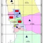 |
Figure 1: A map of Jeddah city showing different locations of Flies collection
|
Collected flies were immobilized after they were kept in the deep freezer for 15 to 20 minutes. After preservation adult flies were morphologically characterized and classified with the help of available authentic keys. (Akbarzadeh et al 2015; Al ahmadi and Salem et al 1999, Al Ghamdi and Alikhan 2015; Alikhan et al 2016,; Dabbour and El Dawy 1981, Irish et al. 2014; Mc Alpine 1989; Nazni et al 2011; Pont 1991; Setyaningrum and Al Dhafer, 2014; Szpila 2012; Verves 2005).
In the present study only salient morphological features of the flies are discussed. The detailed identification key was already published in previous paper (Alikhan et al 2016).
Results and Discussion
According to morphological identification of adult flies the following ten dipteran fly species belonging to six families were recorded as myiasis causing on the basis of reported cases in Saudi Arabia (Table.2). The flies collected during survey were Megaselia scalaris, Musca domestica, Fanniia canicularis, Sarcophaga haemorrhidalis (Bercaea cruentata); Parasarcophaga ruficarnis, Wohlfahrtia nuba , Chrysomya marginalis, chrysomya albicep, Chrysomya megacephala, and Oesteris ovis. The present prevalence of each fly species was recorded (Table.1). The highest prevalence was recorded for the common house fly Musca domestica (67.6%). Most of the species were collected from slaughter houses (50%) and animal farms (39%). (Table.1).
Table 1: Showing number of Flies collected by different Traps and their percentage
| Family
Name |
Species
Name |
Malaise trap | Yellow Trap | Swipe Net | Bait Trap | Total
No. Of flies |
Total percentage |
| Phoridae | Megasellia
scalaris |
43 | 134 | 0 | 29 | 206 | 7.721% |
| Muscidae | Musca domestica | 383 | 442 | 610 | 369 | 1804 | 67.616% |
| Fanniidae | Fanniia canicularis | 11 | 24 | 0 | 0 | 35 | 1.311% |
| Sarcophagidae | Sarcophaga haemorrhoidalis | 53 | 49 | 5 | 126 | 233 | 8.733% |
| Sarcophagidae | Parasarcophaga ruficarnis | 10 | 16 | 2 | 9 | 37 | 1.386% |
| Sarcophagidae | Wohlfahrtia nuba | 8 | 6 | 1 | 16 | 31 | 1.161% |
| Calliphoridae | Chrysomya albicep | 101 | 81 | 23 | 71 | 276 | 10.344% |
| Calliphoridae | Chrysomya megacephala | 7 | 1 | 0 | 4 | 12 | 0.449% |
| Calliphoridae | Chrysomya
marginalis |
13 | 4 | 2 | 7 | 26 | 0.974% |
| Oestridae | Oestrus ovis | 0 | 0 | 6 | 2 | 8 | 0.299% |
| TOTAL | 629 | 757 | 649 | 633 | 2668 | ||
| Percentage | 23.57% | 28.37% | 24.32% | 23.72% |
Table 2: Showing total number of flies and their percentage collected from different locations
| Family
Name |
Species
Name |
Abattoirs
Collection |
Animal farm
Collection |
Fish & veg.
Market Collection |
Animal Carcases
Collection |
Total
No. Of flies |
Total percentage |
| Phoridae | Megasellia
scalaris |
56 | 62 | 6 | 82 | 206 | 7.721% |
| Muscidae | Musca domestica | 980 | 766 | 32 | 26 | 1804 | 67.616% |
| Fanniidae | Fanniia canicularis | 18 | 11 | 6 | 0 | 35 | 1.311% |
| Sarcophagidae | Sarcophaga haemorrhoidalis | 106 | 15 | 59 | 53 | 233 | 8.733% |
| Sarcophagidae | Parasarcophaga ruficarnis | 18 | 10 | 4 | 5 | 37 | 1.386% |
| Sarcophagidae | Wohlfahrtia nuba | 14 | 5 | 8 | 4 | 31 | 1.161% |
| Calliphoridae | Chrysomya albicep | 120 | 49 | 79 | 28 | 276 | 10.344% |
| Calliphoridae | Chrysomya megacephala | 6 | 3 | 2 | 1 | 12 | 0.449% |
| Calliphoridae | Chrysomya
marginalis |
13 | 6 | 4 | 3 | 26 | 0.974% |
| Oestridae | Oestrus ovis | 3 | 5 | 0 | 0 | 8 | 0.299% |
| TOTAL | 1334 | 932 | 200 | 202 | 2668 | ||
| Percentage | 50% | 34.93% | 7.50% | 7.57% |
Following are the salient morphological diagnostic characteristics and habitats of the species collected.
Family: Phoridae
Megasellia scalaris (Loew, 1866) (Coffin fly or scuttle fly)
Brownish small fly which is also named as scuttle fly due to its sudden and rapid movements. The adult fly may be easily recognised by “humpbacked” thorax and laterally compressed hind femur. The fore margins of the wings are thickened, wing veins are parallel to each other and without cross veins. One segmented antenna with long aristae.
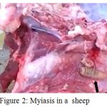 |
Figure 2: Myiasis in a sheep
|
The fly maggots are found near rotten meat, vegetable remains, trash containers, abattoirs, over animal carcases and near poor sanitary conditions. (it causes human accidental myiasis in wounds, intestine, eyes, respiratory system and urino-genital system).
Table 3: Cases of human and animal myiasis recorded from Saudi Arabia IN HUMANS
| Family | Species | Type of Myiasis | Reported By | Region |
| Phoridae | Megasellia
scaleris |
Urinary | Wakid,MH 2008 | Jeddah |
| Sarcophagidae | Wohlfahrtia magnifica | Aural
Scalp |
Al Jbar,I 2015
Al Badri et al 2016 |
Al Ahsa
|
| Sarcophagidae | Sarcophaga sps. | Open Diabetic wound | Zaglool et al 2013 | Makkah |
| Calliphoridae | Cardylobia anthropophaga | Cutaneous | Omar & Abdallah 1992
Afifi et al 2015 |
Al Asir
Al Baha |
| Oestridae | Dermatobia hominis | Cutaneous“ | Al Otaibi2016
Akhtar et al 2000 Hiba Radwan2015 |
Al Khobor
Al Asir Taif |
| Oestridae | Ostris ovis | Opthalmo myiasis | Kenway et al. 2014 | Al Asir |
| IN ANIMALS | ||||
| Oestridae | Ostris ovis | Nasopharyngeal | Al Ahmad et al. 2000
Banja & Madbouly 1981 Bosly 2013 |
Riyadh
West Jazan |
| Oestridae | Cephalopina titillator |
Nasopharyngeal |
Fatani &Hilali 1994
Ghandour, 1988 Banja & Madbouly 1981 Buttiker & Zumpt 1982 |
Al Ahsa
West West Riyadh |
| Calliphoridae | Chrysomya bezziana | Dermal | Al Ahmad et al 2001,
2006 |
Riyadh |
| Calliphoridae | Chrysomya albicep | Dermal | Al Ahmad 2001 | Riyadh |
| Calliphoridae | Chrysomya megacephala | Dermal | Ramdan & El Bihari 1980 | Hafuf |
| Sarcophagidae | Wohlfahrtia magnifica | Dermal | Al Ahmad 2001 | Riyadh |
Family: Muscidae
Musca domestica (Linnaeus, 1758) (House fly)
Thorax with 4 black stripes on greyish background, abdomen with yellowish spots on the anterior lateral sides below the wings and a dark midline. Antennae are short and 3 segmented, anal cell in the wing is closed, 4th vein angled with discal cell.
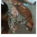 |
Figure 3: Myiasis in Human
|
It lives close to humans and contaminate the food, feed and breed in garbage and animal faeces. (It is an agent of accidental myiasis of intestine and urino-genital system).
Family: Fanniidae
Fanniia canicularis (Linnaeus, 1761) (Lesser house fly)
The thorax is brown-grey with less distinct three black brown stripes, abdomen yellow in colour, eyes are with white border in male, head silvery in colour, halters are yellowish, the 4th vein not angled but straight, anal vein short.
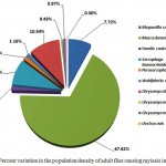 |
Figure 4: Percent variation in the population density of adult flies causing myiasis in Jeddah
|
High number of Fanniia found in poultry farms and animal sheds. Deposit eggs on decaying organic matter such as cow dung, human and poultry faeces.
(It causes accidental myiasis of intestine and urino-genital system)
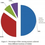 |
Figure 5: Percentage of flies causing myiasis collected from different locations of Jeddah
|
Family: Sarcophagidae
Sarcophaga haemorrhoidalis (Fallen,1817) (red-tailed flesh fly)
Thorax with 3 dark longitudinal stripes, antennal arista is plumose at the base and bare at the tip; antenna and palpi are greyish black with white hair on gennae. Abdomen with grey black checker board. Sixth tergite of female is red in colour, separated with a row of strong bristles at the edge.
Flesh flies usually attracted to rotten meat, fish, vegetables, colonize the corpses, animal and human faeces.
Larviparous (Lay larvae) (Produce myiasis on necrotic or dead flesh in both humans and animals.).
Parasarcophaga ruficarnis (Fabricius,1794) (flesh fly species)
Can be identified by white hairs on gennae below eyes, antenna and palpi orange in colour. Breed both in faeces and dead bodies.
(breeds on flesh causing myiasis or carrion and faeces, also a vector for diseases).
Wohlfahrtia nuba (Wiedemann,1830) (spotted flesh fly)
Greyish with three black round spots at the end of each abdominal segment, antennal arista bare or with very short hairs, larviparous. Breed on the open wounds of the camel and causes severe myiasis.
(Causes severe myiasis in Camel it is a secondary invader of wounds.)
Family: Calliphoridae
Chrysomya albicep (Wiedemann,1819) (Blow fly species)
Body metallic green with narrow dark lines on the rear edge of each abdominal segment; anterior thoracic spiracle white/pale yellow, antennal arista plumose, head with silvery hair.
(Female deposit egg on the decomposing tissues or in the wounds of living in animals and in humans and causes a specific myiasis in wounds, sores and nasal cavities.)
Chryasomya megacephala (Fibricius,1794) (oriental latrine fly)
Large red eyes with metallic blue/green body, buccal area and gennae with orange setae. Anterior thoracic spiracle black/brown. Lower calypter white at the base and the rest is dark.
(Causes myiasis in animals and humans resulting into economic losses in cattle and fish industries all over the world.)
Chrysomya marginalis (Wiedemann,1830)
Anterior wing margins dark, anterior spiracle white in colour. Greater ampulla with thick hairs dorsally. Lower calypter with dense hairs.
Family: Oestridae
Oestrus ovis (Linnaeus, 1758) (Sheep nasal bot fly)
Short stout and dark grey body with black spots at the abdomen. Head and legs are dull yellow.
Female fly deposit eggs in the nostrils of the camel or sheep. Usually found where sheep camel or goats are reared.
(Responsible for myiasis of skin, mucous membranes of mouth, eyes or nasal cavities both in animals and humans.)
Most of the reports on myiasis in animals & humans from Saudi Arabia are about the case studies. ( Afifi et al, 2015; Al Otaibi et al. 2016; Al Badri, 2016; Al Ahmad et al, 2006; ). Many myiasis causing flies collected by different workers from other parts of the country such as, Cordylobia anthropophaga, Dermatobia hominis, Wohlfahrtia magnifica, Cephalopina titillator, Chrysomya bezziana etc., may be present in Jeddah region but could not be trapped during this study. Some of the workers studied the identification of forensic important flies from Jeddah (Al Ghamdi and Alikhan, 2015, Al shareef H. 2016) but there is no record of myiasis causing flies from this region.
Conclusion
Present study is important because it provides a list of flies causing myiasis to animals and humans in this part of the kingdom and responsible for the economic losses and agony. The flies may be different taxonomically but they affect animals and humans in the same way and their ecology and habitats also similar.
The easy method of identification is needed for the veterinary & health workers to control the myiasis causing flies and to reduce the economic loss and human sufferings.
Acknowledgement
The authors are indebted to the Al Amana Public Health Laboratory, Jeddah for providing the facilities to carry out this research work. We are also thankful to all our colleagues for their kind help and suggestions during this study.
References
- Afifi M.A, Jiman-Fatani A.A, Alsiny F.I, Anshasi W.S. A new focus of autochthonous transmission of Cordylobia anthropophaga in Saudi Arabia. Journal of Microscopy and Ultrastructure. 2015;3(2):82-5.
CrossRef - Akbarzadeh k, Wallman J, Sulakova H, SDzpila K. Species identification of Middle Eastern blow flies ( Diptera: Calliphoridae) of forensic importance. Prasitol Res. 2015;114:1463-1472.
CrossRef - Akhter J, Qadri S.M, Imam A.M. Cutaneous myiasis due to Dermatobia hominis in Saudis. Saudi medical journal. 2000;21(7):689-91.
- Al Ahmad A.M, Al Dawood A.S and kheir S.M. Seasonal activity of flies causing myiasis in livestock animals using sticky traps baited with swarm lure-4 in Riyadh region, Saudi Arabia. Scientific Journal of King Faisal University ( Basic and applied sciences). 2006;7:107-119.
- Alahmed A. M. Incidence of myiasis in sheep caused by Chrysomya bezziana in Saudi Arabia.J. King Saud Univ. 14: Agric. Sc. 2001;(2):109-112.
- Al Ahmadi A.Z and Salem M.M. Entomofauna of Saudi Arabia, Part 1: Check list of Insects. Academic Publishing & Press. King Saud University.
- AlBadri R, Harbi A.l.T, Tonnsi A, Almatary A, Hassanein R. Cutaneous Myiasis in a Child Scalp Caused by Wohlfahrtia magnifica (Diptera: Sarcophagidae): A Case Report. MOJ Clin Med Case Rep. (2016). 1999;4(3): 00093. DOI:15406/mojcr.2016.04.00093
- AlJabr I, Myiasis A. a Rare Cause of Earache; Case Reports in Otolaryngology Article ID. 2015;219529:3.
- Al-Shareef. Species Identification of Forensic Important Blowflies (Diptera, Calliphoridae) that occur in Jeddah, Kingdom of Saudi Arabia. Glob J Nurs Forensic Stud. 2016;1:3.
- Amoudi M.A , Diab F.M, Abou-Fannah S.S.M. The occurrence of Megaselia scalaris (Loew) ( Diptera: Phoridae) in saudi Arabia with some aspects on the life history and distribution in Riyadh Province. J King Saud Univ.Sci. 1989;1-2:43-51.
- Amoudi M.A. New records of some Sarcophagid flies with distribution of all known flesh flies (Diptera: Sarcophagidae) of Saudi Arabia. J. of Egyptian Society of Parasitology. 1993;23(1):297-304.
- Alikhan et al. Public health and veterinary important flies (order: Diptera) prevalent in Jeddah Saudi Arabia with their dominant characteristics and identification key. Saudi Journal of Biological Sciences. 2016. http://dx.doi.org/10.1016/j.sjbs.2016.08.
- Al-Ghamdi, Alikhan et al. Characterization of forensically important Nacrophagus Flies ( Diptera) of Jeddah, Saudi Arabia. Advances in Environmental Biology. 2015;9(8):58-71.
- Alotaibi S, Al shahrani D, Alzomor O. Cutaneous Myiasis in Saudi Infant: A Rare Case Report IMJH. 2016;2(9);8-12.
- Badawi A. Arthopods of Medical and veterinary importance in the kingdom of Saudi Arabia. King Saud Uni. Press Riyadh. 1994.
- Areej O, Bakhraibah. Prevalence and organ distribution of larval Oestrus ovis (Diptera : Oestridae), Cysticercus tenuicollis and Echinococcus granulosus (Cestoda : Taenidae) in slaughtered sheep and goats at Jeddah in Saudi Arabia. 2016.
- Banja A & Madbouly M.M. Field and laboratory observations on three dipterous larvae causing myiasis in the western region of Saudi Arabia. B ulletin Faculty of Science, King Abdulaziz University. 5:77-80.
- Bosly A.H. (2013) Seasonal prevalence of Oestrus ovis L. (Diptera:Oestridae) larvae in infested sheep in Jazan Region,Saudi Arabia; J. Parasitol. Vector Biol. 1981;5(5):66-71.
- Boorman J. Insects of Saudi Arabia Culicoides (Diptera: Ceratopogonidae) of the Arabian peninsula with notes of their medical & veterinary importance, Fauna of Saudi Arabia. 1989;10:160-225.
- Buttiker W & Zumpt F. Myiasis in domestic animals. Fauna of Saudi Arabia. 4:520-524.
- Dabbour A.I. “Short note on dipterous flies in western and central regions of Saudi Arabia”.Journal of Agriculture Research, Riyadh University. 1979;1982;4: 81-83.
- Dabbour A.I. Note on dipterous flies in western and central regions of Saudi Arabia”. Journal of Faculty of Science, Riyadh University. 1979;10:117-119.
- Dabbour A.I and Dawy E.l .M. Morpholigical and classification studies of some dipterous flies in the kingdom of Saudi Arabia. Agriculture Research Centre Bulletin. 1981.
- Dabbour A.I, Hammad S.M. Insect and Animal pests and their control in the Kingdom of Saudi Arabia. Uni. Libraries, King Saud Univ. Riyadh (In Arabic). 1982.
- Francesconi F and Lupi O. Myiasis. Clinical Microbiology Reviews . 2012;25(1).
CrossRef - Fatani A, Hilali m. 1994 – Prevalence and monthly variations of the second and third instars of Cephalopina titillator (Diptera: Oestridae) infesting camels (Camelus dromedarius) in the Eastern Province of Saudi Arabia.
- José G.B.D, Allen C.G.H, Rademaker M. Human myiasis in New Zealand: imported and indigenously-acquired cases; the species of concern and clinical aspects. NZMJ. 2010;123(1322):1175 8716.
- Ghandour A. M. Health Hazards in Humans and Animal Caused by Imported Livestock Diseases in Saudi Arabia. Fauna of Saudi Arabia. 1988;9:468-476.
- Hall M.J.R. Screwworm flies as agents of wound myiasis; World animal Review, Special issue” New World Screwworm; Response to an emergency” R.D.S. Branckaert (Ed). 1991;52.
- Hall M.J.R and Wall R. Myiasis of humans and domestic animals. Adv. Parasitol. 35:257–334.
- Hiba M.R. (2015) Myiasis. Trop Med Surg . 1995;3(4):1000197.
- Irish S, Lindsay T, Wyatt N. Key to adults of Afro-tropical species of the genus Chrysomya Robineau-Desvoidy (Diptera: Calliphoridae). African Entomol. 2014;22:297–306.
CrossRef - James M.T. The flies that cause myiasis in man. United states department of agriculture: Miscellaneous publication No. 631. Washington, DC: US Government Printing Office. 1947.
- Kamal A.S. comparative study of thirteen species of sarcophagus Calliphoridae and Sarcophagidae (Diptera): Bionomics. Ann, Entomol .Soc. Am. 1958;51:261-271.
CrossRef - Kenway M.A, Al ashry H.A, Shobrak M. Synanthropic flies of Asir Province, Southwest of Saudi Arabia. J. of Ento and Acro Res. 2014;46:3.
- Khyat R.M. A case report on oral myiasis in Saudi Arabia. Saudi.Dent.J. 2002;14:140-2.
- Majed H.W. A Laboratory-Based Study for First Documented Case of Urinary Myiasis Caused by Larvae of Megaselia scalaris (Diptera: Phoridae) in Saudi Arabia. 2008.
- Mashaly A. A preliminary study of forensically important necrophagous Diptera in Riyadh, Saudi Arabia. Conf. 8th Int. Cong. Of Dipterology, Postdam, Gremany. 2014.
- Mc Alpine J.F. Phylogeny and classification of the Muscomorpha. In: Mc Alpine J.F, Wood D.M (eds,) Manual of Nearctic Diptera 3. Research Branch, Agriculture Canada Monograph. 1989;32:1397-1518.
- Morsy T.A, Fayad M.E, Salama M.M.I, Sabry A.A, Seroagi E.A.O.M and Abdullah K.F. Some myiasis producers in Cairo and Giza abattoirs. J Egypt Soc Parasitol. 1991;21(2):539-546.
- Nazni W.A, Jeffery J, Heo C.C, Chew W.K, Lee H.L. Illustrated keys to adult flies of forensic importance in Malaysia. Institute for Medical Research, Kuala Lumpur. 2011.
- Omar M.S and Abdullah R.E. Cutaneous myiasis caused by tumbu fly larvae, Cordylobia anthropophaga in South-western Saudi Arabia. Trop. Med. Parasitol. 1992;43:128-9.
- Patton W.S. Some notes on Indian Calliphorinae III Chrysomyia megacephala Fabr. Bulletin of Entomological Research. 1922;13(1):109-113.
CrossRef - Patton W.S and Cushing E.C. Studies on the higher dipteral of medical and veterinary importance: a revision of the genera of subfamily Calliphorinae based on a comparative study of the male and female terminalia. Tropical Medicine and Parasitology. 1934;28:123-130.
CrossRef - Pont A.C. A review of Faniidae and Muscidae of Arabian Peninsula. Fauna of Saudi Arabia. 1991;12:312-365.
- Ramadan R.O and Bihari E.S. Dermal myiasis in animal farm in El Hofuf Area. Fourth Symposium on the biological aspects of Saudi Arabia. Saudi Biological Society, Faculty of Science, University of Riyadh. 1980; March 10-13(1980):85.
- Setyaningrum H & Al Dhafer H.M. The calliphoridae the blow flies ( Diptera : Oestroidea) of Kingdom of Saudi Arabia. Egypt Acad. J. Biolog. Sci. 2014;7(1):49-139.
CrossRef - Sundharam J, Al-Gamal M. Myiasis in Saudi Arabia. Annals of Saudi medicine. 1994;14(4):352.
CrossRef - Szpila K. Key for identification of European and Mediterranean blowflies (Diptera, Calliphoridae) of medical and veterinary importance – adult flies. In: Gennard D (ed) Forensic entomology, an introduction, II edition. Willey-Blackwell. 2012;P77–81 + plates 5.1–5.9.
- Verves Y.G. A catalogue of Oriental Calliphoridae (Diptera). Dipterological Res. 2005;16:223–310.
- Zaglool D.A, Tayeb K, Khodari Y.A, Farooq M.U. First case report of human myiasis with Sarcophaga species in Makkah city in the wound of a diabetic patient. J Nat Sc Biol Med. 2013;4:225-8.
CrossRef - Zumpt F. Myiasis in man and animals in the Old World: A textbook for physicians,veterinarians and zoologists. London: Butterworths. 1965.

This work is licensed under a Creative Commons Attribution 4.0 International License.


