

Manuscript accepted on : 22 December 2016
Published online on: --
Plagiarism Check: Yes
Beisenova Raikhan Rymbaevna1, Khanturin Marat Rashytovich1, Manekenova Kenzhekyz Boranbaevna2, Mustafa Rauan Sergazykyzy1 and Syzdykova Nazym Kosarbekovna3
1Department of Management and Engineering in the field of environmental protection, L.N. Gumilyov Eurasian National University, Astana, Republic of Kazakhstan.
2Department of Pathological anatomy, Astana Medical University, Astana, Republic of Kazakhstan.
3Department of methods of teaching mathematics and computer science, E.A.Buketov Karaganda State University, Karaganda, Republic of Kazakhstan.
Corresponding Author E-mail: bigozha@gmail.com
DOI : http://dx.doi.org/10.13005/bbra/2441

ABSTRACT: Environmental influence of polycyclic aromatic hydrocarbons observed not only on the general state of the organism, but also the influence on the nervous system in particular. Also consider compensation mechanisms when exposed to polycyclic aromatic hydrocarbons. Anabolic agents through restorative and bio-stimulating effect, can have a beneficial effect in the treatment of almost any disease, and therefore the range of medical indications for the use of anabolic steroids is constantly expanding.
KEYWORDS: Ecdyphyt, morphological characteristics of brain hemisphere sections; Hematoxylin and Eosin stain (H & E) the impact of polycyclic aromatic hydrocarbons; morphological and functional changes in the brain tissue;
Download this article as:| Copy the following to cite this article: Rymbaevna B. R, Rashytovich K. M, Boranbaevna M. K, Sergazykyzy M. R, Kosarbekovna S. N. Morphological Characteristics of Experimental Encephalopathy Under Influence of Toxic Polycyclic Aromatic Hydrocarbons with their Correction. Biosci Biotech Res Asia 2017;14(1). |
| Copy the following to cite this URL: Rymbaevna B. R, Rashytovich K. M, Boranbaevna M. K, Sergazykyzy M. R, Kosarbekovna S. N. Morphological Characteristics of Experimental Encephalopathy Under Influence of Toxic Polycyclic Aromatic Hydrocarbons with their Correction. Biosci Biotech Res Asia 2017;14(1). Available from: https://www.biotech-asia.org/?p=20662 |
Introduction
Stability, hydrophobicity and other physical and chemical properties of polycyclic aromatic hydrocarbons, organic compounds make them sustainable in an environment that allows them to be toxic to all living beings.1
Another group of genotoxic and carcinogenic compounds, nitropolyarenes, may be formed from PAH in emissions from various sources, in particular, diesel engine exhausts combined with the presence of nitrogen oxides.2 Although it is clear that PAHs are carcinogenic, it is important to note that not all strains of mice exposed to these agents develop tumors, but PAHs also have significant interactions with the immune system and benzo[a]pyrene, and 3-methylcholanthrene are all contact allergens when applied to the skin.3
PAHs constitute the largest class of potential carcinogens, to which mutagenic activity is also ascribed. Numerous data illustrate the distribution of concentration of PAHs in the atmosphere in areas of smoke emission and in the vicinity of the large urban centers. Because of their mutagenic or carcinogenic properties PAHs are a risk to human health.4 PAHs also exposure to neurotoxicity and changes in cognitive function. In vitro experiments including animal and human cells report cell death, cell deformities, or decreased cell activity when cells are exposed to PAHs (pyrene and anthracene or benzo(a)pyrene) for a few hours.5 Also urinary polycyclic aromatic hydrocarbons were associated with adult hypertension, heart attack and cancer, although the causality cannot be established.6 Can we find some protective function of body from aggressive environment what human made.
In recent years, medicine is increasingly trying to deal with all sorts of diseases using more natural products or drugs on herbal basis. This is in particular due to the side effect of many modern medicines. A number of studies in our country began to try Ecdyphyt drug as an anabolic agent and remedy for tuberculosis. This drug is used as one of the most promising groups of herbal medicines. It has anabolic, adaptive and tonic effect. The use of these funds is overdue with a wide range of Diseases associated with impaired protein anabolism, general exhaustion of the body, weight loss, asthenia, growth retardation, violation of compensatory and adaptive processes.7 Also this drug is used for the prevention of various kinds of harmful effects on production. For our study, we chose Ecdyphyt drug, because it is a herbal anabolic practically non-toxic, well tolerated, has very few contraindications.8
Material and Methods
The experiments were conducted on laboratory outbred rats. Adult males weighing 250-320 grams. During experiments, animals were maintained under similar conditions as before the experiment. Water and food were given without limitation. Temperature T = 21-22˚S relative humidity, light regime day and night. Animals were housed 5 animals in each cage in accordance with sanitary rules.
The experiment used 30 laboratory rats. Which were divided into three groups of 10 in each group. The first group of the control group, the second group received a dose of gasoline inhalation (LC50 values (10,000 ppm / 7H) 7 days exposure for 4 hours. The third group received gasoline dose inhalation (LC50 (10,000 ppm / 7H) 7 days exhibit for 4 hours and received Ecdyphyt dose according to the dosage for humans 120 μg as a correction. Ecdyphyt diluted in one ml of water and was administered orally by catheter after air seeding gasoline vapors.
To study the pathological changes in the brains of experimental rats, the cerebral hemispheres were removed during autopsy. Sagittal section of the cerebral hemispheres were separated on the plate slices of thickness 0.5 cm.
After labeling, the brain tissue was fixed in 10% formalin solution neutralized with chalk for 3 days. Then the experimental data was carried out through solutions ascending concentrations of ethanol (concentration of 70˚-80˚-90˚). Incubated in equal ratios of 96˚ ethanol and chloroform (solution of an equal amount of chloroform and heated paraffin). Defatted and dehydrated material thus impregnated in two batches of pure paraffin homogenized in a thermostat at a temperature of 56º C.9 Then embedded in paraffin blocks in special plastic cassettes.
Histological sections 5-7 µ obtained on the automated rotary microtome then deparaffinized in two portions of ethyl alcohol 96˚, rinsed in water and subjected to hematoxylin-eosin staining and methylene blue method of Nissl.10
Microscopy and microphotography was performed in hardware complex Leica ICC50 HD
When taking a picture of detectable histological changes on the digital camera used lenses and eyepiece x10: x10, x16, x20 and x40.
Morphometric methods of investigation
For the purpose of objectification morphological studies produced morphometric study.
To compare the degree of effectiveness of toxic encephalopathy correcting with Ecdyphyt, produced morphometric analysis of the number of nerve cells with signs tigrolysis, shrunken neurons and-shadow cells in the cerebral cortex.
Thus obtained was subjected to quantitative statistical analysis by calculating the average statistical indicators with an average error (M ± m).
Results
Morphological changes in the brain tissue were examined after 24 hours from the beginning of the experiment.
Histological studies showed that the correction toxic encephalopathy Ecdyphyt , has a definite influence on the severity of dyscirculatory, dystrophic-degenerative and reactive hyperplastic processes developing in acute toxic inhalation of gasoline vapors.
Thus, the observed lower severity of symptoms of acute disorders of blood circulation in the vessels of the microvasculature.
Noted moderate hyperemia of blood vessels, lowers high permeability of the walls of microvessels, which was reflected in the absence of focal diapedesis hemorrhages in the brain of rats. This reduces the severity of symptoms pericellular edema and perivascular edema phenomenon persisted (Figure 1).
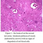 |
Figure 1: The brain of rat the second test series. Moderate plethora of vessels (indicated by arrows) with no signs of diapedesis of red blood cells. Perivascular edema symptoms persist. H & E stain. Increasing x 160.
|
The cytotoxic effects of inhalation of gas fumes were less pronounced. Significantly reduces the symptoms of acute swelling, deformation and rough cytolysis neurons and cells of astrocytic glia. (Figures 2, 3).
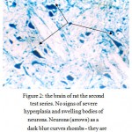 |
Figure 2: The brain of rat the second test series. No signs of severe hyperplasia and swelling bodies of neurons. Neurons (arrows) as a dark blue curves rhombs – they are not increased in size. Painting by Nissl. Increased x200.
|
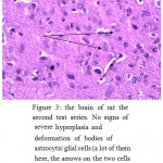 |
Figure 3: The brain of rat the second test series. No signs of severe hyperplasia and deformation of bodies of astrocytic glial cells (a lot of them here, the arrows on the two cells shown an example). H & E stain. Increased x160.
|
Glial-cell responses were maintained. However, glial-cell complexes differ in their structure. Thus, the more pronounced effects were perivascular gliosis, satellitosis and phagocytic activity of glial cells (Figures 4, 5, 6).
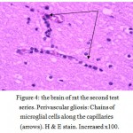 |
Figure 4: The brain of rat the second test series. Perivascular gliosis: Chains of microglial cells along the capillaries (arrows). H & E stain. Increased x100.
|
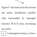 |
Figure 5: The brain of rat the second test series. Satellitosis: satellite cells surrounded by damaged neurons. H & E stain. Increasing the X400. A) is damaged neurons, it has a smudged core and visible cytoplasmic red. B)it – gliocytes (dark blue tight balls), which has had clung to the damaged neurons.
|
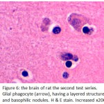 |
Figure 6: The brain of rat the second test series. Glial phagocyte (arrow), having a layered structure and basophilic nodules. H & E stain. Increased x200.
|
It is also determined by the signs of the formation of glial scars in the form of growths as a cord microglial cells in an area of hemorrhagic damage to the brain tissue (Figure 7).
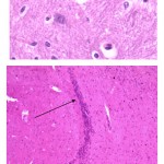 |
Figure 7: The brain of rat the second test series. Glial scar formation – indicated by the arrow (dark blue streak curve of fine microglial cells) in the area of the brain tissue damage. H & E stain. Increased x100.
|
In addition, there is a development of the productive cell reactions around the vessels with the participation of microglial cells and lymphocytes (mikrovaskular cells).(Figure 8).
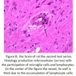 |
Figure 8: The brain of rat the second test series. Histology productive mikrovaskular (arrow) with the participation of microglia cells and lymphocytes. (In the center of the figure the vessel, its wall is thick due to the accumulation of lymphocyte cells (very small powdered blue dots) and around the vessel – microglial cells). H & E stain. Increased x 160.
|
In order to evaluate the effectiveness of neuroprotective effect of Ecdyphyt, contact was made morphometric assessment of volume fraction of neurons with signs of irreversible damage to the brain tissue of rat control and experimental groups. (Table 1).
Table 1: Morphometric parameters of volume fractions (%) of irreversible damage to neurons in the cerebral cortex of rats in the experimental groups.
| Signs of neuronal damage | Control
|
Inhalation of polycyclic
aromatic hydrocarbons |
Inhalation of polycyclic
aromatic hydrocarbons and correcting with Ecdyphyt |
| Tigrolysis | 0,11±0,04 | 2,59±0,46 | 1,45±0,21 |
| Corrugation | 1,32 ±0,09 | 4,96±0,22 | 2,98 ±0,14 |
| Cell – shadows
|
0,09±0,02 | 3,95±1,04 | 1,32±0,09 |
Discussion
Due to the fact, that the Ecdyphyt drug has anabolic, adaptogenic and tonic properties, it’s used in the production of chronic intoxication, which contributes to the improvement of the clinical picture of the general condition, reduction of symptoms and normalization of laboratory values.11
The drug mechanism of action on the body based on the effects of phytoecdysteroids that non-specifically enhance protein synthesis in the body, have a positive effect on nitrogen metabolism.
Given the above data, we note the relative stabilization of metabolic processes in the cerebral hemispheres of rats.
Conclusions
Thus, the background correction with Ecdyphyt experimental toxic encephalopathy, developing inhalation of polycyclic aromatic hydrocarbons vapors, reduces vascular permeability and activity of cytotoxic processes, inhibits the proliferative activity of microglial cells, ependymal of the lateral ventricles and meningothelial lining of the pia mater. However, there is stimulation of inflammatory cell response to the development of micro vasculitis.
References
- Pirozhkova A. A. PAHs The impact on the world and man. Youth Science and Technology Gazette . Bauman Moscow State Technical University. 2014;77-51038. ISSN 2307-0609. http://sntbul.bmstu.ru/doc/732357.html.
- Khesina A. Y. Urban Air Pollution by Carcinogenic and Genotoxic Polyaromatic Hydrocarbons in the Former USSR., Laboratory of Carcinogen Screening Methods, Cancer Research Center Russian Academy of Medical Sciences, Moscow, Russia. 1994;102(4);49-53.
- Craig A. E., Athar M., Karen A. T., Rothaupt D., Xu H.,And Mukhtar H. Susceptibility to the biological effects of polyaromatic hydrocarbons is influenced by genes of the major histocompatibility complex. Natl. Acad. Sci. USA, Immunology. 1998;(95):14915–14919.
- Gworek B., Klimczak K., Kijen´ M. s. The Relation between Polyaromatic Hydrocarbon Concentration in Sewage Sludge and Its Uptake by Plants: Phragmites communis, Polygonum persicaria and Bidens tripartita. Institute of Environmental Protection., National Research Institute, University of Life Sciences, Department of Soil Environment. Sciences, Warsaw. 2014;9(10):1-9.
- Elizabeth A. B., Juarez-Colunga E., James K., William G. L. B., Serdar B. Biomarkers of Exposure to Polycyclic Aromatic Hydrocarbons and Cognitive Function among Elderly in the United States. National Health and Nutrition Examination Survey. 2016;1-18.
- Shiue I. Are urinary polyaromatic hydrocarbons associated with adult hypertension heart attack and cancer. USA NHANES. 2016;(23):3971–3977.
- Таbriz S. The efficiency of phytopreparation Ecdyphyt in complex treatment of tuberculosis. Almanac of Modern Science and Education. Tambov. 2010;3(34):83-85.
- Kapyseva N., Bakhtiyarova K S., Kustova E. A., Urazalieva N. T., Mahmudova L. H. Post-Exercise Effect of Anabolic Drug on Blood Cell Apoptosis. European Researcher. 2013;38(1-1):5-10.
- Korzhevsky D., Gilyarov E. Basics of histologic techniques. Moscow Special Literature. 2010;96.
- Avtandilov G. G. Fundamentals of quantitative pathological anatomy. Moscow Med. 2002;239.

This work is licensed under a Creative Commons Attribution 4.0 International License.


