

Manuscript accepted on : 18 January 2017
Published online on: --
Plagiarism Check: Yes
Molecular Detection of Mungbean Yellow Mosaic India Virus (MYMIV) Infecting Soybean in Madhya Prades
R. S. Marabi1, Deepti B. Sagare1, S. B. Das1, A. K. Bhowmick1 and H. Noda2
1Department of Entomology, College of Agriculture, JNKVV, Jabalpur (MP), India.
2Japan International Cooperation Agency (JICA), Tsukuba, Japan.
Corresponding Author E-mail: rsmarabi78@rediffmail.com
DOI : http://dx.doi.org/10.13005/bbra/2448

ABSTRACT: Detection of MYMIV infecting soybean was carried out during Kharif 2016 using DNA-A (cp) and DNA-B specific molecular markers. Among ten leaf samples collected from different villages, nine were found to possess virus irrespective of the density of whiteflies in concerned region. One sample from Tigan village was found to be virus free even though it was having yellow mosaic disease (YMD) symptoms and whitefly density in an area was moderate. Deficiency of micronutrients could be a reason for YMD like symptoms in virus free plant. There was no correlation between the density of whiteflies and presence of virus in host plant. Molecular markers can be successfully used for detection of MYMIV infecting soybean.
KEYWORDS: YMD; MYMIV; Molecular Markers; Soybean; Whitefly
Download this article as:| Copy the following to cite this article: Marabi R. S, Sagare D. B, Das S. B, Bhowmick A. K, Noda H. Molecular Detection of Mungbean Yellow Mosaic India Virus (MYMIV) Infecting Soybean in Madhya Prades. Biosci Biotech Res Asia 2017;14(1). |
| Copy the following to cite this URL: Marabi R. S, Sagare D. B, Das S. B, Bhowmick A. K, Noda H. Molecular Detection of Mungbean Yellow Mosaic India Virus (MYMIV) Infecting Soybean in Madhya Prades. Biosci Biotech Res Asia 2017;14(1). Available from: https://www.biotech-asia.org/?p=21532 |
Introduction
Soybean is the main Kharif crop of the Madhya Pradesh state of India. The present area under soybean in the state is 6.31 million ha with production of 5.24 million tonnes and productivity 831 kg/ha (Anonymous 2016). Productivity of soybean is less than the potential yield of recommended varieties. Among different biotic factors yellow mosaic diseases (YMD) are major constraints on the productivity of legume crops in India. In Central India, YMD of soybean and blackgram is caused by Mungbean Yellow Mosaic India Virus (MYMIV) (Usharani et al., 2004; Malathi et al., 2005; Girish et al., 2005; Ramesh et al., 2013). MYMIV possesses bipartite single-stranded circular DNA genomes viz., DNA-A and DNA-B (Qazi et al., 2006 and Brown et al., 2015). MYMIV is transmitted by the whitefly, Bemisia tabaci (Govindan et al., 2014) and infect the legumes such as soybean, blackgram, greengram etc.
Recent advancement of molecular techniques is useful for clarifying epidemiology of MYMIV. In particular, detections of MYMIV by polymerase chain reaction (PCR) from the plants and whiteflies can be a key technology. Molecular markers are developed for DNA-A and DNA-B genome of MYMIV and are being used to detect viruliferous whitefly using PCR in recent past (Accotto and Sardo, 2010). Very recently molecular differentiation and rapid detection of MYMV and MYMIV from soybean leaf has been reported by Ramesh et al, (2016). Therefore the current study was carried out to detect MYMIV infection in soybean using molecular markers.
Materials and Methods
Plant Materials
Soybean leaf samples were collected from different villages of Jabalpur district of Madhya Pradesh state, having variable density of whiteflies (Table 1) during Kharif 2016.
Table 1: Soybean samplescollected from different villages and whitefly density
| Village | Date of Collection | Crop age | Density of Whitefly |
| Kuwakheda | 20.08.2016 | 40-45 days after sowing (Vegetative stage) | High*** |
| Kalapatha | 20.08.2016 | High*** | |
| Malakheda | 20.08.2016 | Moderate** | |
| Kevlari | 20.08.2016 | High*** | |
| Karhaiya | 22.08.2016 | Moderate** | |
| Mukanwara | 22.08.2016 | Moderate** | |
| Katrabelkheda | 22.08.2016 | Moderate** | |
| Tigan | 24.08.2016 | Low* | |
| Bargi | 24.08.2016 | Moderate** | |
| Sihora | 24.08.2016 | High*** |
*<5 whiteflies/plant; **6-10 whiteflies/plant; ***>11 whiteflies/plant
DNA Isolation and PCR Amplification
DNA from soybean leaf samples was isolated using DNeasy Plant Mini Kit (Qiagen) from the plants showing YMD symptoms. Molecular markers were designed for DNA-A (cp) and DNA-B genomes of MYMIV (Table 2). PCR was carried out with genomic DNA using molecular markers in Bio-Rad Thermal cycler. The reaction was carried out in 25 µl volumes, which contains 1.0µl (25ng) of soybean genomic DNA, 1.0µl (2.5pmole) of forward and reverse primers each, 1.0µl (2.0mM) of dNTPs, 1.0µl of Taq buffer (10X), 1.0µl of MgCl2 (25mM) and 1 units of Taq polymerase. All the chemicals and plasticwares used were obtained from Genei and Tarsons respectively. PCR Programme (Table 3) was standardized to carry out amplification with DNA-A and DNA-B genome specific primers. The amplified products were resolved on 1.0% agarose gel and visualized under Syngene gel documentation system.
Table 2: Molecular markers and their sequences
| Molecular Marker | Sequence | Amplicon size (bp) |
| DNA-A (cp) | Forward: ACACGGATCCGTTGCATACACAGGATTTG | 750 |
| Reverse: ACACGAGCTCCTCTACCCCGATATCGAATG | ||
| DNA-B | Forward: AGCCTATGACACCGTCAAGAGGA | 541 |
| Reverse: CGCCGGGACAACGGCATAT |
Table 3: PCR programme
| Steps followed in
Thermal cycler |
Temperature in ºC for one cycle | Time
for one cycle |
| Marker | DNA-A(cp)/DNA-B Specific | |
| Step 1 | 94ºC | 1 min. |
| Step 2 | 94ºC | 20 sec. |
| Step 3 | 56ºC | 20 sec. |
| Step 4 | 72ºC | 1 min. |
| Step 2 – Step 4 are repeated for 30 cycles | ||
| Step 5 | 72ºC | 3 min. |
| Step 6 | Hold at 15ºC until ready to load onto gel | |
Results and Discussion
Soybean leaf samples collected from ten different villages, possessing YMD symptoms were subjected to PCR amplification using DNA-A (cp) and DNA-B specific primers. Among ten samples, nine were found to possess both DNA-A (cp) and DNA-B genome of MYMIV (Figure 1). In these nine villages there was variability in whitefly density. Villages viz., Malakheda, Karhaiya, Mukanwara, Katrabelkheda, Tigan, Bargi, where whitefly density was low/moderate; the leaf samples of these villages were found to possess virus i.e. DNA-A (cp) and DNA-B +ve. This indicates that most of the whiteflies present in those areas were viruliferous and sufficient to transmit MYMIV in soybean.
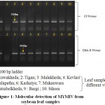 |
Figure 1: Molecular detection of MYMIV from soybean leaf samples
|
One leaf sample collected from Tigan village was found to be virus free (Figure 1) even though YMD symptoms were present on the leaves and whitefly density was moderate in the area. This indicates that whiteflies present were non-viruliferous hence there was no presence of virus in the soybean leaf, morever the YMD like symptoms which appeared on leaves might be due to other reasons such as deficiency of micronutrients. Further no correlation was observed between the whiteflies density and presence of virus in the soybean host plant (Figure 2).
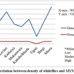 |
Figure 2: Correlation between density of whiteflies and MYMIV in soybean
|
The present study supports the previous studies carried out by Accotto and Sardo (2010); Naimuddin et al (2011) and Ramesh et al (2016) where molecular markers were used for rapid detection of Begomoviruses, MYMIV and MYMV/MYMIV, respectively.
Conclusion
Molecular markers can be successfully used for detection of MYMIV infecting soybean. Deficiencies of micronutrients could be a reason for YMD symptoms in virus free plant. There is no correlation between the density of whiteflies and MYMIV in host plant.
Acknowledgement
Authors are thankful to Director of Research, Jawaharlal Nehru Agricultural University, Jabalpur and Japan International Cooperation Agency (JICA), Japan for providing funds to carry out research under project for maximization of soybean production in Madhya Pradesh.
References
- Accotto G. P and Sardo L. Transovarial Transmission of Begomoviruses in Bemisia tabaci. In book: Bemisia: Bionomics and Management of a Global Pest. 2010;339-345.
- Agricultural Statistics at a Glance – 2015. Government of India Ministry of Agriculture & Farmers Welfare, Department of Agriculture, Cooperation and Farmers Welfare Directorate of Economics and Statistics. 2016;123-125.
- Brown J. K., Zerbini F. M., Navas-Castillo J., Moriones E., Ramos-Sobrihno R and Jose C. E. S. Revision of Begomovirus taxonomy based on pair wise sequence comparisions. Archives of Virology. 2015;160:1593-1619.
CrossRef - Girish K. R and Usha R. Molecular characterization of two soybean infecting begomoviruses from India and evidence for recombination among legume-infecting begomoviruses from South-East Asia. Virus Research. 2005;108:167-176.
CrossRef - Govindan K., Nagarajan P and Angappan K. Molecular studies on transmission of mungbean yellow mosaic virus (YMV) by Bemisia tabaci in Mungbean. African Journal of Agricultural Research. 2014;9(38):2874-2879.
CrossRef - , Akram M and Pratap A. First report of natural infection of Mungbean Yellow Mosaic India Virus in two wild species of Vigna. New Disease Reports. 2011;23:21.
- Malathi V. G., Surendranath B., Naghma A and Roy A. Adaptation to new hosts shown by the cloned components of Mungbean Yellow Mosaic India Virus causing cowpea golden mosaic in northern India. Canadian Journal of Plant Pathology. 2005;27(3):439-447.
CrossRef - Qazi J., Manssor S., Amin I., Awan M. Y., Briddon R. W and Zafar Y. First report of Mungbean Yellow Mosaic India Virus on mothbean in Pakistan. Plant Pathology. 2006;55:818.
CrossRef - Ramesh S. V., Chouhan B. S., Gupta G. K., Ramteke R., Chand S and Husain S. Molecular diversity analysis of coat protein gene encoded by Begomoviruses and PCR assay to detect Yellow Mosaic Viruses infecting Soybean in India. British Biotechnology Journal. 2016;12(3):1-10.
CrossRef - Ramesh S. V., Bhaskale R., Admane N., Gupta G. K and Husain S. M. Multiply primed rolling circle amplification of Yellow Mosaic Virus genome from infected soybean in central India region divulges it as Mungbean Yellow Mosaic Indian Virus-(sb) and its implications for RNAi mediated virus resistance. World Soybean Research Conference-IX (WSRC-IX); Durban, South Africa. 2013.
- Usharani K. S., Surendranath B., Haq Q. M. R and Malathi V. G. Yellow Mosaic Virus infecting soybean in Northern India is distinct from the species infecting soybean in Southern and Western India. Current Science. 2004;86:845-850.

This work is licensed under a Creative Commons Attribution 4.0 International License.


