

Manuscript accepted on : 04 March 2017
Published online on: --
Plagiarism Check: Yes
Image Segmentation Using Mrf Novel Level Set Method
K. Srilatha, S. Pavithra, Neenu Vincent, S. Venkatesh, A. S.Vigneshwar and Medapati Jahnavi Lakshmi
Department of Electronics and Communication engineering Sathyabama University, Tamilnadu, Chennai, India.
Corresponding Author E-mail: 1srilatha169@gmail.com
DOI : http://dx.doi.org/10.13005/bbra/2463

ABSTRACT: The most difficult task in the segmentation of the image is to obtain the segmentation by avoiding almost all the noises within less span of time. The images which are extracted will be from any domain i.e., medical, natural, radar etc. In the proposed method the MRF (Markov random field) algorithm for the process of segmentation is implemented. In this algorithm two methods in parallel fashion are used. The two methods which are used is AMG (Algebraic multi grid) and SFM (Sparse random field).The AMG is used for increasing the time step and SFM will be used in decreasing the computation domain. In the novel level set method it considers that the neighboring pixels also fall in the same region. The noises also will be reduced in this method when it is compared with the existing methods. By using this method, the images of 500*500 sizes also can be segmented. In this technique the number of iterations will be less when compared to the existing systems. The images of many domains can be segmented by using this proposed algorithm i.e., Medical images, noisy images, synthetic aperture radar images (SAR) and natural images. The SAR, natural and noisy images are applicable only in the real-time.
KEYWORDS: Algebraic multigrid (AMG); contour evolution. Novel level set method Markov random field (MRF); sparse random field (SFM);
Download this article as:| Copy the following to cite this article: Srilatha K, Pavithra S, Vincent N, Venkatesh S, Vigneshwar A. S, Lakshmi M. J. Image Segmentation Using Mrf Novel Level Set Method. Biosci Biotech Res Asia 2017;14(1). |
| Copy the following to cite this URL: Srilatha K, Pavithra S, Vincent N, Venkatesh S, Vigneshwar A. S, Lakshmi M. J. Image Segmentation Using Mrf Novel Level Set Method. Biosci Biotech Res Asia 2017;14(1). Available from: https://www.biotech-asia.org/?p=22161 |
Introduction
The fast and strong image segmentation against the errors i.e., noises an algorithm known as Markov Random Field (MRF) in which two methods namely Algebraic multigrid (AMG) and Sparse field method (SFM) are used J. Shenet al.1 The algebraic multigrid technique is used to increase the time step and sparse field method is used to reduce the domain computation. This MRF algorithm assumes that the neighboring pixels fall in the same category while the existing method doesn’t have any relation with the neighboring pixels. The other advantage of this technique is that the number of iterations can be reduced. The MRF technique is also used to segment the images of high density i.e., images of 500*500 sizes. The existing method uses the edge-based and region based models in which the edge detection will be done to the particular image K.Srilatha et al.8 The image segmentation is complex to separate the tissues of the brain which are present inside. In this method that complexity will be reduced in separating the tissues of the brain image by separating the foreground and the background S.Melissa, K.Srilatha et al.2 The iterations which are simultaneously conducted will separate the tissues containing in brain. The graphs for particular segmentations also will be obtained after executing it in the MATLAB. The total segmenting process is done by using the MATLAB software by using MATLAB R2010b installed in a computer with 3.30-GHz CPU and 4-GB memory. The noises will be reduced by using the different filters while segmenting the images. In the existing systems the median filter is used and in proposed system other filters are used to reduce the maximum set of the noises. The block diagram consists several blocks which undergo in the processing of the image for the need of segmentation. The input image will be given initially and the image will be sent to the initialization where it will obtain the pre-processing like filtering and the further process detailed in the proposed system.
The major contributions of entire survey are summarized as follows:
The MRF algorithm is used with AMG and SMG in parallel fashion
This increases the time step and decrease computation domain
Number of iterations can be reduced
Images of 500*500 can be segmented in 2 sec
Existing Method
The basic task of this existing system is to detect the edges of the image and separating the foreground and background. In this existing system, two types of models will be used which are named to be edge-based models and the region-based models. In this existing system, the segmentation will be independent of the neighbor pixels which in turn obtain many noises in it. The edge based regions depends on the edges of the images like the detection of edge in airplane. The region based models will be dependent on the regions like homogenous regions in which it will be more dependent on the intensity homogenous regions. The simulation result of this existing system is done. The algorithm used in this method is level set algorithm. It is very difficult to apply this level set method and the time consuming will be high in this method. It will limit the length in each and every iteration where it will waste so much of time in limiting. In this existing method, the filters are used to reduce the noises and the filter which is used in existing system is median filter S. Osher et al,4 M. Abdulghafour et al.19 This will almost reduce all the noises but the noises will not be removed 100% accurately. The disadvantages which are contained in the existing system will be overcome by the proposed method.
Proposed Method
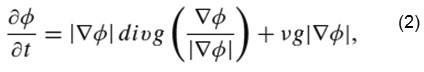
The basic concept used in this proposed method is MRF novel level set method in which the segmentation of the image is done by using two methods at the same time. The two methods which are implemented are 1.Algebric multigrid (AMG) and 2.sparse field method (SFM) X.Lu et al,3 M. Kass et al.5 By using these two methods the complexity will be reduced in segmenting the image. Level set methods implicitly model the planar closed curve C by the zero level set of the level set function φ(x, y, t),
![]()
The edge-based models endow F with detailed expression formula. For example, Caselles et al.6 proposed the geodesic active contour (GAC) model.
In this method, it will assume that the neighboring pixels also fall in the same region hence the noise will be much lesser in this technique. In this the neighboring cells will be encouraged to fall in the same region. The algebraic multigrid technique is used to increase the time step and the sparse field method is used to increase the computation domain. The complexity created by the CFL conditions can be reduced by using these techniques. The image segmentation will be accurate when it’s compared with the previous techniques. AMG doesn’t have any time constraint so it will take less iteration to obtain the image.
Along with the finite difference scheme, 7 it can get the common discretization formula in a matrix-vector form
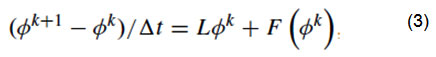
where Lφk = Tinternal , F(φk ) = Texternal + TMRF
Explicitly, as a result of the external term implemented from,8 the signed distance property |∇φ| = 1 [7] can be continued, particularly in a neighborhood nearby the zero level set
![]()
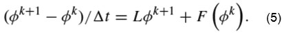
In this proposed method implement the semi-implicit scheme8 and obtain
The proposed system is applied on many different images like noisy images, natural images and synthetic aperture radar images in the real time applications. The medical images of brain are taken as input and the output has been obtained in the paper. By using this technique one more main advantage is that, the image of high density which is of 500*500 pixels can be reduced in this method.
The Gaussian noise which is obtained will be eliminated by using the Gaussian filter. The step by step process is, initially the input image of brain is taken and it will be sent to the initialization block where it will be applied with pre-processing i.e., filtering and then it will be sent to AMG where it will reduce the complexity of CFL conditions. The initialization process will determine the length, breadth, width and the curvature of the image and the sparse field method will reduce the computation domain and the final output will be sent to the output block. In these steps the image will be segmented. The output images can contain up to 220 Iterations in which all the process will be finished within seconds.
The brain images which are taken for the output is shown in the following figure (2) and figure (3). The brain tissues will be separated and the background will be separated from the brain tissues which are segmented. Even the little difference in the tissue can be captured by using the MRF technique by using algebraic multigrid and the sparse field method at the same time.
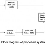 |
Figure 1: Block diagram of proposed system |
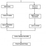 |
Figure 2: Flow Chart of proposed system |
Results and Discussion
The performance of the proposed method has validated in image segmentation trials on a wide range of images, including noisy artificial images,medical images.To conduct a fair comparison, here use the same initialization curve in both the reference methods and the proposed method. Similarly, the parameters of the reference techniques are selected according to the values provided in this proposed method.
Noisy Synthetic Segmentation Image
To experiment the robustness of the proposed method in contradiction of noise, independently add three types of noise salt and pepper noise, Gaussian noise and speckle noise to a synthetic image. The following parameters for the proposed method: λ = 0.92, γ = 0.35, t = 20. Input image shows binary segmentation results using altered methods for the noisy synthetic images which added salt and pepper noise with growing density. It can see that related with GAC, DAC, CV, LBF, AMP and GC, the supremacy of the proposed method is important in terms of segmentation accuracy and speed.
Medical Image Segmentation
Medical image segmentation X. Yang et al,11 Wang et al10 plays an important role in computer vision, such as segmenting the tumor tissues from the computed tomography (CT) image. Though, medical image segmentation also takes the problem of noise interference. The following parameters λ = 0.45, γ = 0.35, t = 30 are set.
From these comparison have the following conclusions. Initially, proposed method can segment objects from a background with relatively accurate shapes. Furthermore, the precision and recollection of our method has higher than those gotten by other comparison baselines. Finally, the proposed method greatly improves the segmentation speed of methods GAC, DAC, CV, LBF, AMP and still retains analogous segmentation efficiency with fast method TCF can GM.
In the future it will be analysed solving these problems by developing the optimization patterns recent approaches used to unravel the proposed MRF embedded energy functional.
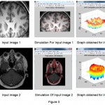 |
Figure 3 |
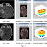 |
Figure 4 |
Table 1: Comparison Of Segmentation Accuracy (Var (F) In Tlwn)With Different Methods In SAR Medical Image Segmentation
| IMAGE | GAC | DAC | LBF | AMP | TCF | PROPOSED |
| IMAGE 1 | 0.46 | 0.22 | 0.6687 | 0.74 | 0.187 | 0.059 |
| IMAGE 2 | 0.69 | 0.28 | 0.326 | 0.87 | 0.198 | 0.06 |
| IMAGE 3 | 0.87 | 0.93 | 0.753 | 0.86 | 0.207 | 0.1089 |
| IMAGE 4 | 0.88 | 0.95 | 0.78 | 0.89 | 0.209 | 0.107 |
Table 2: Comparison Of Execution Time With Different Methods In SAR medical Image Segmentation
| IMAGE | GAC | DAC | LBF | AMP | TCF | PROPOSED |
| IMAGE 1 | 46.89 | 5.32 | 6.54 | 18.23 | 2.18 | 1.51 |
| IMAGE 2 | 50.98 | 6.29 | 5.87 | 13.87 | 1.85 | 1.08 |
| IMAGE 3 | 390.83 | 32.76 | 37.89 | 45.09 | 7.20 | 7.09 |
| IMAGE 4 | 200.98 | 53.98 | 53.87 | 79.09 | 12.09 | 6.17 |
Conclusion
The image segmentation is enormously used in the computer vision . The MRF technique which is used in this proposed method provides the segmented image of the medical images and its is also applicable to Synthetic Aperture Radar(SAR) , Noisy images and Natural images in concurrent application . The medical images are taken for the segmentation in which the tissues classified the brain will be segmented by specific number of iterations by using the MRF technique which combines both Algebraic multi grid(AMG) and Sparse Field method(SFM) which decreases the time step and increase the computation. The neighboring pixels of the will be cheered to fall in the same region.In future the chance of attaining the noises however the segmentation will be less matched to the existing system .
References
- Du S. Y., Wang W and Li X. Lazy random walks for super pixel segmentation,IEEE Trans. Image Process. 2014;23(4):1451–1462.
CrossRef - Melissa., Srilatha K. A novel approach for pigmented epidermis layer Segmentation and classification. International Journal of Pharmacy & Technology. 2016;8(1):10449-10458.
- Lu and Li X. Group sparse reconstruction for image segmentation. Neurocomputing. 2014;136:41–48.
CrossRef - Osher and Sethian J. A. Fronts propagating with curvature dependent speed: Algorithms based on Hamilton–Jacobi formulations. J. Comput. Phys. 1988;79(1):12–49.
CrossRef - Witkin K. A and Terzopoulos D. Snakes: Active contour models. Int. J. Comput. Vis. 1988;1(4):321–331.
CrossRef - Xu and Prince J. L. Snakes shapes and gradient vector flow. IEEE Trans. Image Process. 1988;7(3):359–369.
- Kimmel C. R and Sapiro G. Geodesic active contours. Int. J. Comput. Vis. 1997;22(1):61–79.
CrossRef - Srilatha. Multifocus Image Fusion Using Improved Dual Tree Complex Wavelet Transform and Discrete Optimization Method. Journal of Engineering and Applied Sciences. 2014;9(10-12):414-421.
- Li C., Gui X. C and Fox M. D. Level set evolution without re-initialization: A new variational formulation. in Proc. IEEE Comput. Soc. Conf. Comput. Vis. Pattern Recognit. 2005;430–436.
- Gao W. X., Tao D and Li X. A nonlinear adaptive level set for image segmentation. IEEE Trans. Cybern. 2014;44(3):418–428.
CrossRef - . Gao Y. X., Li J and Han B. A shape-initialized and intensity adaptive level set method for auroral oval segmentation. Inf. Sci. 2014;277:794–807.
CrossRef - Gao X., Wang B., Tao D and Li X. A relay level set method for automatic image segmentation.IEEE Trans. Syst. 2011;41(2):518–525. Man, Cybern. B, Cybern.
CrossRef - Clarke L. P., Velthuizen R. P., Phuphanich S., Schellenberg J. D., Arrington J. A.,Silbiger M. MRI: stability of three supervised segmentation techniques. Magn Reson Imaging. 1993;11(1):95–106.
CrossRef - Bandettini P. A., Wong E. C., Hinks R. S., Tikofsky R. S and Hyde J. S. Time course EPI of human brain function during task activation. Magnetic Resonance in Medicine. 1992;25:390–397.
CrossRef - Liao P. S.,Chen T. S and Chung P. C. A fast algorithm for multilevel thresholding. J. Inf.Sci. Eng. 2001;17(5):713–727.
- Bhowmik S., Datta V. A Survey On Clustering Based Image Segmentation. International Journal Of Advanced Research In Computer Engineering & Technology. 2012;1(5).
- Verma O. P.,Hanmandlu M.,Susan S., Kulkarni M and Kumar P. J. A simple single seeded region growing algorithm for color image segmentation using adaptive thresholding” IEEE International Conference on Communication Systems and Network Technologies (CSNT). 2011;500-503.
CrossRef - El Emary I. M. M. On the Application of Artificial Neural Networks in Analyzing and Classifying the Human Chromosomes. Journal of Computer Science. 2006;2(1):72-75.
CrossRef - Senthilkumaran N and Rajesh R. A Study on Edge Detection Methods for Image Segmentation. Proceedings of the International Conference on Mathematics and Computer Science (ICMCS-2009). 2009;I:255-259.
- Abdulghafour M. Image segmentation using Fuzzy logic and genetic algorithms. Journal of WSCG. 2003;11(1).

This work is licensed under a Creative Commons Attribution 4.0 International License.


