

Manuscript accepted on : 30 September 2016
Published online on: --
Plagiarism Check: Yes
Isolation and Characterization of Rhizobacteria from Mustard Field
M. S. Bochalya, Anil Kumar, A. S. Rathi, Rakesh* and Kushal Raj
Department of Plant Pathology CCS Haryana Agricultural University- Hisar.
Corresponding Author E-mail: punia.rakesh98@gmail.com
DOI : http://dx.doi.org/10.13005/bbra/2395

ABSTRACT: An in vitro study were carried out in the Department of Plant Pathology, CCS Haryana Agricultural University to isolate and identify different rhizobacterial isolates present in mustard field (cv RH-30). Samples of soil, rhizoplane and rhizosphere were collected from previously grown mustard fields of pathological research farm. The rhizobacterial isolates were isolated on King’s ‘B’medium by serial dilution of samples. These isolated rhizobacterial isolates were identified by following standard protocols. Results indicated that in mustard field three species of genus Bacillus, three species of genus Rhizobium, two species of genus Pseudomonas, one species of genus Arthrobacter and one species of genus Micrococcus were found. All three species of genus Bacillus having same morphological, physiological and biochemical characters except colony colour (white and yellow), colony thickness (large, medium and small raised), colony smoothness (smooth and rough), colony arrangement (lobate, entire and waxy) bacteria arrangement (pair and chains), citrate utilization test (+ve and -ve), oxidase test (+ve and -ve) and acid production test (+ve and -ve). Whereas Rhizobium genus are different in colony colour (white and pink), colony thickness (large and medium raised), bacterial morphology (medium, long and big rods), bacteria arrangement (single and groups), indole production test (+ve and -ve), methyl red test (+ve and -ve), citrate utilization test (+ve and -ve), oxidase test (+ve and -ve), starch hydrolysis test (+ve and -ve)and cellulose hydrolysis test (+ve and -ve). Two species of genus Pseudomonas is different in colony colour (yellowish white and creamy white), colony appearance (flat and convex), bacterial morphology (small and medium rods) and methyl red test (+ve and -ve). After identification these rhizobacterial isolates were maintained on King’s ‘B’medium for further studies like their possible role in induction of systemic resistance against Sclerotinia sclerotiorum causing stem rot in mustard alone or in combination with different non-conventional chemicals (salicylic acid, acetyl salicylic acid, indole butyric acid, indole acetic acid, zinc sulphate, magnesium sulphate and carbendazim fungicide at 10, 50 and 100 ppm). Effect of these isolated rhizobacterial isolates and non-conventional chemical on biochemical composition and enzymatic activity will also be tested.
KEYWORDS: Rhizobacteria; Mustard; Rhizoplane; Rhizosphere; Pseudomonas; Bacillus; Rhizobium
Download this article as:| Copy the following to cite this article: Bochalya M. S, Kumar A, Rathi A. S, Rakesh R, Raj K. Isolation and Characterization of Rhizobacteria from Mustard Field. Biosci Biotech Res Asia 2016;13(4). |
| Copy the following to cite this URL: Bochalya M. S, Kumar A, Rathi A. S, Rakesh R, Raj K. Isolation and Characterization of Rhizobacteria from Mustard Field. Biosci Biotech Res Asia 2016;13(4). Available from: https://www.biotech-asia.org/?p=16964 |
Introduction
Indian mustard (B. juncca) is a major crop of oilseed Brassica, which fulfils nearly 29.2 per cent of national oilseed crop outputs [1]. Among rapeseed-mustard growing countries in the world, India ranks third in area and production after China and Canada [2]. Rapeseed-mustard crop in our country is cultivated over an area of 6.3 million ha with a production of 7.4 million tonnes with an average yield of 1176 kg/ha [2]. Despite considerable increase in productivity and production, a wide gap exist between yield potential and yield realized at farmer’s field, which is mainly due to biotic and abiotic stress. The destructive diseases of oilseed brassica include those caused by fungi, bacteria and viruses. Among them, stem rot incited by Sclerotinia sclerotiorum (L.) de Bary is the most serious fungal disease that causes maximum damage [3]. The diseases is gaining importance in the raya growing areas, which may lead to disastrous crop failure as the disease incidence up to 80% has been recorded in some parts of Haryana and Punjab [4, 5]. Suppression of many soil borne pathogens by the rhizobacterial strains in various crops has been well documented [6]. Besides, pathogen infection, several chemicals and plant growth promoting rhizobacteria are known to induce resistance against several pathogens in many plants [7]. The role of foliar spray and micro-injection of rhizobacterial strains viz., P. fluorescence and P. aeruginosa, on chickpea cv. avrodhi after challenge inoculation with S. sclerotiorum and reported increase in PAL activity compared to the untreated control in both the treatments [8]. A strain of B. subtillis (NJ-18) with broad antimicrobial activity was screened in the field, for control of Sclerotinia stem rot of rape, fermentation of NJ-18 at 1.0 X 10⁷cfu / ml significantly reduced disease incidence and severity; control by NJ-18 was as high as 77.1 per cent and was comparable with control by 46 per cent dimethachlon and better than control by 50 per cent carbendazim at 750 g ai / ha and concluded that strain NJ-18 of B. subtillis is a promising biological control agent and should be further studied and tested for control of Sclerotinia stem rot of rape, and other diseases [9]. The induced systemic resistance in potato against P. infestans using inducer chemicals (salicylic acid, benzothiadiazol, fosetyl-AI, calcium chloride, ethylene, hydrogen peroxide, potassium hydrogen phosphate) and bio-agents (P. fluorescence, T. viride and T. hamatum) and reported that minimum disease severity (2.0%) was recorded in case of plants sprayed with SA @ 100 μg/ml followed by BTH @ 100 μg/ml. They also reported that maximum increase (52.4%) in PO and chitinase activity in SA treated plants. Comparatively, lower induction of resistance was observed with the biocontrol agents tested [10].
Material and Methods
An in vitro study was carried out in the Department of Plant Pathology, CCS Haryana Agricultural University to isolate and identify different rhizobacterial isolates present in mustard field (cv. RH-30). Samples of soil, rhizoplane and rhizosphere were collected from previously grown mustard fields of pathological research farm. Bacterial isolates were grown at 30±1°C for 24 h on nutrient agar medium slants/plates. The bacterial cultures were examined for various morphological, biochemical and physiological characteristics as per procedure described in Bergey’s Manual of Determinative Bacteriology [11]. The inter-relationship of the microorganisms in each section of Bergey’s Manual is based on characteristics such as morphology, staining reactions, nutritional and cultural characteristics and biochemical tests for specific metabolic end products. After isolation, purification and identification, these isolates were maintained on King’s ‘B’ medium for further studies. The composition of King’s ‘B’ medium is given below.
Peptone – 20 gm
Glycerol – 10 ml
K2HPo4 – 1.5 gm
MgSo4 _ 1.5 gm
Bacteriological Agar – 15 gm
Distilled Water – 1000 ml
Cultural and morphological characters
Bacterial isolates were grown on King’s ‘B’ medium/NA slant/plate for 24 h at 30±1°C. Each isolate was examined for (i) Colony morphology, (ii) Pigment production, (iii) Gram’s reaction, (iv) Bacterial cells morphology, (v) Bacterial cells arrangements and (vi) Sporulation. Bacterial isolates were classified as Gram +ve or –ve with Gram’s differential staining method.
Biochemical characters
The biochemical tests conducted for characterization of the bacterial isolates were following: (i) Indole production (ii) Methyl red test (iii) Voges- Proskauer reaction (iv) Citrate utilization test (v) Oxidase test (vi) Catalase production (vii) Acid production (viii) H2S production (ix) Starch hydrolysis and (x) Cellulose hydrolysis. The details of above mentioned methods described are given below.
Indole production test
Peptone broth was inoculated with the test bacterium and incubated at 30±1°C for 24 h. Following incubation, 0.2 ml of Kovac’s reagent was added to 5 ml of the culture medium. A cherry red colour in the alcohol layer indicated a positive reaction.
Methyl red (MR) test
The tubes containing Methyl red – Voges-Proskauer (MR-VP) broth were inoculated with test cultures and incubated at 30±1°C for 48 h. Following incubation, 5-6 drops of methyl red solution were added. A positive reaction was indicated by bright red colour at pH 4.2. Yellow or orange colour indicated a negative reaction while a weekly positive test was indicated by red-orange colour.
Voges – Proskauer (acetoin production) test
The MR-VP broth was inoculated with the test culture and incubated at 30±1°C for 48 h. Then 1 ml of 40 per ecnt KOH (having 1% kreatine) and 3 ml of a 5 per cent solution of alpha-naphthol in absolute ethanol was added to it. A positive reaction was indicated by the development of pink colour in 2-15 minutes which become crimson in 30 min. The tubes were shaken intermittently to ensure maximum aeration.
Citrate utilization test
Simmons’ citrate medium (a modification of Koser’s medium with agar and indicator) slants were prepared and sterilized after dispensing the medium in different test tubes. To minimize the nutrient carryover, the cells were rinsed with sterile distilled water before transferring to Simmons’ citrate agar slants and then incubated for 96 h at 30±°C. A positive reaction shows a blue colour on the streak of growth. Retention of original green colour and no growth on the line of streak indicated a negative reaction. Bromothymol blue tests green when acidic (pH 6.8 and below) and blue when alkaline (pH 7.6 and higher). Only those cultures that show a pH shift of the medium to alkaline conditions were considered positive.
Oxidase test
The cultures were grown in Luria Bertani medium. Then Whatman’s No. 1 filter paper strip was moistened with a freshly prepared 1% solution of N, N, N’, N-tetramethyl-p-phenylenediaminedihydrochloride (Wurster’s reagent). It was immediately rubbed with speck of culture using a platinum loop. Appearance of intense deep purple blue colour within 5-10 seconds indicated a positive reaction. While in delayed reaction, coloration appeared in 10-60 seconds and no colour change or light pink coloration indicated the absence of oxidase activity.
Catalase test
Bacterial cultures were grown for 24 h on Luria Bertani medium slants. Then a loopful of bacterial growth was taken on the slide and a few drops of 3% H2O2 were added. A positive catalase reaction was indicated by effervescence due to production of oxygen as a result of breakdown of H2O2..
Acid production
A loopful of freshly grown cultures was spotted on tryptone agar medium plates containing bromocresol purple dye. The plates were incubated at 30±1°C for 48-72 h. The change in the colour of the dye from purple to yellow around bacterial colonies indicated acid production.
H2S Production
Tubes containing 9.0 ml of medium broth were prepared. Tubes were inoculated with the bacterium in duplicates, using an un-inoculated one as control. Lead acetate strip was hanged with cotton plug in each tube. Broth tubes were incubated at 30±1°C for 48-72 h and then examined for the change of colour of the strip. The darkening of lead acetate strip is caused by the formation of lead sulphide (PbS). Due to production of PbS, strip colours changed from white to black indicating production of hydrogen sulphide.
Starch Hydrolysis
A loopful of the freshly grown culture was spotted on starch minimal medium plates. Plates were incubated at 30±1°C for 48-72 h. Amylase-producing bacteria were scored by flooding the surface of the starch agar plates with Gram’s iodine. A clear zone around bacterial colony indicated hydrolysis of starch.
Cellulose hydrolysis
A loopful of freshly grown culture was spotted on plates of solidified Deckman’s medium. Plates were incubated at 30±1°C for 3-4 days. The plates were examined after every 24 h for the clearance zone around the bacterial growth.
Results and Discussion
Results in Table 1 indicated that in mustard field three different species of genus Bacillus, three species of genus Rhizobium, two species of genus Pseudomonas, one species of genus Arthrobacter and one species of genus Micrococcus was found. All three species of genus Bacillus having same morphological, physiological and biochemical characters except colony colour (white and yellow), colony thickness (large, medium and small raised), colony smoothness (smooth and rough), colony arrangement (lobate, entire and waxy), bacteria arrangement (pair and chains), citrate utilization test (+ve and -ve), oxidase test (+ve and -ve) and acid production test (+ve and -ve). Whereas species of Rhizobium genus were different in colony colour (white and pink), colony thickness (large, and medium raised), bacterial morphology (medium, long and big rods), bacterial cell arrangement (single and groups), indole production test (+ve and -ve), methyl red test (+ve and -ve), citrate utilization test (+ve and -ve), oxidase test (+ve and -ve), starch hydrolysis test (+ve and -ve) and cellulose hydrolysis test (+ve and -ve) (Figure 1). Species of genus Pseudomonas is different in colony colour (yellowish white and creamy white), colony appearance (flat, and convex), bacterial morphology (small and medium rods) and methyl red test (+ve and -ve). Results are agreement with earlier studies conducted on bean, carnation [12, 13, 14]. Suppression of many soil borne pathogens by the rhizobacteria strains in various crops has been well documented [6]. These rhizobacterial isolates can be utilized for suppression of soil borne pathogens in mustard crop.
Table 1: Isolation and characterization of rhizobacterial isolates from mustard soil, rhizoplane and rhizosphere
| Sr. No. | Morphological, physiological and biochemical characters | Isolate 1 | Isolate 2 | Isolate 3 | Isolate 4 | Isolate 5 |
| 1 | Colony morphology | Non Gummy, white, small, raised, rough, entire
|
Gummy, white, large, raised, smooth, entire | Non gummy, creamy white, medium, raised, smooth, entire | Gummy, white, large, raised, smooth, entire
|
Non gummy, yellowish white flat, medium, smooth,entire |
| 2 | Pigment production | – | – | – | – | – |
| 3 | Gram reaction | +ve | -ve | -ve | -ve | -ve |
| 4 | Bacteria morphology | Big rods | Long rods | Cocci | Big rods | Small rods |
| 5 | Bacteria arrangement | Pairs and chains | Singly and groups | Pairs | Singly | Singly |
| 6 | Endospore position | Central | – | – | – | – |
| 7 | Indole production | – | – | – | – | – |
| 8 | Methyl red test | – | – | – | – | – |
| 9 | Voges-Proskauer reaction | – | – | – | – | – |
| 10 | Citrate utilization | – | – | – | + | – |
| 11 | Oxidase test | + | – | + | + | + |
| 12 | Catalase test | + | + | + | + | + |
| 13 | H2S production | – | – | – | – | – |
| 14 | Starch hydrolysis | – | – | – | – | – |
| 15 | Cellulose hydrolysis | – | – | – | – | – |
| 16 | Acid production test | + | – | – | – | – |
| 17 | Probable genus | Bacillus
(Soil) |
Rhizobium
(Rhizoplane) |
Micrococcus
(Rhizoplane) |
Rhizobium
(Rhizoplane) |
Pseudomonas
(Rhizosphere) |
| Sr. No. | Morphological, physiological and biochemical characters | Isolate 6 | Isolate 7 | Isolate 8 | Isolate 9 | Isolate 10 |
| 1 | Colony morphology | Non gummy, light reddish, large, raised, smooth, lobate | Non gummy, creamy white, medium, convex, smooth, entire | Gummy, pink, medium, raised, smooth, entire
|
Non gummy, yellow, medium, raised, smooth, wavy | Non gummy, white, large raised, rough, lobate |
| 2 | Pigment production | – | – | – | – | – |
| 3 | Gram reaction | -ve | -ve | -ve | +ve | +ve |
| 4 | Bacteria morphology | Small rods | Medium rods | Medium rods | Big rods | Small rods |
| 5 | Bacteria arrangement | Pairs | Singly | Singly | Pairs | pairs |
| 6 | Endospore position | – | – | – | Central | Central |
| 7 | Indole production | + | – | + | – | – |
| 8 | Methyl red test | – | + | + | – | – |
| 9 | Voges-Proskauer reaction | – | – | – | – | – |
| 10 | Citrate utilization | + | – | + | + | + |
| 11 | Oxidase test | + | + | + | – | + |
| 12 | Catalase test | + | + | + | + | + |
| 13 | H2S production | + | – | – | – | – |
| 14 | Starch hydrolysis | + | – | + | – | – |
| 15 | Cellulose hydrolysis | – | – | + | – | – |
| 16 | Acid production test | – | – | – | – | – |
| 17 | Probable genus | Arthrobacter
(Soil) |
Pseudomonas
(Rhizosphere) |
Rhizobium
(Rhizoplane) |
Bacillus
(Soil) |
Bacillus
(Soil) |
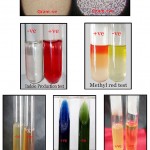 |
Figure 1: Isolation and characterization of Rhizobacterial isolates |
Acknowledgement
We thanks to Dr. S. S. Karwasara Head, Department of Plant Pathology for providing facilities and member of my advisory committee for their kind help in critical reading of the manuscript and encouragement.
References
- Nashaat, N.I., Kumar, A. and Patil, V.D. (2007). Indo-UK collaboration on rapeseed-mustard research for rural livelihoods development.Outlooks on Pest Management.18: 209-12.
CrossRef - Anonymous, (2013). Directorate of Rapeseed-Mustard Research, www. drmr.res.in
- Kolte, S.J. (1985). Diseases of annual edible oilseed crops, vol. II, Rapeseed-mustard and sesame diseases. CRC Press Inc. Boca Raton, Florida, 135 pp.
- Kang, I.S. and Chahal, S.S. (2000).Prevalence and incidence of white rot of rapeseed and mustard incited by Sclerotinia sclerotiorum in Punjab. Plant Dis. Res., 15:232-233.
- Sharma, S. Yadav, J.L. and Sharma, G.R. (2001).Effect of various agronomic practices on the incidence of white rot of Indian mustard caused by Sclerotinia sclerotiorum. Mycol. Plant Pathol.,31:83-84.
- Vidyasekaran, P., Sethuraman, K., Rajappan, K. and Vasumathi, K. (1997). Powder formulations of Pseudomonas fluorescensto control pigeon pea wilt. con., 8:166-171.
- Rohilla, R., R.L., Singh, R.L., Singh, U.S., Ramji Singh, R., Duveillar and Singh, H.B. (2001) Recent advances in the management of plant diseases using chemicals. Indian J. Plant Pathol.19:1 and 2.
- Basha, S.A. and Chatterjee, S.C. (2007b).Effect of PGPR on Sclerotinia sclerotiorum infection through elicitation of phenylalanine ammonia lyase in chick pea.Indian Phytopathol.,60(3): 313-316.
- Yang, D., Wang, B., Wang, J., Chen, Y. and Zhou, M. (2009). Activity and efficacy of Bacillus subtilis strain NJ-18 against rice sheath blight and Sclerotinia stem rot of rape. Cont., 51: 61–65
CrossRef - Kumar, S., Thind, T.S., Bala, A. and Gupta, A.K. (2010).Induced resistance in potato against Phytophthorainfestans using chemicals and bio-agents.Plant Dis. Res., 25(1):12-18.
- Holt, J.G., Kreig, N.R. and Sneath, P.H.A. (1994).Bergeys Manual of Determinative Bacteriology.19th edition, Williams and Wilkins, Baltimore.
- Alstorm, S. (1991). Induction of disease resistance in common bean susceptible to halo blight bacterial pathogen after seed bacterization with rhizosphere J. Gen. Appl. Microbiol., 37: 491-501.
CrossRef - Van Peer, R., Niemann, G.J. and Schippers, B. (1991). Induced resistance and Phytoalexin accumulation in biological control of Fusarium wilt of carnation by Pseudomonas Strain WCS 417 R. Phytopathol., 81: 728-734.
CrossRef - Wei, L., Kloepper, J.W. and Tuzun, S. (1996). Induced systemic resistance to cucumber disease and increase plant growth by plant growth promoting rhizobacteria under field conditions. Phytopathol., 86: 221-224.
CrossRef

This work is licensed under a Creative Commons Attribution 4.0 International License.


