

Manuscript accepted on : 20 February 2016
Published online on: 17-03-2016
Olga Kriger1, Andrei Lisitsyn2, Alexander Prosekov1
1Federal State-owned Budgetary Educational Institution of Higher Vocational Education “Kemerovo Institute of Food Science and Technology”, 47 Stroiteley Boulevard, 650056 Kemerovo, Russia. 2Federal State-owned Budgetary Scientific Institution “The Gorbatov's All-Russian Meat Research Institute (VNIIMP)” 26 Talalikhina str., 109316 Moscow, Russia.
DOI : http://dx.doi.org/10.13005/bbra/2007

ABSTRACT: Molecular weight distribution of prion proteins in meat, blood, processed blood products, gelatin, milk, cheese has been studied in a paper. The obtained electrophoregrams of blood plasma samples indicate the presence of two fractions of blood protein in the range of 30 to 40 kDa. The relative abundance of these proteins in the blood plasma contains 22.06 % of the total, which are 2.15 g/100 g of blood plasma. Electrophoretic separation of gelatin industrial samples, which is obtained by partial hydrolysis of collagen extracted from bones, hides and skins, cores and tendons of cattle, shows a high degree of product purification. The light fractions of proteins are not detected. The results obtained indicate that the soluble protein fraction of meat and blood plasma has a high degree of infectivity against pathogenic form of prion protein.
KEYWORDS: prions; light fractions; electrophoresis; blood plasma
Download this article as:| Copy the following to cite this article: Kriger O, Lisitsyn A, Prosekov A. Characteristics of The Molecular Weight Distribution of The Prion Protein Fractions in Blood and Milk Processing Products. Biosci Biotech Res Asia 2016;13(1) |
| Copy the following to cite this URL: Kriger O, Lisitsyn A, Prosekov A. Characteristics of The Molecular Weight Distribution of The Prion Protein Fractions in Blood and Milk Processing Products. Biosci Biotech Res Asia 2016;13(1). Available from: https://www.biotech-asia.org/?p=7285 |
Introduction
Prions (Proteinaceous infectious particles) is a special class of pure protein, containing no nucleic acids, infectious agents that cause serious diseases of the central nervous system in humans and some higher animals (the so-called slow infection) [1].
Prion diseases is a group of transmissible neurodegenerative diseases of animals and humans. These diseases have a long incubation period, but quickly progress after clinical onset of the disease. All prion diseases are deadly and currently there are no effective methods of treatment [2].
The study of prions and the diseases caused by them is a new and rapidly developing field of biomedical research that in recent years has acquired an important scientific and practical significance [3].
Practical interest is linked, first of all, to the outbreak of epizootic bovine spongiform encephalopathy in the United Kingdom, as well as to the identification, mainly in the UK, young people with Creutzfeldt-Jakob disease), and proof of the possibility of transmission of this disease to people through eating meat products derived from infected animals [4].
Also, one of the ways of human infection with BSE are neurosurgery, transplantation of tissues, the appointment of hormones derived from the infected donor with undetected Creutzfeldt-Jakob disease (iatrogenic cases of Creutzfeldt-Jakob disease) [5].
Cases of prion diseases cause the need to incorporate the growing likelihood of prion diseases in humans and animals.
According to statistics in Russia with a population of approximately 150 million Creutzfeldt-Jakob disease annually affects about 150 people. However, consideration of these patients being hard: the cases are not detected and are not registered [6].
Constantly growing theoretical interest in the problem due to the results of molecular biological studies of the structure of prion proteins allowed to collect and organize a large extent of a significant material on the structure, function and accumulation of infectious agents in infected humans and animals [7].
Exactly the results of molecular biological studies of the prion proteins structure provided the basis to identify new directions for further approaches to the diagnosis and treatment of prion diseases.
Methods
The objects of research were meat, blood, processed blood products, gelatin, milk, cheese.
When performing the work standard, conventional and original research methods were used.
Determination of the molecular weight distribution of the protein fractions. Molecular weight distribution of peptides and proteins in the resulting hydrolyzates has been evaluated by protein electrophoresis method of Laemmli [8]. 0.1% of sodium dodecyl sulfate with denaturing polyacrylamide gel (12% separating and 4% of a focusing) were used for protein separation. Electrophoresis was performed on a single electrode buffer with 0.1% of sodium dodecyl sulfate at 15 mA. The gel was stained with 0.2% of Coomassie R250 (prepared with glacial acetic acid) at an elevated temperature for 7-10 minutes, then washed three times with distilled water.
Viewing and photographing of gels were performed on a UV transilluminator TCP-20M («Vilber Lourmat», USA) at a wavelength of 312 nm radiation. Storing and processing of the data have been carried out by gel documenting system Vitran-Photography [9].
Determination of total protein in the samples has been performed by the method of measuring the mass fraction of total nitrogen and protein in meat, meat products and protein-containing foods on the Dumas combustion method. This procedure is certified by the Federal State Unitary Enterprise “All-Russian Research Institute of Metrological Service” (FSUE “VNIIMS”) (Certificate of attestation № 41-09 from August 17, 2009).
In terms of absolute content of nitrogen in the sample and the sample mass, the weight fraction of nitrogen has been calculated (automatically). Then, the mass fraction of the protein was calculated using conversion factors specific to each product (Table. 1). For the measurement result, the arithmetic mean of the results of two parallel measurements was taken [10].
Table 1: Characteristics of the device «Rapid N cube»
| The measuring range of nitrogen, mg | 0.1 – 200.0 |
| Maximum permissible absolute standard deviation of measurements, mg | 0.01 – 0.2 |
| Analysis time (depending on the material and its mass), min | 4 |
| Power consumption, kW, not more | 1.8 |
| Voltage of supply, V | 120/230 |
| The frequency of the alternating current, Hz | 50/60 |
| Dimensions, mm, no more than | 480х550х550 |
| Weight, no more than | 65 |
| Ambient temperature, ° C | 15 – 35 |
The method is based on the high-temperature combustion of the sample in a stream of carrier gas CO2 dispensing oxygen. All the nitrogen-containing compounds in the combustion products, passed through a plurality of reaction tubes, are converted into molecular nitrogen, which is a carrier gas, supplied to the measuring cell thermal conductivity detector (TCD). The detector produces a signal (peak area on the chromatogram) proportional to the nitrogen content in the sample.
The sample was weighed before the start of the analysis, and its weight is transferred to the database management program. The weighing result is recorded up to 0.1 mg. Conversion factors are presented in table 2.
Table 2: Conversion factors
| Name of the sample | The estimated percentage of total nitrogen, % | Factor of recalculation, K |
| Beef stew | 3.31 | 6.25 |
| Canned meat | 13.91 | 5.78 |
| Pate beef | 1.05 | 5.7 |
| Beef fat | 1.06 | 5.64 |
| Lean beef | 1.07 | 5.62 |
| Animal protein | 4.64 | 4.69 |
The molecular weight distribution of proteins in the sites have been assessed using the method of Laemmli protein electrophoresis. For the separation of proteins, denaturing polyacrylamide gel (12% separating and 4% focusing) with 0.1% of sodium dodecyl sulfate was used. Phoresis was performed at a single electrode buffer with 0.1% SDS at 15 mA. The gel was stained with 0,2% Coomasie Brilliant Blue R250, colorant prepared on glacial acetic acid, at elevated temperature for 7-10 minutes, then washed three times with distilled water. Viewing and photographing of gels was performed with a UV transilluminator TCP-20M («Vilber Lourmat», USA). Storing and processing of the data have been carried out by gel documenting system «Doc-It LS».
Before electrophoresis, samples were incubated in the presence of sodium dodecyl sulfate. Calibration of the gel was performed using a set of marker protein production SibEnzyme, comprising twelve highly purified recombinant proteins of molecular weight from 10 to 250 kDa, which, after electrophoresis on a polyacrylamide gel and fixation Coomasie Brilliant Blue R-250, form discrete bands. For a quantitative estimation protein content of the fractions held calibration proteins of human serum albumin with known concentrations. Approximation of the calibration curve was carried out by a polynomial of the third order, and the correlation coefficient is 1.0.
Results
Sample preparation has been carried out by diluting liquid samples in distilled water so that the protein content of the gel pocket did not exceed 5 micrograms per 20 l of a solution. In the present case we managed to achieve the best possible distribution of the protein fractions (Figure 1).
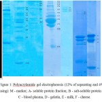 |
Figure 1: Polyacrylamide gel electrophoresis (12% of separating and 4% of a focusing): M – marker; A- soluble protein fraction; B – salt-soluble protein fraction, C – blood plasma, D – gelatin, E – milk, F – cheese. |
Results of denaturant electrophoresis in the samples are presented in Table 3.
Table 3. Total protein content and distribution of the fractionated proteins
Table 4 shows the values of the total amount of protein in the samples of products.Table 4. Total protein content quantified in samples
| Name of the sample | The number of samples, units | Total protein, g / 100 g | The number of protein fractions, units in the range | ||
| 15-30 kDa | 30-40 kDa | 40-250 kDa | |||
| Water-soluble fraction | 20 | 6.83 | 1 | 2 | 6 |
| Salt-soluble fraction | 20 | 8.78 | 0 | 0 | 6 |
| Stroma fractions | 20 | 3.90 | 0 | 0 | 2 |
| Blood plasma | 20 | 9.73 | 1 | 2 | 8 |
| Gelatin | 10 | 84.32 | 0 | 0 | 3 |
| Milk | 20 | 3.02 | 6 | 0 | 2 |
| Cheese | 20 | 21.57 | 4 | 0 | 1 |
| The object of study | Weight, mg | The total nitrogen content, % | Factor of recalculation | Total protein, % | Measurement error,
±δ,% |
| Whole milk | 195.60
192.40 |
0.576
0.558 |
4.64 | 2.63 | 0.31 |
| Blood serum | 188.12
180.72 |
2.370
2.374 |
6.25 | 14.83 | 0.89 |
| Whole blood | 214.50
254.80 |
3.462
3.571 |
6.25 | 21.97 | 1.32 |
| Cheese | 189.70
229.60 |
3.328
3.506 |
4.64 | 15.85 | 0.95 |
| Lean beef | 126.50
110.00 |
3.222
3.347 |
5.62 | 18.46 | 1.11 |
| Water-soluble fractions | 97.40
96.50 |
0.558
0.557 |
5.62 | 6.83 | 0.41 |
| Salt-soluble fractions | 136.30
142.25 |
1.710
1.658 |
5.62 | 8.78 | 0.53 |
| Stroma fractions | 186.80
171.20 |
0.675
0.679 |
5.62 | 3.90 | 0.23 |
Assaying for prion protein is the following:
1) Antigen (prion protein) has been added by 100 ul to the wells of polystyrene 96-well plate and incubated at 37 °C for 60 min. To select the optimal conditions for antigen adsorption on plastic antigen incubation is conducted with 100 l per well at concentrations of 0.5; 1; 2.5; 5; 10; 25; 50 ug / ml for 30, 60, 90, 120, 150, 180 minutes. Unbound material is removed from the wells simply by shaking followed by washing three times (wash buffer: 50mmol / l Tris, 150 mmol / l NaCl, 0.5 ml / liter Tween 20).
2) Next, blocking of the unoccupied plastic sites, making PBS solution, containing bovine serum albumin in the wells with 100 ul per well for 30 minutes has been carried.
3) After a removal of the blocking solution (by washing), to determine the adsorbed material,100 ul of biotinylated monoclonal antibody has been introduced into the wells to the pathogenic prion protein 15V3 and incubated for 2 hours at 18 ° C.
4) The unbound antibody has been removed by wells washing three times with PBS solution, containing 1 ml / l Tween (Tween) and 3 times with PBS, containing 15 g / l of bovine serum albumin.
5) Preparation of DNA reporter. Streptavidin-biotin complex was selected as a link between the antibody and DNA reporter.
One molecule of streptavidin is composed of four identical subunits, capable of interacting with four molecules of biotin, it can be used as a linking molecule between the biotin-containing compounds. In this case, the tail of biotinylated DNA and streptavidin acts as a bridge linking the two molecules containing biotin residues.
Preparation of Conjugates (DNA antibodies and biotin) is carried out quite easily and is accompanied by minimal modification of their immunological activity.
Recombinant streptavidin has been pre-incubated for 45 min at 4 °C with biotinylated reporter DNA at a molar ratio of 1 : 02. As a complex result of streptavidin DNA was then added to the wells and incubated for 30 min at room temperature.
5) Wells were washed 5 times with PBS, and 10 times with distilled water and then subjected to PCR.
During the course of the study it was found that the stroma fractions and salt-soluble proteins of no light fractions, are consistent with the literature data, while just two protein fractions sized from 30 to 40 kDa (the size range of the normal prion protein) are contained in the water-soluble fraction.
The relative content of protein fractions in weight from 30 to 40 kD in the soluble fraction of proteins of meat is 18.19% of the total, is 1.25 g / 100 g meat.
The obtained electrophoregrams of blood plasma samples indicate the presence of two fractions of blood protein in the range of 30 to 40 kDa. The relative abundance of these proteins in the blood plasma of 22.06% of the total, is 2.15 g / 100 g of blood plasma.
Based on the data presented in Table 4 we can conclude that the highest content of total protein in the samples is as follows: Whole blood is 21.97 g / 100 g lean beef is 18.46 g / 100 g, water-soluble fraction is 6, 83 g / 100 g, salt-soluble is 8.78 g / 100 g, stromal fraction is 3.90 g / 100 g.
To select suitable antibody for the responses against the pathogenic prion proteins antibodies offered by manufacturers were analyzed. Due to high homology interspecies marked for PrP protein, are particularly suitable antibody for which conjugates of peptides.
Thus, during the analysis, a murine monoclonal antibody 15V3 (Rrionics Company) was selected, which is obtained using 3 different sequences (epitope) of human PrP peptide: 15b3-1 includes residues 142-148 GSDYEDR (YY); 15b3-2 residues 162-170 YYRPVDQYS; 15b3-3 residues 214-226 CITQYQRESQAYY.
15V3-1 3-2 and 15 are associated with β-sheets, which accumulate in the PrP Sc and 15V3-3 recognizes amino acid residues near the C-terminus.
15B3 is an antibody that specifically recognizes aberrantly folded protein PrPsc, instead of the normal molecule PrP (PrP C). The specificity of the antibody, which was developed by the founders of Prionics in 1997, was confirmed in a Biasini study.
15V3 antibodies are able to detect PrPSc in the brain homogenates, without the need for the action of proteinase K. This is important for its use in the blood.
It was experimentally demonstrated that PrPSc 15V3 reacts with human, bovine, sheep, deer, mice and hamsters, but does not react with normal prion PrPC. Therefore, 15V3 may be used as detection antibody for the analysis.
Conclusion
Electrophoretic separation of gelatin industrial samples, which is obtained by partial hydrolysis of collagen derived from bones, hides and skins, cores and tendons of cattle, shows a high degree of purification of the product. The light fractions of proteins are not detected.
The results obtained indicate that the soluble protein fraction of meat and blood plasma, have a high degree of infectivity against pathogenic form of prion protein.
References
- Borisov L.V. Medical microbiology, virusology, immunology. – M., 2001.
- Grigor’ev V. B. — Prion diseases of humans and animals. – Questions of Virology, V.49(№ 5), Pp.4-12, 2004 (review)
- Zuev V.A., Zavalishin I.A., Rojhel’ V.M. Prion diseases of humans and animals: A Guide for Physicians. – M., 1999.
- S. Shkundina, M. D. Ter-Avanesyan. Prions. Advances Biological Chemistry, V. 46, 2006 (review)
- Pokrovskij V.I., Kiselev O.I. Molecular basis of prion diseases. // Bulletin of the Russian Academy of Medical Sciences. – 1998. – № 10.
- Pokrovskij V. I., Kiselev O. I., Cherkasskij B. L. — Prions and prion diseases // Bulletin of the Russian Academy of Medical Sciences, 2004, 384 P., ISBN 5-7901-0038-4
- Shlegel’, G. General Microbiology: translation from German. / G. Shlegel’.- M.: Peace, 1987.- 567 P. 273.
- Shults, G.E. Principles of the structural organization of proteins / G.E. Shults, R.H. Shirmer.– M: Peace, 1982.– 354 P.
- Jaroslav Petr, DrSc. Phony a ustni dutina. Progresdent, 2004, № 2, P. 12-16
- Prion biology and diseases, edited by S.B. Prusiner, Cold Spring Harbor, NY, 1999, ISBN 0-87969-547-1

This work is licensed under a Creative Commons Attribution 4.0 International License.


