

Manuscript accepted on :
Published online on: 12-12-2015
Alexey Aleksandrovich Shumkov, Almaz Mullayanovich Hanov, Timur Rizovich Ablyaz, Evgeniy Aleksandrovich Morozov, Polina Nikolaevna Kilina, Dmitry Olegovich Pustovalov
Perm National Research Polytechnic University, 29, Komsomolsky Ave, Building A, Office 208, Perm, Russia 614990
ABSTRACT: Cranio-maxillo-facial surgeons need accurate and objective information about the structure, shape and size of various types of traumatic defects for evaluating clinical picture of a disease, planning and preparation for future surgeries. Computer tomography is often used for obtaining such information; it makes it possible to obtain high-precision three-dimensional images of various structures in the human body. However in order to obtain complete and objective information, especially about the nature of traumatic damages and for their effective treatment, namely, for the use of an implant instead of a lost or a damaged organ, it is desirable to have a real three-dimensional model. Consequently, the relevant task is fast and accurate conversion of data obtained by computer tomography into a three-dimensional computer model, and then into a solid model of the lost or damaged organ. It is often impossible to manufacture individual implants of complex shape. In manufacturing implants, rapid prototyping technologies (RP - technologies) were used. The technology is based on initiating the reaction of polymerization using digital video projectors. This technology makes it possible to obtain individual implants of complex shape without using additional tools, by the results of cone-beam computer tomography. By the results of tomography, 191 images of the object were selected. The 3D-DOCTOR software suite was used to build a three-dimensional computer model; the images were merged into cross-sections of the will-be 3D model. The modes of building an implant of man's jaw from cross-linked photopolymer material SI500 have been experimentally determined. A possibility has been established to obtain individual geometrically-complex implants using the rapid prototyping technology, where two digital video projectors are used for initiating the reaction of polymerization.
KEYWORDS: implant; rapid prototyping; cone-beam computer tomography (CBCT); photopolymer material
Download this article as:| Copy the following to cite this article: Shumkov A. A, Hanov A. M, Ablyaz T. R, Morozov E. A, Kilina P. N, Pustovalov D. O. Using the Technology of Layer-By-Layer Synthesis of Photopolymer Material During the Manufacturing of Medical Prototype Implants. Biosci Biotech Res Asia 2015;12(2) |
Introduction
Until recently, X-ray examination has been the only reliable and fast method of obtaining information about internal structure of human organs, presence of fractures, foreign objects, and status of transplants and implants (Chavladze, 2002). However, a conventional X-ray photograph, being a two-dimensional image of the object, does not convey all peculiarities of its shape, surface relief pattern, distorts its true dimensions, and therefore does not always provide complete information necessary for diagnostics and patient treatment. Computer tomography provides wide possibilities, making it possible to obtain high-precision three-dimensional images of various structures in the human body (Rabukhina, 2006). However, obtaining complete and reliable information, especially about the nature of traumatic injuries and their effective treatment, requires having a real three-dimensional model of the damaged organ or object (Sykes, 2004). The widespread acceptance of digital technologies in design (CAD), simulation and analysis (CAE), and mechanical processing (CAM) stimulated development of RP technologies (rapid prototyping). Digital 3D technologies opened unique capabilities to reproduce most complex spatial forms, objects and mechanisms (Zlenko, 2003). Modern methods of rapid prototyping make it possible to rapidly and quickly solve the problem of making copies of computer models created with the use of three-dimensional design software (Popat, 1998). Adoption of digital description of objects – CAD, followed by appearance of CAD and RP technologies, has revolutionized such areas as medicine, archaeology and forensics. RP technologies based on photo-polymerization of photosensitive resin can be divided into: SLA – stereolithography, PolyJet – spraying drops of resin with a spraying head, and exposure to UV (ultraviolet) lamps, DLP (Direct Light Projection) – exposure to UV lamps using a DLP-mask, MJM (Multi Jet Modeling) – multi-jet spraying of resin and exposure. Currently, plastic models are most widely used in cranio-maxillo-facial surgery, in preparation and planning surgeries for removing innate and post-traumatic defects, and in implants manufacturing (Winder, 2005).
The systems of implants that are currently produced and used in medicine are unified, i.e. implants are available in different sizes, but of the same shape. This imposes serious limitations on the use of implants in complicated cases (Tverskoy, 2012), namely, increases the time of surgery due to the need to fit the implant, and leads to forming stress concentration zones that reduce strength of the implant. The solution could be the use of complex-shaped implants designed and manufactured using modern technologies of rapid prototyping (Abramov, 1998).
Therefore, the urgent task is fast and accurate conversion of the data obtained by measuring machines, tomographs and other means for contact and contact less diagnostics of objects, into their physical copies (E. V. Kotsuba, 1996).
This work is aimed at studying the methods of obtaining copies of a human jaw fragment by the results of computer tomography, using the method of stereolithography based on initiation of the reaction of polymerization using digital video projectors (Digital Light Processing).
Methodology
The work shows an experimental study of obtaining a plastic prototype of a human jaw by the results of cone-beam computer tomography (CBCT) using the method of stereolithography.
The experimental studies included the following stages:
- Patient examination with a Planmeca ProMax 3D X-ray cone-beam computer tomograph.
- Pre-processing the results of tomographic examination in order to obtain an ordered set of CT images (axial slices) in the form of files in DICOM format.
- Converting the CT images into a three-dimensional computer model in STL format.
- Manufacturing a plastic copy of a jaw fragment using the stereolithography method based on the Digital Light Processing technology from the Envisiontec Company.
This research was based on obtaining an experimental plastic sample from photopolymer material SI500 in a stereolithographic machine, according to the results of cone-beam computer tomography (CBCT) of the human jaw.
In this work, the Planmeca ProMax 3D installation was used for obtaining digital images. The basic size of the picture area of Ø80 x 80 mm is optimal for most images, since it covers the whole dentition area, and provides clear view of both upper and lower jaws (Sarment, 2003). The X-ray system of Planmeca ProMax 3D uses a modern flat panel that makes it possible to obtain high-precision images for three-dimensional reconstruction, without distortion. Unlike image brightness boost sensors where outdated vacuum cathode ray tubes (CRT) and multi-stage focusing are used, flat panels of sensors ensure single-step reading of images without geometric distortion and without loss of sensitivity. In order to obtain clear and reliable three-dimensional images with limited radiation load on the patient in the Planmeca ProMax 3D X-ray installations, cone beam is used in the 3D tomography technology (CBVT). Scanning with cone beam is an ideal method of obtaining images of exactly the maxillofacial area, since this method uses a pyramid-shaped beam for scanning the entire studied area in one semicircular pass, unlike medical computer tomography, where many axial slices are used with many passes over the complete circle (Baumgaerte, 2009). Three-dimensional images are merged using computer software into single cylindrical image for viewing. In course of scanning, each image is formed with a short X-ray pulse, rather than continuous exposure. Full scanning time for one 3D area is 18-26 sec, but the actual exposure time is only 3 seconds. This technology can considerably reduce the radiation load on the patient, and ensures stroboscopic X-ray effect, which, in combination with slight rotation during scanning (the minimum angle is only 200°), virtually eliminates aliasing, contributing to exceptional image quality. The reconstructed three-dimensional image consists of millions of voxels. These voxels are isotropic; this ensures exact measuring in the 1:1 scale, and preservation of geometric relationships throughout the image. Extremely small size of the voxels ensures obtaining detailed images with high resolution without aliasing (Kulakov, 2003).
For obtaining high-quality three-dimensional images, scanning with a cone-beam tomograph was performed with certain process parameters. Process parameters are shown in Table 1.
Table 1: Parameters of the scanning process.
| Number of frames | 401 |
| Voltage (kV) | 90 |
| Current (mA) | 10 |
| Exposure time (sec) | 12.118 |
| Patient irradiation level
DAP (mGy/cm²) |
1094.2 |
The results of tomographic examination were pre-processed in the Planmeca Romexis Viewer 3.6.0 R software suite. The software is intended for viewing the results of scanning at a tomographic installation, editing images, measuring damaged areas, and importing images into software modules intended for converting into a three-dimensional model.
The 3D-DOCTOR software module was used for converting the results of tomographic scanning into a three-dimensional model. The program allows image processing of the examined tomogram, merging cross-sections of the will-be 3D model, and saving the results in the STL format.
The rapid prototyping EnvisionTec Perfactory Xede system was used to build a prototype from the photopolymer material. The main difference of this system from classical stereolithography is shifting away from the laser scheme for initiating polymerization reaction, and replacing it with several digital video projectors that use Digital Light Processing (DLP). The scheme of stereo-lithographic machine operation with the use of DLP technology is shown in Figure 1.
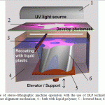 |
Figure 1: The scheme of stereo-lithographic machine operation with the use of DLP technology: 1 projector; 2-photomask; 3 – polymer alignment mechanism; 4 – bath with liquid polymer; 5 – lowered base; 6 – the cured polymer model |
The element of projectors using DLP technology is a Digital Micromirror Device, or DMD, i.e., a matrix of hard mirrors made of aluminum alloy with high reflectance; the mirrors are mounted to the substrate through which the movable plates are connected to the base of the matrix. At opposite angles of the mirrors, electrodes connected to the memory cells are placed. Under the action of electric field, the substrate with a mirror takes one of the two positions that differ by 20°, due to the limiters placed on the base of the matrix. These two positions correspond to reflection of the incoming light flux into the lens and light absorber, respectively. The area of each mirror of the matrix is 16 μm or less, the distance between the mirrors is about 1 μm. The brightness of the projected image is adjusted by changing the time that the mirror stays in different positions. In course of building the model, the so-called “mask” of each of the current cross-sections of the model is formed, which is projected onto the working platform through a system of DMD elements (mirrors) using a high brightness projector. Thus, while laser stereolithographic machines (SLA) use the “point” principle of illumination, the Envisiontec machines use the “surface” principle, i.e., the entire surface of the layer is exposed. This explains very high speed of building models – an average of 25 mm/h in height, with layer thickness of 0.05 mm. When the 25 µm step is used, the models have virtually no layer «steps», which are characteristic for all technologies of layer-by-layer synthesis (Jacobs, 1995). This makes it possible to obtain products of high surface quality with roughness of up to Ra0.1, and dimensions accuracy up to 0.1 mm. Forming and irradiating each layer with visible light occurs relatively fast. This explains high speed of building the models, averaging 1 cm per hour in height, in 50 mm increments. The material of supports is the same as the main material, namely, acrylic cross-linked photopolymer. The effective working area of building, and thickness of building layer are adjusted by changing the lenses in the optical system. The feature of the XEDE series is that, unlike other technologies, it uses continuous movement of the platform down at slow speed, rather than discrete/incremental movement. Models require further processing – removing supports, rinsing in alcohol and material depolymerization. All technologies based on photo-polymerization (SLA – stereolithography, PolyJet) feature high-precision and high-quality models; however, the dimensions of the mold are unstable, especially under the light, when they are removed from the working area at the end of the building process. However, dimensional instability can be worked around by subsequent depolymerization in a UV camera, which will provide additional hardness and strength to the model.
The main parameters controlled in the process of building a solid experimental prototype of patient’s jaw from photopolymer material SI500 at the EnvisionTec Perfactory Xede installation are shown in Table 2.
Table 2: Parameters of the build process
| Layer thickness
(µm) |
Supports thickness (µm) | Supports height
(mm) |
Duration of prototype cross sections exposure (ms) | Time of supports
exposure (ms) |
| 50 | 250 | 4 | 13,000 | 40,000 |
The scheme shown in Figure 2 was used for prompt manufacturing plastic copies of patient’s jaw fragments. The result of patient examination with a cone-beam computer tomograph was converted into a set of 401 tomograms with 0.600 mm increments, which were entered into a personal IBM-compatible computer in DICOM format. The obtained tomograph images were converted into a three-dimensional computer model in the STL format, using the 3D – DOCTOR software; STL format is the input format for software developed for the Envisiontec Perfactory XEDE stereolithography installation.
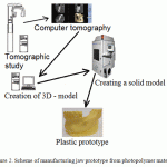 |
Figure 2: Scheme of manufacturing jaw prototype from photopolymer material |
Results
The Planmeca Romexis Viewer 3.6.0 R software was used for processing the results of research with a cone-beam computer tomograph. A three-dimensional image is simultaneously displayed in four different views: Sagittal (red, x-axis), Coronal (green, y-axis), Axial (blue, z-axis), three-dimensional rendering (3D rendered view) (Figure 3).
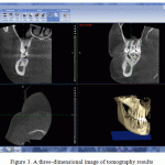 |
Figure 3: A three-dimensional image of tomography results |
Axial view was used for selecting images as the basis for building a 3D model. A fragment of the lower jaw was chosen as the area for building a 3D model. Images were chosen with the snapshot of axial slices. The chosen photographs of the lower jaw fragment cross sections in the amount of 191 images were then saved in JPEG format for subsequent file transfer.
The dentition images obtained with CT were processed, photographs of axial slices of lower jaw fragment in JPEG format were selected, and a digital model was built on their basis with the help of the 3D DOCTOR software suite. The first stage of image processing is segmenting the selected zones of interest with creating a 3D object (Figure 4).
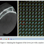 |
Figure 4: Marking the fragment of the lower jaw with a marker |
Taking into account the fact that each point of the tomogram has its own brightness that depends on how optically dense the area of the organ or tissue is, it is possible to separate the segments of the image with the same brightness or optical density by setting the threshold value of optical density. Such a segment will be a fragment of the lower jaw. The threshold value for segmentation is selected so that the pixels with intensity above the threshold include only the most optically dense part of points in the dentition picture. In this case, it is the bone of jaw. In the next stage, the image segmented by the threshold value is marked according to the fragments of the lower jaw. The result of segmentation is an object of a fragment of the lower jaw (Figure 5).
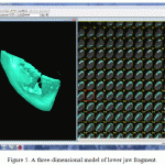 |
Figure 5: A three-dimensional model of lower jaw fragment |
If the obtained three-dimensional model is of insufficient quality, the program makes it possible to replace markers, or change the threshold of segmentation. Since the density of bone tissue in some parts can be quite low, specific parts of the jaw may be not displayed after the first stage of segmentation. Therefore, in order to determine the true size of the jaw, it is necessary to expand its initial model, indicating the least low segmentation threshold. The use of two-stage segmentation with various thresholds in image processing makes it possible to accurately reconstruct the jaw bone without loss of data (Naumovich, 2013). The obtained three-dimensional model of the lower jaw fragment is saved in STL format. The computer model cannot be final for “growing” plastic copies in a stereolithographic machine, as it will have various aliases caused by defects in scanning and selecting bone tissues (D’Urso, 1999).
Further, the computer model in STL format was processed in the Magics12 program from the Materialize Company. Model processing included: removing aliases generated in the process of creating the three-dimensional model, positioning in the workspace of the Perfactory XEDE stereolithographic machine from the Enviziontec Company, and generating support elements (Figure 6). After editing, the examined object is a hollow model with the volume of 26.8 cm³, with the following dimensions by the axes: x = 81.2 mm; y = 79 mm; z = 40.3 mm.
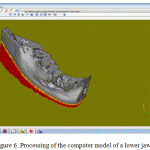 |
Figure 6: Processing of the computer model of a lower jaw fragment |
The solid experimental prototype of lower jar fragment was created at the Perfactory XEDE stereolithographic machine from the Envisiontec Company. The installation makes it possible to manufacture plastic copies of computer models with dimensions of 220х280х380 mm and thickness of the polymerizable layer of 0.025 – 0.01 mm. The computer model of human jaw fragment was made of cross-linked photopolymer material SI500. The manufactured experimental model of human lower jaw fragment is shown in Figure 7.
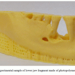 |
Figure 7: The experimental sample of lower jaw fragment made of photopolymer material SI500 |
For the purpose of high detailed elaboration of the profile of the experimental copy of the lower jaw fragment, the built-up increment of 0.05 mm was used, with exposure time for each subsequent layer of 13 seconds. The process of creating the experimental prototype took 4 hours. This mode of building ensures dimensional accuracy, and minimum surface roughness of the plastic copy.
Discussion
The methods of modern stomatological treatment keep on improving, due to the introduction of three-dimensional modeling computer technologies, and prompt production of exact plastic copies of lost or defective organs using rapid prototyping technologies (Roginsky, 2000).
The model of lower jaw fragment made using the rapid prototyping technology is the most convenient form of visualizing information obtained using the cone-beam computer tomography. A plastic copy makes it possible to diagnose complex bone deformations more accurately, and provides the possibility to foresee complications during surgery. A photopolymer model makes it possible to accurately determine size, shape and location of a bone defect, and to define required size of the implant (Arkharov, 1999). The use of photopolymer models in planning maxilla-facial surgery will considerably reduce duration of surgery due to availability of a prepared implant.
Rather high accuracy of models allows measuring and surgery planning, with the knowledge that the resulting model will accurately match the real state of the bone tissues (Kermer, 1998).
The experimental model obtained from the tomography results showed that the studied object was a rather complex formation with many internal cavities and various optical densities in various locations. In one case, with a lower threshold, some areas may be merged, and with a higher threshold, one area may be presented by several areas. Correct definition of boundaries of the inspected object is possible with the knowledge of the mechanism of tomographic scanning of specific types of objects and presenting their images on a tomogram (Kermer et al, 1998). After defining the boundaries of the object, the manufacturing accuracy of the model is determined by the number of image points, and by the number of layers in the source data (Evseev, 2005). If the distance between the layers is 1mm, the surface of the plastic model will have the same accuracy, but the model will have expressed “steps” between its layers (Andersson, 1987). The average error in manufacturing models does not exceed 0.8 mm. With that, the error of the model depends on the resolution of the tomograph, and on slice thickness, which is 1 mm in modern scanners (Bianchi et al., 1997). Consequently, there is a need for smoothing the surface of the model of the examined object. Stereolithography that is based on initiating the polymerization reaction with digital video projectors makes it possible to obtain higher accuracy. This is due to the fact that the package for three-dimensional positioning and «slicing» the computer model into layers used in the Perfactory XEDE installation makes it possible to vary layer thickness regardless of CT scan results.
The use of CT scanning results with slice thickness of 0.06 mm made it possible to further reduce errors in the model. The used methodology of two-step segmentation in processing axial slices images of lower jaw fragment made it possible to reduce aliases on the surface of the computer model formed in course of tomographic examination (Naumovich, 2013).
Due to the high precision of the material of the plastic copies made by rapid prototyping technologies, and the possibility to reconstruct the results of tomographic studies using modern software, it is possible to allocate main areas of using the rapid prototyping technology in implantology (Arver, 1994):
- improving visualization of anatomical details;
- planning surgery, namely, calculating and sizing of the implant;
- selecting necessary parts for reconstructive surgery; and
- using the plastic copy as a master model for manufacturing individual metal implant;
Conclusion
The performed studies have shown high efficiency of manufacturing plastic implants using the technology of stereolithography for deformed or completely lost bone tissues of the maxillofacial organs. The distinctive feature of this methodology, namely, obtaining an exact copy of lower human jaw fragment, is the use of medical diagnostic equipment together with the rapid prototyping system.
It has been found that for obtaining high-quality computer models it is necessary to perform two-phase segmentation of the object, since the density of bone tissue in some parts of the object may be low, which can lead to defects of the contour of the “grown” plastic copy. The data obtained by segmentation of the studied area make it possible to pinpoint the areas of aliases resulting from cone-beam computer tomography, which makes it possible to edit the 3D computer model.
In course of the study, the optimal growing conditions for stereolithographic machine have been experimentally determined, namely the time of exposing layers of the supports, which is equal to 40 seconds, and the time of the basic model exposure equal to 13 seconds. In order to reduce the “step” effect in the plastic copy of the lower jaw fragment, thickness of each polymer layer was reduced to 0.05 mm. This made it possible to obtain high-quality experimental plastic sample without the need for additional machining of the surface.
The main disadvantage of this rapid prototyping technology is the limited choice of materials, namely, biocompatible materials (Antonov, 1998).
Basing on the results of the research, it seems possible to develop in the future a technology for manufacturing biocompatible implants that mimic bone and soft tissues by using photopolymer compositions for rapid prototyping systems based on initiating the polymerization reaction with video projectors, depending on intensity of light of various colors. This will make it possible to better visualize tissues in relation to the bone.
References
- Abramov, S.S., Boldyrev, N.N., Evseev, A.A. et al. (1998). Manufacturing plastic copies of three-dimensional objects by tomographic data. Optic Engineering, 1(13), 45–49.
- Antonov, E.N., Evseev, V.A., Topolnitskiy, O.Z. et al. (1998). Forming bioactive mineral-and-polymer composites using the method of laser stereolithography. Optical Engineering, 1(13), 55-60.
- Arkharov, S.L. (1999). Studying efficiency of CT and other radiology examinations in planning dental implantation (pp. 12), thesis of Candidate of Medical Sciences. Kemerovo.
- Evseev, A.V., Kamaev, S.V., Kotsyuba, E.V., et al. (2005). Modern Computer biomodeling and laser stereolithography. Laser – information and laser technologies: Collected works of IPLITRAN (pp. 119-130). Moscow: Intercontact. Science.
- Zlenko, M. (2003). The rapid prototyping technology – layer-by-layer synthesis of physical copy based on a 3D CAD model. CAD/CAM/CAE Observer, 2(11), 2-9.
- Kulakov, A.A., Rabukhina, N.A., & Adonina, O.V. (2003). Pre-surgery x-ray examination of patients in case of dental implant surgery in distal parts of the upper jaw. In the Proceedings of the all-Russian Conference “Prevention of major dental diseases”, Moscow (pp. 76–79).
- Naumovich, S.S., Naumovich, S.A., & Goncharenko, V.G. (2013). Segmentation of computer tomograms of dentition. The Journal of Medicine, 3, 89-87.
- Rabukhina, N.A., Golubeva, G.I., & Perfilyev, S.A. (2006). Spiral computed tomography in diseases of the maxillofacial area (pp. 226). Moscow.
- Roginsky, V.V., Evseev, A.V., & Kotsyuba, E.V. (2000). Laser stereolithography, a new method of biomodeling in cranio-maxillo-facial surgery. Children Stomatology, 3-4, 92-95.
- Tverskoy, M.M., Petrov, L.N., Aladin, A.S., & Zharinova, A.S. (2012). Computer technology for manufacturing medical implants using the layer-by layer method of sintering. Bulletin of the SUSU, 23, 64-69.
- Shavladze, Z.N., Nalapko, V.I., Rabukhina, N.A. et al. (2002). Using radiographic methods in dental implantology. Dentistry, 6, 34–37.
- Andersson, L., & Kurol, M. (1987). CT scan prior installation of osseointegrated implants in the maxilla. Journal of Oral and Maxillofacial Surgery, 16, 55.
- Arver, J.F., Barker, T.M. et al. (1994). Maxillofacial Biomodelling. British Journal of Oral & Maxillofacial Surgery, 32, 276–283.
- Baumgaertel, S, Palomo, J.M., Palomo, L., & Hans, M.G. (2009). Reliability and accuracy of cone-beam computed tomography dental measurements. Am J Orthod Dentofacial Orthop, 136, 19-25.
- Bianchi, S.D. (1997). The validation of stereolithographic anatomical replicas: the authors’ own experience and a review of the literature. Radiol.Med.(Torino), 94(5), 503-510.
- D’Urso, P.S., Barker, T.M., & Earwaker, W.J. (1999). Stereolithographic biomodelling in cranio – maxillofacial surgery: a prospective trail. Journal of Cranio-Maxillofacial Surgery, 27, 30-37.
- Jacobs, P.F. (1995). Stereolithography and Other RP&M Technologies (pp. 451). Dearborn, MI: Society of Manufacturing Engineers.
- Kermer, C. (1998). Preoperarive stereolithographic model planning for primary reconstruction in craniomaxillofacial trauma surgery. Journal of Cranio-Maxillofacial Surgery, 26(3), 136-139.
- Kermer, C., Rasse, M., Lagogianis, G. et al. (1998). Colour stereolithography for planning complex maxillofacial tumour surgery. Journal of Cranio-Maxillofacial Surgery, 26:6, 360-139.
- Popat, A.H. (1998). Rapid prototyping and medical modeling. Phidias Rapid Prototyping in Medicine, 1, 10-12.
- Sarment, D.P., Sukovic, P., & Clinthorne, N. (2003). Accuracy of implant placement with a stereolithographic surgical guide. The International Journal of Oral & Maxillofacial Implants, 18, 571-577.
- Sykes, L.M., Parrott, A.M., Owen, C.P., & Snaddon, D.R. (2004). Applications of rapid prototyping technology in maxillofacial prosthetics. The International Journal of Prosthodontics, 17: 454-459.
- Winder, J., & Bibb, R. (2005). Medcial rapid prototyping technologies: state of the art and current limitations for applications in oral and maxillofacial surgery. Journal of Oral and Maxillofacial Surgery, 63, 1006-15.
(Visited 269 times, 1 visits today)

This work is licensed under a Creative Commons Attribution 4.0 International License.


