

Manuscript accepted on :
Published online on: 14-12-2015
Aleksey Efimovich Kobityansky and Alexander Olegovich Trofimov
The Perm National Research Polytechnic University, 29, Komsomolsky Ave., Perm, 614990, Russia
ABSTRACT: Designing and creating devices for rehabilitation of elbow joints is an actual and important problem. The purpose is to create a methodology for designing a mobile and relatively simple device that allows recovery of motor abilities of elbow joints, followed by its subsequent manufacturing using the method of rapid prototyping. The article considers various approaches to synthesis of such designs. Basing on the block diagrams, a number of devices have been offered. The methodology and the algorithm of synthesizing one of selected designs have been considered. The proposed device was synthesized on the basis of selecting mechanical analogues of the elements of elbow joints. A calculation scheme was developed and a three-dimensional model was created in the ASCON KOMPAS 3D V15 environment. Strength and deformation were calculated in the SolidWorks Simulation environment. Several loads and cross-sections relative shear curves were calculated, and patterns of deformation of each element of the device were obtained. A method of manufacturing the device using the process of rapid prototyping was proposed. This methodology allows to complete designing and engineering synthesis of the rehabilitative device that can be applied in medical practice and as an exercise machine in health groups and in some sports.
KEYWORDS: elbow joint; synthesis; prototyping; rehabilitation; device
Download this article as:| Copy the following to cite this article: Kobityansky A. E, Trofimov A. O. The Method of Structural and Technological Synthesis of a Device for Rehabilitation of the Elbow Joint Based on 3D Modeling and the Process of Rapid Prototyping. Biosci Biotech Res Asia 2015;12(2) |
Introduction
There are many patients who require treatment of elbow joint contractures. Such contractures are the result of forearm dislocations, intra-and-juxta-articular fractures, injuries of the ligamentous-capsular apparatus, over-stress diseases (epicondylitis, tenosynovitis, myofascial syndromes, other enthesopathies), and shoulder and forearm fractures, which require prolonged immobilization after injuries till adhesion of the bone fragments (Kapandji, 2009). In accordance with the Decree of the Ministry of Health of the Russian Federation (Program of modernization of Public Health System in the Russian Federation dated 08.11.2010 N 29-1/10/2-10191), the task of creating rehabilitation centers and cabinets to be equipped with modern home-made equipment, including, rehabilitation of elbow joints was set. According to the statistical data, intra-articular fractures of elbow joints lead to disability in 9% of cases. For example, in the Perm Territory, about 180 elbow joint surgeries are made in children and 160 – in adults. It should be noted that surgical treatment is used for the most severe damages of the joint. According to the same statistics, elbow joint damages account for 40 to 50% of all injuries of the musculoskeletal system, resulting in incapacitation, material and moral costs (Khanov A. M. et al., 2013).
The main methods of treating elbow joint injuries are shown in Figure 1:
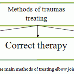 |
Figure 1: The main methods of treating elbow joint injuries |
Elbow joint work out is a part of regenerative medicine and includes rehabilitation after upper extremities injuries, and after surgery on muscles, tendons, joints (Figure 1). The need to rehabilitate upper extremities is caused by the fact that elbow joints perform vital functions. These joints are a kind of connecting “bridge” between the shoulder and the wrist. Treating its injuries is associated with prolonged lack of movement in the injured area, which in turn causes restricted mobility (contracture), muscle atrophy, poor blood circulation and lack of normal metabolism in upper extremities. Elbow joints are considered most “difficult” to work out due to their special anatomy and functions. Therefore, the absence of measures aimed at restoring upper limbs leads to limitation of their function, causing poorer quality of life. Indications for working out elbow joints after injuries and surgeries are: immobilization with plaster after bone fractures; surgery on tendons, muscles and nerves; surgery on bones; surgery on joint and endoprosthesis replacement. One of rehabilitation methods is mechanotherapy that is based on using special devices and constructions that ensure appropriate treatment with mechanical motion. This method is one of the most important elements in rehabilitation after joint traumas and surgery. The purpose of mechanical therapy is improving the mobility in the joint after surgery, recovering the maximum range of motion in it, exercising muscles, improving blood circulation and microcirculation in the tissues of the joint. In course of mechanical therapy, movement in the elbow joint can be active, when the hand is moved by the patient himself, and passive, when the movement is ensured by the apparatus. Patient’s joint is “getting used” to moving, and it is easier for the patient to perform movement himself. The moving joint send signals to the brain; these signals make the central nervous system “understand” that the joint is “working” (Parkhotnik, 2007). The feature of mechanical therapy is that in some cases the patient can independently perform assigned exercises using a specific apparatus without participation of medical staff, and in some cases – under supervision. In course of mechanical therapy, the patient may at any time readjust settings, features and modes of the rehabilitation procedure, that is, measure load, amplitude, rhythm, number of movements and a number of other factors more precisely. Based on the problems that arise in case of musculoskeletal system injury, the program of modernizing the Health Care System in the Russian Federation includes a number of tasks, where the concept of development of the rehabilitation system has been shaped. Implementation of such a program is possible due to creating special medical equipment, including mobile, ergonomic, technologically advanced and multifunctional devices for rehabilitation of elbow joints.
Methodology
The subject of the methodology of designing and synthesis is a device for elbow joint rehabilitation based on structural analysis, elements of design and 3D modeling, including production of the device prototype.
- The structural design model and the mathematical model for elbow joint rehabilitation
In the first phase of the design, mechanical equivalents of elbow joint elements were identified (Kapandji, 2009). As an example, Figure 2 shows several such mechanical analogues and their three-dimensional models:
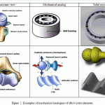 |
Figure 2: Examples of mechanical analogues of elbow joint elements |
In accordance with Figure 2, it is possible to represent elbow joint elements in the form of the following mechanical analogy: a ball bearing that ensures rotation with one degree of freedom; a sphere and coil on one axis for the shoulder bone, and a capitated elevation. In this case, one degree of freedom is ensured for flexion/extension of the inner part of the joint, and two degrees of freedom – flexion/extension and axial rotation of the outer part of the joint. Thereby, it is possible to proceed to forming the structure of the designed device.
Based on mechanical analogs of elbow joint components, the variants of block diagrams of mechanisms that implement its basic movements have been reviewed (Artobolevsky, 1988). Structural schemes have been proposed that mimic mechanical movement of the joint that are shown in Figure 3:
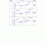 |
Figure 3: Block skeleton diagrams of the designed device |
Each of these schemes, except for the first, implements all movements of the elbow joint. It should be noted that block skeleton diagrams 2 and 3, while providing the same degree of freedom, are structurally implemented with kinematic pairs of different classes. So, in case 2a ball joint with a pin is used, while in Figure 3, flat hinges of the fifth class are used, which simplifies the design itself. This makes it possible to adjust the length of the links and ensures ease of assembling the designed construction.
According to the results of the analysis, the device for elbow joint rehabilitation is based on the scheme that corresponds to the third variant of Figure 3. A design scheme (Figure 4) and corresponding mathematical model have been formed for this structure.
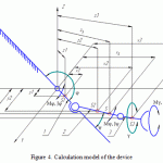 |
Figure 4:Calculation model of the device |
1, 2, 3 are moving parts of the device design.
φ, ψ, γ are rotation angles of the forearm, relative to the shoulder, in planes XZ, XY, YZ, respectively. With that, angle φ characterizes flexion/extension of the elbow joint; ψ is the value of angular displacement of forearm axis relative to the shoulder; γ is the angle of forearm pronation/supination.
S1, S2, S3, S are positions of the centers of mass of respective links and the entire system.
xS, yS, zS are coordinates of the center of mass of the entire device system.
xi, yi, zi (i=1-3) are the coordinates of the centers of mass of the ith link and the entire system.
Mφ, Mψ, Mγ are the moments of forces acting on the links of the device when its appropriate elements are moving.
Jφ, Jψ, Jγ are the moments of inertia of respective links.
When the elbow joint is operating, there exists a relationship between the flexion-extension motion and individual displacement of forearm axis relative to the shoulder (Kapandji, 2009). If we assume that these motions are proportional, the following condition is adopted:
Ψ = k – φ ¸
where k is the coefficient of proportionality.
As a result of mathematical transformations the following mathematical model of the elbow joint motion has been obtained:
The last three ratios, in the form of restrictions, shall be based on anatomical features of human hand, and φ0, ψ0, γ0 characterize the initial position of forearm axes (Kapandji, 2009). 145°, 15°, 175° are the average constraints on the corresponding angles values, and 160°, 20°, 180° are their limit values (Parkhotic, 2007). In relations (1), c is the stiffness coefficient of the elastic element, which can be introduced into the block skeleton diagrams, simulating loading on the flexion/extension motion, due to restrictions. This makes it possible to implement conditions for active rehabilitation of the joint.
Upon obtaining relations (1), the elbow joint and the device were, with some error, taken as a combination of three bodies: forearm cradle – as part of a truncated hollow cylinder; the lower arm in the form of a truncated cone, and the wrist is in the form of a spherical surface.
The parameters of mass and geometric characteristics were determined in accordance with the methods of technical mechanics (Frolov et al., 1987) (Anuriev, 2003) (Dunaev and Lelikov, 1998). Their values were found for corresponding reference materials for certain age categories of patients (Parkhotic, 2007).
Thus, the presented ratios make it possible to estimate and identify the main parameters of the device for elbow joint rehabilitation that are necessary for manufacturing.
Solid modeling of parts of the device for elbow joint rehabilitation
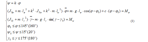
Solid modeling is performed in accordance with the algorithm shown in Figure 5.
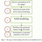 |
Figure 5:The stages of solid modeling of the elbow joint rehabilitation device |
Schematic design of the elbow joint rehabilitation device
At the first stage (Unit 1 in Figure 5) x-ray photographs of the elbow joint that had been obtained at the Department of Traumatology were analyzed. This photograph and the results of ratios calculations (1) are the basis for choosing the main parameters of the designed device.
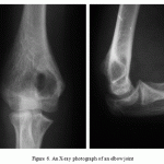 |
Figure 6: An X-ray photograph of an elbow joint |
After that, preliminary design of the device was made based on the selected parameters (Unit 2 in Figure 5). The main structural parts of the device are the following: elbow hinge, hinge for shifting forearm axis relative to the shoulder, rotating arc, shoulder support.
A sketch of the elbow joint in the initial position and during extension of the elbow is shown in Figure 7. It should be noted that its main unit may be a roller bearing which is exposed to axial load and torque. Due to the unit geometry, restrictions of the elbow joint flexion-extension angle φ (Figure 4).
 |
Figure 7: A sketch of the device of the elbow hinge |
Since there is really a correlation of the flexion-extension motion with simultaneous displacement of the axis of the forearm characterized by the coefficient k from relations (1), it is possible to implement such a scheme, the sketch of which is shown in Figure 8. Here displacement of the forearm axis during elbow flexion-extension is achieved using ball bearings or bushings.
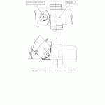 |
Figure 8:Sketch of a hinge for forearm axial displacement relative to the shoulder |
In order to ensure forearm rotation motion, regardless of other motions, a rotational arc may be used, the sketch of which is shown in Figure 9. In this case, the forearm is rotated around its axis within the predetermined range of rotation angles γ (Figure 4) due to the geometry of the structure.
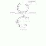 |
Figure 9: Rotation arc sketch |
The support in the form of lodgment (Figure 10) is attached to the rack of the device with a special mechanism that makes it possible to adjust support height. In turn, the rack of device is directly connected to the elbow joint.
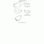 |
Figure 10: Shoulder support sketch |
Thus, with regard to Figures 7 through 10, the sketch of the general view of the entire device is shown in Figure 11.
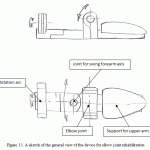 |
Figure 11: A sketch of the general view of the device for elbow joint rehabilitation |
Forming three-dimensional models of device elements and their calculation
Based on the sketches and the results of geometric calculation (Burdakov, 1986), three-dimensional models of components of the elbow joint rehabilitation device were created (Unit 3 in Figure 5). These models were generated using the ASCON KOMPAS 3D V15 software suite (Gerasimov, 2012) with regard to the selected parameters of assemblies and subassemblies (Mineev, 2008). This software suite also makes it possible to create 3D models of parts (Kuprikov et al., 2009) (Prokhorenko, 2004), and generate technical documentation based on these models. Design of machine-building and instrument-making products imposes high demands on the tool used. The possibilities of the system ensure designing machine-building products of any complexity in accordance with cutting-edge design methods (Bolshakov et al., 2011). The system has tools for working using the “top-down” method, or the down-going design methodology, as well as the already familiar “bottom-up” method. (Goldschmidt, 2007). 3D models of the main components of the device are shown in Table 1.
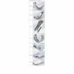 |
Table 1: 3D models of the main components of the device for elbow joint rehabilitation |
In the next stage of design (unit 4 in Figure 5) by means of mathematical modeling were used to calculate the strength, stiffness and relative deformation of elements and nodes of the device. The basis of mathematical modeling was the method of finite elements (Alyamovsky, 2004) implemented in the SolidWorks Simulation software suite (Alyamovsky, 2004). This suite has a lot of opportunities and, in addition to the above-mentioned transactions, makes it possible to simulate motion and other procedures. Examples in Tables 2-3 are fragments of calculations for the rotation arc and unit responsible for forearm rotation.
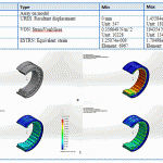 |
Table 2: Result of “Rotation arc” part strength calculation |
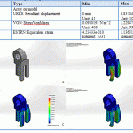 |
Table 3: Result of “Pivot arm” part strength calculation |
Thus, with regard to the strength and deformation characteristics of the structural elements, a three-dimensional model was generated, which can be manufactured using the method of rapid prototyping, in particular.
Synthesis of the prototype of the elbow joint rehabilitation device
The device prototype was manufactured using the method of rapid prototyping (Morozov, 2005) (Ferry et al., 2010). The basis for carrying out such works was a solid 3D model obtained by modeling. The technological process of manufacturing the prototype of this device involved the use of stereolithography (Ferry et al., 2010) at the Envision Tec Perfactory XEDE installation.
Prototyping was implemented in accordance with the algorithm shown in Figure 12.
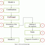 |
Figure 12: Device prototyping stages |
Materialise Magics (EnvisionTec, 2015) software was used for editing each unit of the model, i.e., for generating the supports system for each composite model. In accordance with the scheme (Figure 12), the main operation steps of the process should be highlighted:
- 3D models of the device components are loaded.
- The merge function is launched to automatically remove all self-intersections in order to avoid support intersecting the surface of the other models.
- The FixWizard function is used to compensate for low-quality edges, gaps and other errors.
- Models are oriented for manufacturing.
- Supporting structures are created.
- The seaming operation is used to automatically repair defective edges.
- The products with supporting structures are obtained.
The material is liquid photopolymer SI-500. The model is divided into layers, and then elements of the device are grown layer by layer with the use of the Perfactory RP (EnvisionTec, 2015) software product. The entire prototyping process consists of periodically repeated actions. The duration of polymer layer exposure, supports exposure, thickness of the supports, etc. are set here. Upon completion of the manufacturing process, supports are removed from the resulting product. If necessary, it is additionally polymerized. It is important to note that the material of the construction may be stainless steel, titanium alloys and other materials. All parts and components of the device for elbow joint rehabilitation created using the method of prototyping are shown in Figure 13
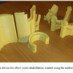 |
Figure 13: Parts of the device for elbow joint rehabilitation created using the method of rapid prototyping |
Results
The design of the device for elbow joint rehabilitation was obtained using assembly prototyped parts. One of the variants of such a device is shown in Figure 14.
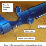 |
Figure 14: The prototype of elbow joint rehabilitation device |
The main stages of the device operation include preparation and operation itself. Preparation includes the following.
- Shoulder support is adjusted to a required length, namely the length of patient’s shoulder, with an adjustable mating of 2 parts that are parts of this support (Figure 14).
- Using elastic bands (not shown), the shoulder of the patient is fixed on the support.
- The necessary length of the active part of the device is set by adjusting 2 mating parts details: support of rotation arc and the unit responsible for forearm rotation relative to the shoulder. In this case, patient’s forearm is fixed in the rotation arc with elastic bands (not shown).
Direct operation of the device is ensured by kinematic connection of the flexion/extension motion of the elbow and forearm axis displacement relative to the shoulder. All anatomical features are observed with regard to the structure of the elbow joint. The supination-pronation motion occurs independently of other motions of elements of the device due to peculiarities of the design (Figure 14). This motion occurs due to rotation of the radial bone and imaginary rotation of the elbow bone. It should be noted that due to the design, the supination/pronation motion is possible without flexion/extension. A detachable elastic element mounted in the elbow joint (not shown) generates the load required for active rehabilitation process. Thus, the designed and manufactured device ensured all basic motions of the elbow joint.
Possible perspectives of using the device for elbow joint rehabilitation are shown in Figure 15.
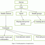 |
Figure 15: Possible perspectives of using the device |
Discussion
At present, various devices for mechanical therapy for rehabilitation of the musculoskeletal system are actively developed. In a number of works (Yatsun and Tarasova, 2011) (Yatsun and Rukavitsyn, 2012) (Yatsun and Tarasova, 2013) theoretical approaches to synthesis of such devices have been provided. In article (Yatsun and Tarasova, 2013) the scheme and design of such a device was proposed. Calculations have been made for determining trajectories of device units in general. However, this design does not ensure all motions of human hand, but only flexion/extension of the elbow joint. The pronation/supination motion and changing position of the forearm axis during extension are not taken into account. Besides, the design is complicated due to the considerable number of moving parts. (Steven Paul Toddes Jr., 2003) shows a design of the device that restores the function of muscular fibers in upper limbs in case of central nervous system diseases. The design is stationary, not mobile, and does not ensure all major movements of the elbow joint. Fairly efficient is the design shown in (Tariq Rahman and Whitney Smple, 2004), which makes it possible to ensure all movements of the human hand. However, the device contains peripheral devices: external power supplies, control panel, and control unit. Such a design has limited mobility and lacks ease of operation. Stationary, expensive and difficult to operate devices include designs shown in works (Yatsun and Tarasova, 2013) (United States Patent Rahmanctal, 2004). The design presented in (United States Patent Rahmanctal, 2004) consists of two parts connected by a hinge. Each part both has one degree of freedom and includes a 4-link hinge and an elastic element that generates equilibrium force that corresponds to the weight of the main parts of the structure. The device is stationary, not mobile, and does not ensure all major movements of the elbow joint. The design of a rehabilitation device shown in (Yatsun and Tarasova, 2013) makes it possible to ensure both types of motion (flexion/extension and pronation/supination) at the same time. For operation it needs a drive. The design is stationary, bulky, and not mobile.
Thus, despite a number of studies, the field of rehabilitation lacks attention to the methods of designing and manufacturing mobile, technological and inexpensive devices for elbow joint rehabilitation.
Conclusion
According to the results shown in the article, the following conclusions can be made:
- A method of synthesizing a device for elbow joint rehabilitation has been developed.
- Structural design schemes and mathematical model have been proposed.
- On the basis of built three-dimensional models and solid modeling, a technological process has been developed, and a prototype device for elbow joint rehabilitation has been manufactured.
- The design of the device ensures treatment and rehabilitation for elbow joints, features mobility, adaptability, and ease of use and maintenance.
- The use of the device will reduce the lack of medical equipment of this type, and will make it possible to solve the problem of medical care in the field of traumatology, as well as to address some socioeconomic challenges.
Further research will be focused on optimization of the design of the elbow joint rehabilitation device and its wide use in medical practice.
References
- Alyamovsky, A.A. (2004). SolidWorks/COSMOSWorks Engineering analysis by the finite element method (pp. 275). MDK Press.
- Anuriev, V. I. (2003). Handbook of the mechanical engineer (2003, Vol. 3). Moscow: Mechanical Engineering.
- Artobolevsky, I. I. (1988). Theory of mechanisms and machines: Textbook for technical college (4th ed., revised and amended, pp. 640). Moscow: Nauka. Chief ed. of phys.-mat. lit.
- Bolshakov, V.P., Bochkov, A.L., & Sergeev, A.A. (2011). 3D modeling in AutoCAD, KOMPAS-3D, SolidWorks, Inventor, T-Flex: a training course (pp. 336). Saint Petersburg: Piter.
- Burdakov, S. F. (1986). Designing manipulators of industrial robots and robotic systems (pp. 264)..
- Gerasimov, A.A. (2012). New possibilities in KOMPAS-3D V13. Tutorial (pp. 288). BHV-St. Petersburg.
- Goldschmidt, M.G. (2007). Methodology of design. Tutorial (pp. 173)..
- Dunaev, P.F., & Lelikov, O.P. (1998). Designing units and parts of machines: Textbook for technical specialized Universities (5th ed., revised and amended). Moscow: Vysshaya Shkola.
- Kapandji, A.I. (2009). Physiology of joints. Volume 1, Physiology of the joints (6th edition, pp. 368). Moscow: Eksmo.
- Kuprikov, M.Y., Maslov, Y.V., Hotina, G.K., & Nikishina, L.B. (2009). Solid modeling of parts in SolidWorks geometric modeling environment (pp. 264). Moscow: MAI-PRINT.
- Mineev, M.A. (2008). CAD. PRO/EngineerWildfire 2.0/3.0/4.0/5.0. Science and technology. Saint Petersburg.
- Morozov, V.V. (2005). Research and development of technological modes of manufacturing castings from burned out models obtained by laser stereolithography, the Moscow State Technical University n.a. N.E. Bauman (pp.161). Moscow.
- Parkhotic, I.I. (2007). Physical rehabilitation after upper extremities injuries (pp. 282).
- Prokhorenko, V.P. (2004). A practical guide (pp. 448). Moscow: “Binom-Press” LLC.
- Frolov, K.V., Popov, S.A., Musatov, A.K. et al. (1987). Theory of mechanisms and machines: Textbook for technical colleges (pp. 496). Moscow: Vysshaya Shkola.
- Khanov, A.M., Kobityansky, A.E., Belokrylov, N.M., Fefilov, D.A., & Trofimov, A.O. (2013). Synthesis and design of equipment for elbow joint rehabilitation. Master’s journal, Mechanic engineering, Perm, 103-109.
- Yatsun, S.F., & Tarasova, E.S. (2013). Kinematic features of elbow joint rehabilitation device movement together with human hand. Journal of Academic and Applied Studies, 4 (Medical Sciences), 59-64.
- Yatsun, S.F., & Tarasova, E.S. (2011). Kinematic analysis of hand movements at the elbow joint during rehabilitation methods of mechanical therapy (pp. 1215-1220). South-West State University, biotechnology and bioengineering.
- Yatsun, S.F., & Rukavitsyn, A.N. (2012). Developing bioengineered mechatronic module for lower limbs exoskeleton.
- Envision Tec. Perfactory® rapid prototyping system. Envision TEC GmbH hardware and software guide (2015, pp. 359). Gladbeck, Germany.
- Melchels, F.P.W., Feijen, J., Grijpma, D.W. (2010). A review on stereolithography and applications in biomedical engineering. Biomaterials, Vol. 31, 24, 6121-6130.
- Melchels, F.P.W., Bertoldi, K., Gabbrielli, R., Velders, A.H., Feijen, J., & Grijpma, D.W. (2010). Mathematically defined tissue engineering scaffold architectures prepared by stereolithigraphy. Biomaterials, 31, 27, 6909-6916.
- Toddes Jr., S.P. (2003). Optimization for commercialization of a two degree of freedom powered arm othosis..
- Rahman, T., Simple, W. (2004). Orthosis device.
- United States Patent Rahmanctal, US 6,821,259 b2. – 2004.

This work is licensed under a Creative Commons Attribution 4.0 International License.


