

Manuscript accepted on :
Published online on: 12-12-2015
Alexey Efimovich Kobityansky, Alexey Vladimirovich Shafranov, Vladimir Sergeevich Beloborodov, Alexander Nikolaevich Mulkov
Perm National Research Polytechnic University, 29, Komsomolsky Ave., Perm, 614990, Russia
ABSTRACT: Designing and creating implants used in medical practice, with regard to requirements and indications is a relevant and an important problem. The purpose is to create methodology for designing and subsequent growing implants with porous structure using the method of rapid prototyping on the example of jaw implants. The article discusses various technological methods for manufacturing implants, and the stereolithography method has been selected for obtaining it. The algorithm and the process of obtaining a 3D model of an implant based on the mathematical apparatus with the use of affine transformations have been proposed. On the basis of the developed software suite that uses mathematical relations, the configuration of the cellular structure has been chosen. Software implementation of the proposed algorithm has been performed, and the technological process of growing the cellular structure prototype by the stereolithography method at the Tec Invision Perfactory XEDE installation has been developed. Some calculation results have been shown along with a prototype of the jaw implant grown in accordance with the 3D models obtained from mathematical modeling. This technique makes it possible to switch to replacing or completely removing jaw defects (e.g., after trauma), if necessary, in maxillofacial surgery and in bone defects recovery.
KEYWORDS: synthesis; implant; cellular structure; mathematical modeling; prototyping
Download this article as:| Copy the following to cite this article: Kobityansky A. E, Shafranov A. V, Beloborodov V. S, Mulkov A. N. Synthesis of Jaw Implants with Porous Structure on the Basis of Mathematical Modeling and the Rapid Prototyping Process. Biosci Biotech Res Asia 2015;12(2) |
Introduction
Currently innovative technologies are widely used in various fields of medical practice (Vasilyuk et al., 2014). Special attention should be paid to the importance of such technologies in recovering bone defects, in dentition recovery and in other cases where bone tissue engineering is used. In particular, damaged bone tissue can be recovered by the organism in the presence of a biologically compatible porous matrix with appropriate architecture. Thus, tissue is built up in the presence of stimuli for bone formation (Barinov and Komlev, 2005). The required matrix can be obtained using different technologies. One such technology is the process of rapid prototyping, when the prototype is obtained using special operations, and this prototype can replace damaged bone tissue (Topolnitsky et al., 2012). Here a possibility exists of creating such implant prototypes from biocompatible materials, for example, photosensitive resin, polycaprolactone, special bioceramic materials based on calcium phosphates, bone cement, polymethyl methacrylate, metals (stainless steel, alloys of titanium, cobalt, chromium) and other (Milovanović and Trajanović, 2007). A number of implants have hybrid structure, which is a monolithic part (the shell), and a cellular structure featuring porosity and a core frame, which consists of intersections (Sirotenko et al., 2010). The advantage of these implants is good fixation at the beginning of the healing process (Marin et al., 2010). In the process of healing, the bone tissue can replicate the geometry of the implant (Khodorenko et al., 2001). Due to the cellular structure, elements of the bone tissue grow through it until the parts of the bone connected to the implant are fused, which is an advantage of the relevant cellular structures (Murr et al., 2010). In course of medical surgery, the use of such jaw implants in some cases requires no additional means of fixation, such as pins (Radkevich et al., 2012).
Currently there are various methods of creating cellular structures, one of which is the technology of rapid prototyping (Stuchilov et al., 2002). In its turn, this technology has various areas, depending on the method of obtaining the prototype. These technologies include extruding the material through a die hole, laser sintering, polymerization due to laser radiation, etc. There are also technologies where three-dimensional models are obtained by impregnating the powder with a binder material, or photocuring without laser (Kuznetsov, 2003).
As an example, we shall outline a number of features of these technologies. The Fused Deposition Modeling (FDM) technology makes it possible to use easily-melting materials that are extruded from a die-hole in the manufacturing process. Along with high performance, it features high inhomogeneity and requires additional processing using putty (Kuznetsov, 2003). The TermoJet technology belongs to the same class. Its advantages and disadvantages are similar to the FDM technology (Kuznetsov, 2003).
There is a powder-based technology for manufacturing three-dimensional models using laser (Selective Laser Sintering), when powder materials are sintered with the possibility to produce complex models. A wide range of materials is used, but this process results in inhomogeneous roughness and geometry limitations (Kuznetsov, 2003). Z-corporation technology is similar to the previous one. Here compositions based on starch and cellulose are used as powder. The product is obtained by impregnating powder with binder. It has low accuracy and low surface quality (Kuznetsov, 2003).
Let’s focus on the Stereolithography technology. In this case, the prototype is obtained by the action of laser beam on photopolymer liquid. According to the predefined program, the laser beam sequentially copies layer contours on the surface of the photopolymer composition, which instantly cures, after which it is lowered into the bath with photopolymer substance to the depth of the cured layer. (Evseev et al., 2005). Then, the photopolymer is smoothed by using the special bar, and the process is repeated. Three-dimensionality of the prototype is formed by laser beam scanning in the horizontal plane, and due to vertical submersion of the device table into the photopolymer. It features relatively high accuracy and ensures high surface quality (Morozov, 2005). Special attention should be paid to the possibility of obtaining sufficiently complex models, both solid and hollow (Evseev et al., 2005).
Despite the need for additional polymerization of the model, this method is in some cases very convenient in manufacturing implants, implants jaw in particular.
Methodology
The object of methodologies prototyping development is cellular structure of an implant of a part of jaw. The technique of implant growing is based on analytical synthesis with the use of mathematical modeling. The algorithm for design and manufacturing the prototype of an implant is shown in Figure 1.
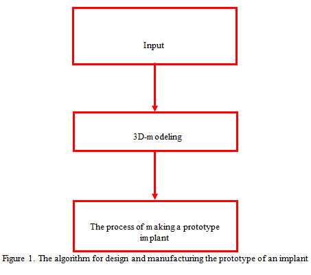 |
Figure 1: The algorithm for design and manufacturing the prototype of an implant |
Synthesis of the cellular structure on the basis of mathematical modeling.
The algorithm of the cellular structure synthesis on the basis of mathematical modeling is shown in Figure 2.
 |
Figure 2: Algorithm of 3D modeling of the cellular structure of the implant |
At the initial design stage, a CT of the jaw is obtained, where dense tissues that basically correspond to its bone sections are identified (Figure 3).
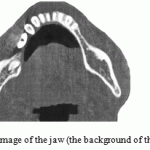 |
Figure 3: Tomographic image of the jaw (the background of the jaw is brighter, lighter) |
Basing on the obtained tomographic images, a 3D model of the jaw is generated using the 3D-doctor software suite (Figure 4).
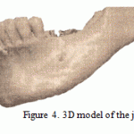 |
Figure 4: 3D model of the jaw |
Here defective sections of the jaw are identified, which are then built-up until no defects are observed, by editing the initial 3D model using the Materialise Magics software product. The problem is reduced to forming a cellular structure of the defective area model. To implement this task, the original 3D model of the missing part of the jaw (Figure 5) is used in the form of a solid three-dimensional body, which later serves as the basis for synthesis of the cellular structure. The body of the cellular structure itself is formed by transferring the missing part of the jaw using the ANSYS software (Zhidkov, 2006).
 |
Figure 5: The model of the missing part of the jaw |
In the ANSYS environment, partitioning grid is built on the model obtained (Figure 6). The ease of using ANSYS is dictated by the ability to use it with variable sizes of partitioning grid elements (Zhidkov, 2006). In building the grid, the maximum and the minimum sizes of its elements are chosen, as well as split method and cell shape (Jaecquesa et al., 2004).
 |
Figure 6: Partitioning grid |
Partitioning is performed by the method of finite elements that form the grid structure of the future implant (SÜLI, 2012). One-dimensional, two-dimensional and three-dimensional elements may be used as the splitting elements (Trudonoshin, 2000). Tetrahedron was chosen as such an element for the maximum versatility and ease of modeling.
As a result of splitting, nodes that are centers of the sides and vertices of the tetrahedron were obtained. This collection of nodes forms a “cloud” of points (Figure 7). Each node is characterized by three coordinates.
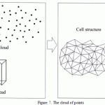 |
Figure 7: The cloud of points |
The cloud of points is the basis for creating bridges – direct, passing through 2 nodes and making the “skeleton” of the created ordered cellular structure (Shevchenko et al., 2012).
Then, from the previously obtained partitioning grid, separate pairs of adjacent points are selected that are united into a single vector that characterized the bridge. Numerical procedures for obtaining bridges are based on using affine transformations: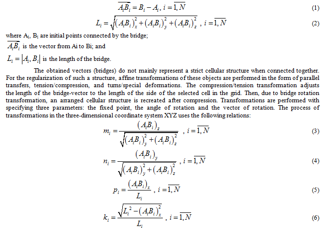
where mi, ni, pi, ki are the coefficients of the matrix of rotations that characterize the values of the cosines and sines of the rotation angles:

The elements of these matrices depend on the coordinates of the points between which the bridge is stretched. Thus, the randomly located and connected bridges, while deforming, but maintaining straightness, form an ordered cellular structure. Transformation occurs around the fixed (stationary) point in a layer-by-layer manner, preserving the structure of each cell by component Z. Consequently, a layer-by-layer cellular structure is formed, with layers rigidly parallel to each other along the Z axis. The XY plane is perpendicular to the end face of the shell of the jaw implant model. New locations of the cellular structure bridge points are shown by the relations:
![]()
where C1 is the point before affine transformations, and C2 is the point after affine transformations.
Coordinates of unit vectors are
Software implementation of the methodology for designing a cellular structure
The model of the bridge, and the resulting cellular structure and description of its models are generated using the software package developed in the process of synthesis in the C# environment using the STL method (Nakov et al., 2013). The essence of the STL method is text description of the geometry of the structure with the use of triangular facets (Dean, 2005). The algorithm and the block diagram of synthesis software implementation are shown in Figure 8.
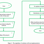 |
Figure 8: The algorithm of synthesis software implementation |
Its main parts implement the following tasks:
- invocation of the modules responsible for operation of the algorithms that contain built-in procedures and functions;
- setting global variables for storing initial and intermediate data used in the algorithm;
- The procedures for running the modeling algorithm.
Except for cases where additional modules are required, calling all other modules is standard, and is automatically written in the text of the program in C# (Nakov et al., 2013).
The edge of the grid element itself contains a set of vertex coordinates, as well as the normal. The normal sets that location of each face of the modeled body. The description text is merged into blocks with the help of keywords that make it possible to correctly interpret the numerical values of the coordinates. To outline the boundaries of the described individual body with its surface, key words “Solid” and “endsolid” are used, which are set in the beginning and in the end of model description. Thus, text block structures that are easy to handle using cycles are formed. To denote the triangular facets oriented with the normals that make up the surface, keywords “facet” and “endfacet” are used. That is, the coordinates of three points and the guiding cosines of the normals that characterize the model facet are defined. The list of points coordinates with regards to their distance from each other is set using keywords “outer loop” and “endloop”, thereby underlying the beginning and the end of the list of coordinates of the facet vertices. The procedures listed above are implemented by blocks 2 and 3 (Figure 8). The list of elements and nodes is read by the same function performed with different arguments:
private List<List<float>>Filetolist(StreamReader sr, string name).
Since in the process of calculations there are unused columns of data, they are deleted, and the text strings are converted into numeric values. The processed list of elements is divided into pairs of nodes. Moreover, nodes that duplicate the previous ones are removed. To do so, the function for processing list of elements is used:
private List<List<float>>pair<(List<List<float>>spisok).
Block 4 makes it possible to read the bridge file in accordance with the procedure that selects a file from the provided list:
private void comboBox1_SelectedIndexChanged(object sender, EventArgs e).
With the help of inter-block operators, repetition of coordinates is eliminated. Block 5 invokes affine transformations and relations (1 – 9) using the following procedure:
private void hollow(List<List<float>>spisok).
It uses coordinates of each pair of points on the basis of the aggregate of the corresponding list and data about the bridge. A number of functions and procedures in this block are complementary. For the sake of ease of using the program, there are comments that make it possible to track and clarify the results of the synthesis stage.
A fragment of the text file of the obtained model is shown in Figure 9.
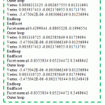 |
Figure 9: Fragment of text file of the model obtained |
Synthesis of a jaw implant with porous structure in the process of rapid prototyping
The cellular structure obtained from the results of mathematical modeling is the basis for manufacturing jaw implant. Finally, the model of the implant is obtained by connecting this cellular structure with the solid shell, the model of which is obtained in the Materialise Magics software suite, and is a thin-walled object with constant thickness open at two ends.
The technological process of manufacturing the prototype of the implant was performed using the method of stereolithography (Ferry et al., 2010) at the Envision Tec Perfactory XEDE installation using liquid photopolymer SI-500. The material is similar to ABS (acrylonitrile butadiene styrene) plastic. In the fabrication process of the prototype, along with those previously discussed, the Perfactory RP software suite is used, which makes it possible to divide the model into layers for subsequent layer-by-layer growing. The technological process consists of cyclically repeated actions, depending on the time of polymer layer exposure, supports exposure time, thickness of the supports and the place of growing on the installation platform. Its main stages are:
- Exposure of the upper layer section of the photopolymer composition.
- Smoothing the top layer with a special rod.
During this sequence, the platform is continuously lowered, therefore next layer is exposed each time with the same level of the photopolymer composition.
3 In the end of this process, the platform with the grown prototype of the implant is raised above the level of the photopolymer composition. Then supports are removed and the product is additionally polymerized in a UV chamber, if necessary.
Results
The main results of synthesizing a part of a jaw implant with cellular structure have been formed as a set of separate stages by the following sequential steps (Figure 10), from the beginning of the research to the final implementation.
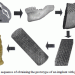 |
Figure 10: The sequence of obtaining the prototype of an implant with porous structure |
With that, the number of the results of the synthesis main stages should be outlined. Thus, in the process of modeling, a 3D cellular model (Figure 11) has been created, which is merged with the shell (Figure 12) obtained using the Materialise Magics software. This shell is a thin-walled element with constant thickness. Due to such merging, the model of jaw implant is formed.
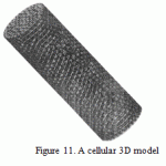 |
Figure 11: A cellular 3D model |
 |
Figure 12: The thin-wall shell |
The result of the process of implant prototype manufacturing is the sample shown in Figure 13.
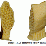 |
Figure 13: A prototype of jaw implant |
If biocompatible materials are used, the resulting structure may be used in real conditions in medical practice. The software product is created in such a way that the structure and the size of selected cells may vary, which makes it possible to perform multivariate synthesis with choosing optimal characteristics in the future. This increases quality and reliability of such cellular structures. Using the proposed method, it is possible to create cellular structures of predefined type and size, which makes it possible to compare their use and, consequently, to synthesize the most technologically advanced of them.
Discussion
Articles (Ferry et al., 2010) describe the method of rapid prototyping, stereolithography. This method describes the ways of using stereolithography in medicine. The properties of the scaffolds are provided, as well as the variants with direct manufacturing of implants in a stereolithographic machine. As an example, experiments with the structure of jaw implants in pigs were mentioned. However, mathematical description and the corresponding modeling of porous structures arranged depending on the shape of the implant have not been shown. In these articles, the authors noted that stereolithography makes it is possible to obtain many important properties of the porous material for medical purpose. The scheme of the stereolithographic process has been shown. It has been mentioned that there are biodegradable materials that are, by their mechanical properties, similar to human bone. Comparison of models with cubic, gyroid and diamond structure, as well as analysis of these structures, regardless of the shape of the implant, have been provided. (Marin et al., 2009) describe the SLS technologies. It has been noted that for connecting human tissues with biomaterials, porosity and pore size are critical. The results of experiments with cellular titanium have been presented, where the cell structure is ordered regardless of the form of the implant. With that, the basic element of the matrix is a hexagon developed in three planes. Boolean operations have been used to achieve the desired shape of the implant. It should be noted that an implant with only one type of structure is considered. Article (Puleo and Nanci, 1999) highlights factors of bone healing and connecting with the implant, and describes the process of bone growth and the need to ensure immobility of the implant. It has been noted that metallic implants are most widespread, due to their mechanical characteristics. Variations of the implant structure (porous structure, structure with guides) have been considered, and attention has been paid to its biochemical modification consisting in the use of some organic substances. From the article it follows that implants with cellular structure are more technologically advanced, since it is this structure that reduces its mobility. It has been pointed out that during the interaction of implant body metal with the organism, the tissues of the latter may be damaged due to the release of metal ions. In our case, the use of stereolithography with exposure of the photopolymer liquid makes it possible to exclude any adverse effects on human body in case of using biocompatible materials.
The analysis shows that in course of synthesizing cellular structures of jaw implant, the system approach that takes into account the relationship between modeling stages and the manufacturing process have not been sufficiently studied. The methods of obtaining final result on the basis of mathematical modeling and related software have also not been sufficiently studied.
Conclusion
According to the results shown in the article, the following conclusions can be made:
- A method for synthesizing cellular structure of jaw implants using a systematic approach has been developed.
- Mathematical models of cellular structure that meet size, shape and configuration requirements have been obtained.
- The software of the process of numerical experiment in automatic mode makes it possible to perform multivariant calculations, and evaluate cellular structure.
- On the basis of the built 3D models of cellular structures, the technological process of growing implant jaw prototype has been developed.
- The proposed method is an effective tool for guided synthesis of cellular structures used in medicine.
The use of this method is also possible in the field of creating cellular implants for replacing various bone structures. Further research will be aimed at optimizing cellular structures with regard to biocompatible materials, calculation of their strength characteristics and their practical use in medical practice.
References
- Barinov, S.M., & Komlev, V.S. (2005). Bioceramics based on calcium phosphates (pp. 204). Moscow: Nauka.
- Vasilyuk, V.P., Straube, G.I., Kocherjuk, S.A., Kosareva, P.V., & Asanovich, M.A. (2014). Experimental substantiation of using innovative technologies in manufacturing implants with cellular structure to replace bone defects of facial skeleton (preliminary results) (pp. 40-52). Family Health – The 21st Century, 2, 40-52.
- Evseev, A.V., Kamaev, S.V., Kotsyuba, E.V., Markov, M.A., Novikov, M.M., Popov, V.K., & Panchenko, V.Y. (2005). Computer biomodeling and laser stereolithography. Collection of papers of IPLIT RAS “Modern laser-information and laser technologies” (Corresponding member of RAS Panchenko, V.Y., & Professor Golubev, V.S., Eds., pp.119-130). Intercontact Science.
- Evseev, A.V., Kotsyuba, E.V., Mayorova, S.A., Brusova, L.A., Brusov, I.P., Perfiliev, S.A., & Adamyan, A.A. (2005). Computer modeling and laser stereolithography in thoracic surgery. Proceedings of the VI Scientific School of Young Specialists “Concentrated Energy Fluxes in Space Technology, Electronics, Ecology and Medicine”, November 21-22, 2005 (Ishkhanova, B.S., & Novikova, L.S., Eds., pp. 112-117). MSU NPRI.
- Zhidkov, A.V. (2006). Use of ANSYS system for solving problems in geometrical and finite element modeling. Learning and teaching aids for the “Information Systems in Mathematics and Mechanics” training program (pp. 115). Nizhny Novgorod.
- Kuznetsov, V. (2003). System rapid prototypes manufacturing and their extensions / CAD/CAM/CAE. Observer, 4(13), 2-7.
- Morozov, V.V. (2005). Research and development of technological modes of manufacturing castings from burned out models obtained by laser stereolithography. (Synopsis of thesis of Candidate of Technical Sciences (05.16.04), pp. 16). Moscow State Technical University n.a. N.E. Bauman, Moscow.
- Radkevich, A.A., Gantimurov, A.A., & Gunter, V.E. (2012). Replacing mandibular defects with implants of porous titanium nickelide. Implants with Shape Memory, 1-2, 18-27.
- Sirotenko, L.D., Mutygullina, E.V., Khanov, A.M., Samusev, I.V., & Bashkirtsev, G.V. (2010). Prediction of physico-mechanical properties of high-porous permeable cell materials ion the basis of structural modeling. Bulletin of the Perm National Research Polytechnic University. Mechanical engineering, materials science, 12, 1, 17-29.
- Stuchilov, V.A., Nikitin, A.A., & Evseev, A.V. (2002). Laser stereolithography in cranio-facial surgery. Electronics: Science, Technology, Business, 4, 44-45.
- Topolnitskiy, E.B., Dambayev, G.C., Khodorenko, V.N., Fomina, T.I., Shefer, N.A., Gunter, V.E. (2012). Tissue reaction to cellular implants made of titanium nickelide after displacing of post-resection defects of anatomical structures in the chest. Bulletin of Experimental Biology and Medicine, 153, 3, 366-370.
- Trudonoshin, V.A. (2000). Introduction into the method of finite element (pp. 20). Moscow: MSTU.
- Khodorenko, V.N., Yasenchook, Y.F., & Gunter, B.E. (2001). Biocompatible porous permeable materials. Biocompatible materials and implants with shape memory (pp. 9-24).
- Shevchenko, A.V., Dudnik, E.V., Zurenko, V.V., & Ruban, A.K. (2012). Highly porous composite material with cellular structure in the ZrO2—Y2O3—CeO2 system. Proceedings of the “Ukrainian Research Institute of refractory materials” n. a. A. S. Berezhnoy”, 112, 103-109.
- Dean Al STL – a format for rapid prototyping. Part I (2005). Output in STL format. CAD/CAM/CAE Observer, 5(23), 64-69.
- Dean Al STL – a format for rapid prototyping. Part II (2005). Real experience in outputting STL files. CAD/CAM/CAE Observer, 6(24), 65-69.
- Melchels, F.P.W., Feijen, J., & Grijpma, D.W. (2010). A review on stereolithography and its applications in biomedical engineering. Biomaterials, Vol. 31, 24, 6121-6130.
- Melchels, F.P.W., Bertoldi, K., Gabbrielli, R., Velders, A.H., Feijen, J., & Grijpma, D.W. (2010). Mathematically defined tissue engineering scaffold architectures prepared by stereolithography. Biomaterials, Vol. 31, 27, 6909-6916.
- Jaecquesa, S.V.N., Van Oosterwycka, H., Murarua, L., Van Cleynenbreugela, T., De Smetb, E., Weversc, M., Naertb, I., & Vander Slotena, J. (2004). Individualised, micro CT-based finite element modelling as a tool for biomechanical analysis related to tissue engineering of bone. Biomaterials, 25, 9, 1683-1696.
- Marin, E., Paussa, L., Fusi, S., & Pressacco, M. (2010). Characterization of cellular solids in Ti6Al4V for orthopaedic implant application: Trabecular titanium. Journal of the Mechanical Behavior of Biomedical Materials.
- Milovanović, Je., & Trajanović, M. (2007). Medical application of rapid prototyping. Mechanical Engineering, 5, 1, 79–85.
- Murr, L.E., Gaytan, S.M., Medina, F., Lopez, H., Martinez, E., Machado, B.I., Hernandez, D.H., Martinez, L., Lopez, M.I., Wicker, R.B., Bracke, J. (2010). Next-generation biomedical implants using additive manufacturing of complex, cellular and functional mesh arrays. Philosophical Transactions A., 2010, 1999-2032.
- Svetlin, N., Veselin, K. et al. (2013). Fundamentals of computer programming with C# (pp. 1122). Faber Publishing, Bulgaria.
- Puleo, D.A., & Nanci, A. (1999). Understanding and controlling the bone-implant interface. Biomaterials, 20, 23-24, 2311-2321.
- SÜLI Endre Lecture notes on finite element methods for partial differential equations (2012, pp. 106). Mathematical Institute University of Oxford.
(Visited 277 times, 1 visits today)

This work is licensed under a Creative Commons Attribution 4.0 International License.


