

Manuscript accepted on : 25 March 2015
Published online on: --
Grigorenko Andrey, Panfilov Leonid, Smirnov Alexander, Starikovskiy Andrey, Rubin Dmitry, Shulga Ekaterina, Sychev Nikolay and Nikolaeva Alexandra
National Research Nuclear University MEPhI (Moscow Engineering Physics Institute), Kashirskoe Highway 31, 115409, Moscow, Russian Federation.
DOI : http://dx.doi.org/10.13005/bbra/2190

ABSTRACT: For the latest time oncological diseases started attracting more attention from the increasing number of the diseased. Radionuclide diagnostics performs gamma-probes destined for radiopharmaceutical local areas identification in human body that permits to identify the diseased cells assembly more accurate. This article covers modern tendencies in the field of such devices development and gives suggestions for new gamma-probe creation, which would excel the existing analogs at functional opportunities and price.
KEYWORDS: Gamma-probe; oncological diseases; sentinel gland; cancer
Download this article as:| Copy the following to cite this article: Andrey G, Leonid P, Alexander S, Andrey S, Dmitry R, Ekaterina S, Nikolay S, Alexandra N. The Existing Gamma-Probes Review for Searching Functional Increase and Complex Improvement Possibilities. Biosci Biotech Res Asia 2015;12(spl.edn.2) |
| Copy the following to cite this URL: Andrey G, Leonid P, Alexander S, Andrey S, Dmitry R, Ekaterina S, Nikolay S, Alexandra N. The Existing Gamma-Probes Review for Searching Functional Increase and Complex Improvement Possibilities. Biosci Biotech Res Asia 2015;12(spl.edn.2). Available from:https://www.biotech-asia.org/?p=12873 |
Introduction
Oncological diseases became real “pest” degradation of the 21st century. Among the reasons of the diseased number increase, there are poor ecology, social habits, wrong life style of modern people, etc. According to average statistic data, oncological diseases take at least 300 thousands of lives yearly in Russia (and this number grows every year). By the World Health Organization (WHO) forecast since 1999 to 2020 oncological diseases will increase two times (new diseased from 10 to 20 mln, fatalities from 6 to 12 mln) [1-2].
One of the reasons for heavy mortality of oncological diseases is untimely diagnostics. It is connected with people’s refuse of regular health survey and lack of special devices for the diseases diagnostics. [3]
Among all the varieties of nuclear medicine problems, the leading place goes to oncological diseases diagnostics by radionuclide methods. A special type of such devices is shown by gamma-probes, destined for local identification of areas with radiopharmaceutical. [4]
Gamma-probes are used in recent medicine for punctual disease sites identification during carcinectomy. The patient is inserted by radiopharmaceutical, accumulating in cancer disease sites, and, according to the element used, and radiating gamma quantum. Then the patient is exposed to complex magnetic resonance tomography (MRT) examination-scanning on the results of which cancer disease sites and organs are shown. During the surgery for accurate identification and proving the tumor existing gamma-probe is used – a small device permitting to fix and targetedly gauge gamma quantum radiation at direct contact with the source of radiation, that lets surgeons to find distinctly the tumor borders during the operation. The device functioning is based on the use of scintillator, radiating light during absorbing of ionization radiation, collimator, silicon photomultiplier matrix and the scheme that convert the signals caught into numerical rating of radiation strength. [5-7]
Nowadays the West market presents intraoperative probes for gamma-radiation registration. Now the market of such devices is in progress and represented generally by American companies such as RMD Instruments, Devicor Medical Products Inc., Care Wise Medical, IntraMedical Imaging – all together they cover more than 95% of the world market. In addition, a French company Eurorad SA and German company World of Medicine can be matched. There are some developments of different companies from Germany, France, Spain, Italy, Czech, Argentina, Australia and Iran. But there are no any Russian developments in the world market. [8-9]
Most of the devices used in the world were developed at the height of the research popularity in this field beginning with the first gamma-probe during the surgery on a sentinel gland in 1993 [10] and further until 2003 approximately. From this time, different gamma-probes were invented, using different principles, materials and technologies at the key components: detector (silicon or scintillator), collimator (ordinary or electronic), and electronics (avalanche photodiode or silicon photomultiplier).
Wireless gamma-probe used permits to increase essentially both speed and examination process convenience. Probes of such a type comparing with the wire ones are not in mass distribution yet and are represented in the market by such products as Neoprobe (Devicor Medical Products, Inc., USA), Node Seeker (IntraMedical Imaging, USA), Gamma Finder (World of Medicine, Germany).
In some technologies described, an optimal alternative in its efficiency was found that is used until now. However, new research is being performed, but the speed of component efficiency increase does not let to infuse new technologies as soon as possible, but wait for improvement that is more serious. Though, progress in the field of production, functionality and ways of microcontroller use, permits to achieve serious gamma-probes efficiency increase, using the latest technologies, at that making it accessible for final buyer.
Let us go into particulars of the offered gamma-probe.
Materials and Methods
The basis of every commercially accessible probe is scintillating or solid detector. However, the developing gamma-probe has some peculiarities that greatly increases the technics level thanks to the use of silicon photomultiplier (SiPM) technologies creation, developed in the National Research Nuclear University MEPhI (NRNU MEPhI).
Gamma-probe is being created together with the Nuclear Medicine Department of the NRNU MEPhI that has been dealing oncological diseases problems for a long time (it is also being supported by many oncological hospitals, rehabilitation centers and other specified institutions). Highly qualified specialized staff working in the field of diagnostics and treatment of oncological diseases also support the developing group.
The described gamma-probe is offered to use for intraoperative diagnostics of sentinel glands and noninvasive diagnostics of interfacial types of cancer.
Let us go to the particulars of the main components of the developing gamma-probe.
Collimator permits to decrease the angle of gamma-quantum fixing. It is presented by a metallic construction; usually it is a cylinder with a hole. The main improvement field is the construction itself, its form and the form of the hole. There are few workings in this field and there is no a certain way to achieve fast quality progress.
Scintillator is the main part of the detector. The material it consists of radiates light at gamma-quantum catching, the spots intensity of which is directly proportional to gamma-radiation. The scientists are searching for the material that would produce more photons but at the same time would not be radioactive itself. In the existing gamma-probes such scintillators as LYSO, CdZnTe, CdTe, CsI(TI) are used, that have the described qualities, though new materials search takes much time and do not give fast efficiency increase.
Silicon photomultiplier is matrix of avalanche photodiodes. It permits detecting and enforcing light flashes of poor intensity (on the level of single photons) and time length of unit-hundred order nanoseconds. The ability to detect the most poor flashes influences on the accuracy of the cancer tumor borders identification, and this is a problem of the specialists’ constant work. The photomultipliers development goes to the side of diodes number increase and all the matrix size decrease. It fully depends on the technical progress size and the progress in this field is connected with common electronics miniaturization. In such a way, there are new more sensitive photomultipliers, comparing with those ones used in widely spread gamma-probes, which would let to improve common efficiency.
Microcontroller is a chip for the received signals processing and interpretation. The elements base used in nowadays gamma-probes generation is not only old but also needs heavy spending for production as the supporting period of microcontrollers using in production, is expired already or expires in the nearest future. In most cases since the latest workings three generations of microcontrollers has been changed. The use of newest research in this field will let not only to add functionality and to increase the convenience of usage, but also to decrease the spending on the production.
Form – in most cases gamma-probe is the main station with the indicator of radiation intensity and probe, connected with it by a wire. This probe registers gamma-radiation. The lack of radiation intensity indicator makes the surgeon often turn their attention from the probe area to the main station screen. Such a practice not only makes the process of the tumor search slower and difficult, but also can lead to the loss of the tumor that has been found already, in case of divert external factors existing, and repeating the search.
Figure 1 schematically shows the detection part of the gamma-probe.
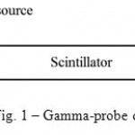 |
Figure 1: Gamma-probe detector.
|
The experimental assembly comprises the following elements:
scintillation detector:
scintillation crystals LYSO (lutetium-yttrium oxyorthosilicate) or LaBr3:Ce;
SiPM MPPC Hamamatsu with sensitive area dimensions of 3mm × 3mm, SiPM developed in NRNU MEPhI, SiPM MAPD-3 (Zecotek) or PMT Hamamatsu H6780-03 series [11,12];
Philips amplifier (Kdef = 20);
line splitter;
delay lines;
integral discriminator;
attenuator (Kdecrease = 0.1 – 1);
former (forms the signal of particular length and form);
QDC (charge-to-digital converter).
All signals were observed on the LeCroy 9350A oscilloscope [13].
Later, SiPM developed in the MEPhI will be referred to simply as SiPM, SiPM produced by Hamamatsu as SiPM Hamamatsu, SiPM MAPD-3 (Zecotek) as SiPM Zecotek, and PMT Hamamatsu as PMT.
The standard laboratory sources were used as gamma radiation sources. Photodiode is powered by an external constant-voltage source (U = 0 ¸ 120 V). The power supply circuit of the photodiode includes a filter capacitor C2 = 2.2 nF. As a light pulse drops on the surface of the photodiode a current pulse occurs, which is transmitted through the clarifier C1 = 2.2 nF to an amplifier. After amplification, the signal is sent to the line splitter, from the output of which it comes to the input of the integral discriminator and the analog input of the QDC. Integral discriminator generates a strobe signal of standard shape and duration (gate), which is fed to the input Gate QDC. QDC digitizes the signal area (charge) in the presence of a strobe pulse, i.e. integration of the signal occurs only during the duration of the strobe. The basic building blocks of the measuring system are made in comparison with NIM (nuclear instrumentation module) standard and the QDC unit with CAMAC (computer automated measurement and control) standard. Computer reads data from the QDC via controller.
It must be remembered that the LYSO crystal has the property of afterglow. Therefore, after using the current scintillator for measurements and before performing the next series, it must withstand a certain time in the dark. During the measurements, however, radiation background of the crystal was measured immediately after getting the spectra of the sources leading to a change in its spectrum.
Conclusion and Discussion
The proposed gamma-probe will not yield to foreign analogues by its functional opportunities. Among its advantages, there are sampling resolution and selectivity. At the use in a surgery room, the device portability plays a special role so as the opportunity to operate it with the help of a small personal computer.
This working will permit greatly increase a patient’s examination efficiency concerning metastasis existence in the organism, forecasting by it heavy forms of oncological diseases.
In the nearest perspectives, the authors plan to finish the stage of Research and Advanced Development for the complex, prepare commercial design for the device, also to organize small-lot manufacturing and develop working-constructing documents.
References
- Oncological diseases statistics. World Medicine Corporation, 2015. URL: http://womco.ru/onkologiya.
- Oncological diseases statistics. URL: http://4lifemd.ru/rezultat_primeneniya/onkologiya/statistika-onkologicheskih-zabolevaniy.
- Davydov G.A. Radionuclide diagnostics in oncology. FGCU “GTsIPK”. URL: http://www.rosoncoweb.ru/library/radiology/005.pdf.
- Gamma Probes. IntraMedical Imaging, 2014. URL: http://www.gammaprobe.com/products/gamma-probes.
- Mihaljevic, A.L.; Rieger, A; Belloni, B; Hein, R; Okur, A; Scheidhauer, K; Schuster, T; Friess, H; Martignoni, M.E. Transferring innovative freehand SPECT to the operating room: First experiences with sentinel lymph node biopsy in malignant melanoma. European Journal of Surgical Oncology, Volume: 40, Issue: 1, Jan. 2014. Pages: 42-48.
- Bellotti, C; Castagnola, G; Tierno, S.M.; Centanini, F; Sparagna, A; Vetrone, I; Mezzetti, G. Radioguided surgery with combined use of gamma probe and hand-held gamma camera for treatment of papillary thyroid cancer locoregional recurrences: a preliminary study. European Review for Medical and Pharmacological Sciences, Volume: 17, Issue: 24, Dec. 2013. Pages: 3362-3366.
- Ikram, M; Akhtar, S; Maseeh-uz-Zaman; Junaid, M; Dhari, T; Ahmad, Z; Hussain, R. Sentinel node localisation using pre-operative lymphoscintigraphy and intraoperative gamma probe in early oral cavity cancer. Journal of the Pakistan Medical Association, Volume: 63, Issue: 8. Aug. 2013. Pages: 976-979.
- Comparison of sentinel gamma probes for 99mTc breast cancer surgery based on NEMA NU3-2004 standard
- NEMA NU3 Performance measurements and quality control guidelines for non-imaging intraoperative gamma probes. 2004.
- Krag DN, Weaver DL, Alex JC, Fairbank JT. Surgical resection and radiolocalization of the sentinel node in breast cancer using gamma probe. Surg Oncol 1993; 2:335–340.
- Mihaljevic, A.L.; Rieger, A; Belloni, B; Hein, R; Okur, A; Scheidhauer, K; Schuster, T; Friess, H; Martignoni, M.E. Transferring innovative freehand SPECT to the operating room: First experiences with sentinel lymph node biopsy in malignant melanoma. European Journal of Surgical Oncology, Volume: 40, Issue: 1, Jan. 2014. Pages: 42-48.
- Ikram, M; Akhtar, S; Maseeh-uz-Zaman; Junaid, M; Dhari, T; Ahmad, Z; Hussain, R. Sentinel node localisation using pre-operative lymphoscintigraphy and intraoperative gamma probe in early oral cavity cancer. Journal of the Pakistan Medical Association, Volume: 63, Issue: 8. Aug. 2013. Pages: 976-979.
- Lecroy 9350A Digital Storage Oscilloscope 500 MHz. Specifications, 2004. URL: http://www.atecorp.com/products/lecroy/9350a.aspx.

This work is licensed under a Creative Commons Attribution 4.0 International License.


